
Photochemical cross-linking of
Escherichia coli
Fpg protein to DNA
duplexes containing phenyl(trifluoromethyl)diazirine groups
Maria Taranenko
1
, Anna Rykhlevskaya
1
, Manana Mtchedlidze
1
, Jacques Laval
2
and Svetlana Kuznetsova
1
1
Laboratory of Nucleic Acids Chemistry, Department of Chemistry, Moscow State University, Moscow, Russia;
2
Groupe ‘Reparation de l’ADN’, UMR 8532 CNRS, Institut Gustave Roussy, Villejuif Cedex, France
Formamidopyrimidine-DNA glycosylase (Fpg protein) of
Escherichia coli is a DNA repair enzyme that excises oxi-
dized purine bases, most notably the mutagenic 7-hydro-
8-oxoguanine, from damaged DNA. In order to identify
specific contacts between nucleobases of DNA and amino
acids from the E. coli Fpg protein, photochemical cross-
linking was employed using new reactive DNA duplexes
containing 5-[4-[3-(trifluoromethyl)-3H-diazirin-3-yl]phe-
nyl]-2¢-deoxyuridine dU* residues near the 7-hydro-8-
oxoguanosine (oxoG) lesion. The Fpg protein was found to
bind specifically and tightly to the modified DNA duplexes
and to incise them. The nicking efficiency of the DNA duplex
containing a dU* residue 5¢to the oxoG was higher as
compared to oxidized native DNA. The conditions for the
photochemical cross-linking of the reactive DNA duplexes
and the Fpg protein have been optimized to yield as high as
10% of the cross-linked product. Our results suggest that the
Fpg protein forms contacts with two nucleosides, one 5¢
adjacent to oxoG and the other 5¢adjacent to the cytidine
residue pairing with oxoG in the other strand. The approa-
ches developed may be applicable to pro- and eukaryotic
homologues of the E. coli Fpg protein as well as to other
repair enzymes.
Keywords: formamidopyrimidine-DNA glycosylase; modi-
fied DNA duplexes; 7-hydro-8-oxoguanosine; 5-[4-[3-(tri-
fluoromethyl)-3H-diazirin-3-yl]phenyl]-2¢-deoxyuridine;
photochemical cross-linking.
Derivatives of nucleic acids containing photolabile car-
bene-generating aryl(trifluoromethyl)diazirine groups are
conveniently used to identify specific nucleic acidÆnucleic
acid and nucleic acidÆprotein interactions [1–5]. These
derivatives have a number of essential merits. First, they
produce highly reactive carbene, which breaks even
aliphatic C–H bonds. Second, the lifetime of carbene is
on a nanosecond timescale. Third, photolysis proceeds at a
relatively high light wavelength (350–360 nm) that does
not cause damage to biological molecules. Finally, these
derivatives may be handled under moderate laboratory
illumination. These reagents have been successfully
employed to investigate RNAÆRNA and RNAÆprotein
interactions in ribosomes [1], and to ascertain
2specific
contacts between DNA and some DNA-recognizing
proteins, such as the restriction-modification enzymes
EcoRII and MvaI [2], recombinant rat DNA polymerase
b[3], the large subunit of human immunodeficiency virus
reverse transcriptase [4], yeast RNA polymerase and
others [5].
Escherichia coli formamidopyrimidine-DNA glycosylase
(Fpg protein) is a DNA repair enzyme that catalyzes
the removal of oxidized purine bases from damaged
DNA and cleaves the DNA strand [6]. 7-Hydro-8-
oxoguanine is the major mutagenic base produced in
DNA by reactive oxygen species that are generated by
cellular metabolism, cell injury and exposure to physical
and chemical oxygen radical-forming agents [7]. It is a
miscoding lesion because it pairs preferentially with
adenine rather than cytosine and induces GC fiTA
transversions in vivo and in vitro [8]. The physiological
function of the Fpg protein is to prevent the mutagenic
action
3of oxoG residues in DNA and to maintain genetic
integrity. Three-dimensional structures of the complexes
formed by Lactococcus lactis,Bacillus stearothermophilus
and E. coli Fpg proteins with abasic DNA duplexes have
recently been obtained using X-ray crystallography [9–11].
However, despite this success, further biochemical data are
still needed to understand the dynamics of the interaction
of the Fpg protein active-site residues with various
substrates. Valuable information can be obtained by using
a variety of cross-linking techniques applicable to nucleic
acidÆprotein systems. Previously, we used chemical cross-
linking to identify specific contacts between E. coli Fpg
protein amino acid residues and DNA phosphate groups
[12]. Here, we use photochemical cross-linking to ascertain
specific contacts between the Fpg protein and the
nucleosides adjacent to oxoG. To achieve this, modified
Correspondence to M. Taranenko, Laboratory of Nucleic Acids
Chemistry, Department of Chemistry, Moscow State University,
Moscow 119899, Russia.
Fax: + 7095 939 31 81, Tel.: + 7095 939 31 53,
E-mail: svetlana@belozersky.msu.ru
Abbreviations:EDC,N-(3-dimethylaminopropyl)-N¢-ethylcarbodi-
imide; Fpg protein, formamidopyrimidine-DNA glycosylase; K
D
app,
apparent dissociation constant for the binding of the Fpg protein to
the modified duplexes; oxoG, 7-hydro-8-oxoguanosine; TFMDPh,
4-[3-(trifluoromethyl)-3H-diazirin-3-yl]phenyl; dU*, 5-[4-[3-(trifluoro-
methyl)-3H-diazirin-3-yl]phenyl]-2¢-deoxyuridine.
(Received 10 December 2002, revised 11 April 2003,
accepted 12 May 2003)
Eur. J. Biochem. 270, 2945–2949 (2003) FEBS 2003 doi:10.1046/j.1432-1033.2003.03662.x
DNA duplexes containing 5-[4-[3-(trifluoromethyl)-3H-
diazirin-3-yl]phenyl]-2¢-deoxyuridine (dU*) residues 5¢
to the oxoG lesion or 5¢to the cytidine residue of the
other strand, forming a base pair with oxoG, were
prepared. To our knowledge, this is the first time that
double-stranded oligonucleotides containing reactive 4-[3-
(trifluoromethyl)-3H-diazirin-3-yl]phenyl (TFMDPh)
groups have been used to study interactions with DNA
repair enzymes.
Materials and methods
Oligonucleotides
Oligonucleotides (1)–(6) and DNA duplexes I–IV, used in
this study, are depicted in Fig. 1. Oligonucleotides (1)–(4),
forming DNA duplexes II–IV, were synthesized using a
standard phosphoramidite procedure in an Applied Bio-
systems 380 B DNA synthesizer, as described by Matt-
eucci et al. [13]. Modified oligonucleotides (2) and (6),
containing oxoG, were prepared using commercial
3¢-phosphoramidite of modified 2¢-deoxyguanosine. Syn-
thesis of modified oligonucleotides (3) and (5), containing
dU*, was performed as described by Topin et al.[2].
Oligonucleotide (5), with a 3¢-terminal phosphate group,
was obtained according to Purmal et al.[14].The
oligonucleotides were 5¢end-labelled with T4 polynucleo-
tide kinase and [c
4-
32
P]dATP following the standard
procedure [15]. The concentrations of oligonucleotides
were determined spectrophotometrically.
Chemical ligation of oligonucleotides
An equimolar mixture of oligonucleotides (1), (5) and (6),
forming nicked DNA duplex I (the total nucleotide
concentration was 10 m
M
), was incubated at 75 Cfor
2 min in 0.05
M
Mes/NaOH buffer, pH 6.0, containing
0.02
M
MgCl
2
, and slowly cooled for 2 h. Then, N-(3-
dimethylaminopropyl)-N¢-ethylcarbodiimide (EDC) was
added to a concentration of 0.2
M
. The reaction was carried
out at 20 C for 72 h in the dark. The ligation product was
isolated by PAGE (20% denaturing gel), followed by
elution with 2
M
LiClO
4
, precipitation with five volumes of
acetone and reprecipitation from 2
M
LiClO
4
byafurther
two precipitations with 10 volumes of acetone.
Gel retardation assay
Binding reactions were performed at 0 C for 5 min. The
incubation mixture (20 l
L
5final volume) contained 25 m
M
Hepes/KOH, pH 7.6, 100 m
M
KCl, 5 m
M
b-mercapto-
ethanol, 2 m
M
Na
2
EDTA, 0.1% (w/v) BSA, 6% (v/v)
glycerol, 50–70 p
M
[
32
P]-labelled DNA duplex expressed
as the oxoG concentration and 0.5–10 n
M
Fpg protein.
Samples were subjected to nondenaturing PAGE (10% gel)
and were visualized by autoradiography. The radioactivity
of gel slices was determined by Cerenkov counting. The
yield of the complex was calculated as the ratio of shifted
band radioactivity to the total radioactivity of the loaded
sample. The apparent dissociation constants were deter-
mined as described by Boiteux et al. [16].
Assays for enzymatic activity
The standard assay (12 lL
6final volume) contained 25 m
M
Hepes/KOH, pH 7.6, 100 m
M
KCl, 5 m
M
b-mercaptoeth-
anol, 2 m
M
Na
2
EDTA, 0.1% (w/v) BSA, 6% (v/v) glycerol,
0.7 n
M
[
32
P]-labelled oxoG-containing duplexes, expressed
as the oxoG residues and 5 n
M
enzyme. The incubation was
performed at 37 C. The reaction was stopped by the
addition of 3 lL
7,8 of formamide dye to 2 lL
7,8 of solution. The
mixture was heated at 90 C for 3 min and loaded onto a
denaturing 20% polyacrylamide gel containing 7
M
urea.
Photochemical cross-linking experiments
The Fpg protein (6 n
M
)and[
32
P]-labelled DNA duplexes I
or II (concentration of 5–10 n
M
per duplex) were incubated
in 20 lL
9of the binding buffer at 0 C for 5 min. To analyse
the photochemical cross-linking reaction, the samples were
placed in microwell plates (Fisher Life Science) and
irradiated with ultraviolet (UV) light (366 nm wavelength)
for 30 min on ice using a high-intensity UV lamp (model
UVGL-58). The reaction progress was followed by 0.1%
SDS/12% PAGE [17] after heating the samples in 0.1%
SDS/2-mercaptoethanol solution at 95 C. The gels were
analyzed by autoradiography and silver staining. Equal
mobilities of the radioactive and the protein-containing
bands indicated covalent attachment of DNA to the
enzyme. The yield of the photochemical cross-linking
reaction was calculated as the ratio of the covalent
conjugate radioactivity to the total radioactivity of the
conjugate and unbound DNA.
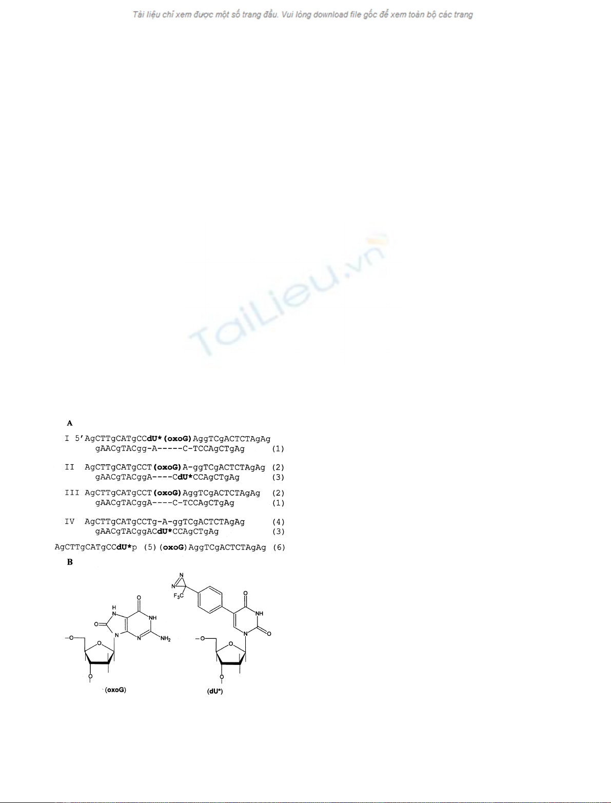
Fig. 1. Structures of (A) oligonucleotides and modified DNA duplexes
and (B) modified nucleosides used in this study. Figures in Roman
indicate the numbers of corresponding DNA duplexes; figures in
Arabic indicate the numbers of corresponding oligonucleotides.
2946 M. Taranenko et al. (Eur. J. Biochem. 270)FEBS 2003
Results and discussion
Design of modified DNA duplexes
E. coli Fpg protein recognizes a hexanucleotide sequence
with oxoG in the middle in the lesion-bearing DNA strand
and specifically binds to it and the oxoG-pairing residue of
the other strand [18]. This residue is thought to be everted
from the double helix during catalysis [19]. We propose that
neighbouring nucleosides are also involved in the formation
of the enzyme–substrate complex. In order to identify
specific contacts between the Fpg protein and the nucleo-
sides located near the oxoG lesion in DNA, modified DNA
duplexes containing dU* residues near oxoG were prepared
(Fig. 1). The dU* residue, bearing a photolabile TFMDPh
group, was introduced into the oxoG-containing strand of a
29/22-mer DNA duplex 5¢to oxoG (duplex I) or 5¢to the
oxoG-pairing cytidine residue of the other strand (duplex
II). DNA duplex III did not contain any dU* residue and
was used to estimate the effect of the TFMDPh group on
DNA duplex binding to the Fpg protein. DNA duplex IV
was similar to duplex II, but contained a guanosine residue
instead of oxoG. This duplex was used to check whether the
binding of DNA duplexes I and II to the Fpg protein was
specific.
A 29-mer oligonucleotide used to prepare DNA duplex I,
and containing both the oxoG and the dU* residues, was
obtained by a template-induced chemical ligation of oligo-
nucleotide (5), carrying a 3¢-end phosphate group, to
oligonucleotide (6), bearing a 5¢-end OH group, as described
in the Materials and methods. The ligation efficiency was as
high as 50%.
DNA duplexes I–IV were formed after annealing of the
corresponding 29-mer oligonucleotides with an equimolar
amount of a 22-mer complementary oligonucleotide.
Binding of the modified DNA duplexes to the Fpg protein
DNA duplexes I–III were tested for binding to the Fpg
protein in order to determine whether it can specifically
recognize dU*-bearing modified DNA duplexes. The
binding was detected by gel-retardation shift assay. We
found that the Fpg protein recognizes and specifically
binds all the tested duplexes with high efficiency. Figure 2
illustrates a single retardation band, which indicates
complex formation between DNA duplex II and the Fpg
protein. The intensity of the retardation band increased
with increasing Fpg protein concentration. The binding
reaction was performed at a low temperature (0 C)
because no significant cleavage was observed in these
conditions.
The apparent dissociation constant, K
D
app, for the
binding of the Fpg protein to the modified duplexes, was
estimated from the gel-retardation data, as described by
Boiteux et al.[16].TheK
D
app values obtained were
1.0 ± 0.2, 1.2 ± 0.3 and 2.0 ± 0.3-n
M
for DNA duplexes
I, II and III, respectively. Thus, the binding efficiency of
the reactive DNA duplexes I and II was similar to the
binding efficiency of DNA duplex III, which has the same
sequence but contains no photolabile TFMDPh group.
The results obtained indicate that introduction of the
TFMDPh group in close proximity to the oxoG residue
has no effect on the recognition and binding of DNA
duplexes by the Fpg protein.
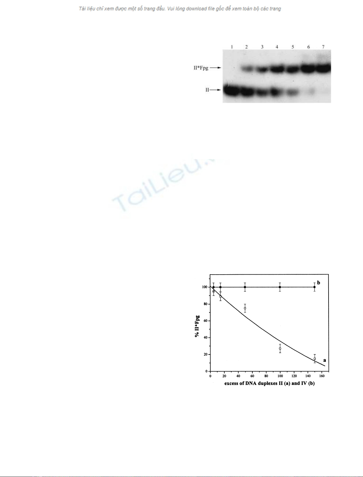
Specificity of Fpg protein binding
The interaction between the Fpg protein and modified
DNA duplexes I and II was shown to be specific by two
independent criteria. First, a 150-fold excess of unlabelled
DNA duplex II almost completely suppressed the binding
of the labelled DNA duplex II (Fig. 3). By contrast, duplex
IV, formed by oligonucleotides (3) and (4) having identical
nucleotide sequences but containing no oxoG, did not
Fig. 2. Binding of DNA duplex II to the formamidopyrimidine-DNA
glycosylase (Fpg protein). Autoradiogram from a gel retardation assay
using 50 p
M
of [
32
P]-labelled DNA duplex II containing a 5-[4-[3-(tri-
fluoromethyl)-3H-diazirin-3-yl]phenyl]-2¢-deoxyuridine (dU*) residue
in the absence (lane 1) or presence of 0.5, 1.0, 2.0, 4.0, 6.0, or 8.0 n
M
of
the Fpg protein (lanes 2–7, respectively). The structure of duplex II is
depicted in Fig. 1; for experimental conditions see the Materials and
methods.
Fig. 3. Suppression of [
32
P]-labelled DNA duplex II binding to the
formamidopyrimidine-DNA glycosylase (Fpg protein) by an excess of
unlabelled DNA duplexes II (a) and IV (b). The binding assay was
carried out with 8.0 n
M
Fpg protein and 50 p
M
[
32
P]-labelled DNA
duplex II containing a 5-[4-[3-(trifluoromethyl)-3H-diazirin-3-yl]phe-
nyl]-2¢-deoxyuridine (dU*) residue in the presence of 5-, 15-, 50-, 100-
and 150-fold excess of unlabelled DNA duplexes II and IV. The
experiment was repeated three times and gave reproducible results.
FEBS 2003 Cross-linking E. coli Fpg protein to DNA duplexes
1(Eur. J. Biochem. 270) 2947


%20--%3e%3cdefs%3e%3cstyle%3e%20.st0%20{%20fill:%20%23fff;%20}%20.st1%20{%20fill:%20%237800fa;%20}%20%3c/style%3e%3c/defs%3e%3cpath%20class='st1'%20d='M117.78,12.18H43.11c2.9,3.47,4.65,7.94,4.65,12.82,0,5.6-2.3,10.66-6.01,14.29h76.02l7.22-13.56-7.22-13.56Z'/%3e%3cg%3e%3cpath%20class='st0'%20d='M53.58,26.17h-.59v-1.46h.59v-4.96h2.83c1.78,0,2.67.94,2.67,2.82v5.76c0,1.87-.89,2.81-2.67,2.81h-2.83v-4.96ZM55.36,21.37v3.34h1.1v1.46h-1.1v3.34h1.01c.61,0,.91-.37.91-1.1v-5.93c0-.74-.3-1.1-.91-1.1h-1.01Z'/%3e%3cpath%20class='st0'%20d='M65.99,31.14h-1.8l-.31-2.07h-2.19l-.31,2.07h-1.64l1.82-11.39h2.62l1.82,11.39ZM65.28,18.04c-.25.46-.51.77-.75.94-.21.15-.47.22-.79.22-.26,0-.57-.07-.92-.22l-.38-.15c-.14-.05-.26-.07-.37-.07-.3,0-.53.18-.71.54l-.91-.68c.25-.46.51-.77.75-.94.21-.14.48-.21.79-.21.26,0,.57.07.92.21l.38.15c.14.05.26.07.37.07.3,0,.53-.18.71-.54l.91.68ZM61.91,27.52h1.73l-.87-5.76-.87,5.76Z'/%3e%3cpath%20class='st0'%20d='M74.53,26.89v1.52c0,1.91-.89,2.86-2.67,2.86s-2.67-.95-2.67-2.86v-5.93c0-1.91.89-2.86,2.67-2.86s2.67.95,2.67,2.86v1.11h-1.69v-1.22c0-.75-.31-1.12-.93-1.12s-.93.37-.93,1.12v6.15c0,.74.31,1.11.93,1.11s.93-.37.93-1.11v-1.63h1.69Z'/%3e%3cpath%20class='st0'%20d='M81.4,31.14h-1.8l-.31-2.07h-2.19l-.31,2.07h-1.64l1.82-11.39h2.62l1.82,11.39ZM75.9,19.2l1.52-1.91h1.71l1.51,1.91h-1.61l-.76-.95-.75.95h-1.61ZM77.32,27.52h1.73l-.87-5.76-.87,5.76ZM83.1,15.99l-1.76,1.91h-1.26l1.17-1.91h1.86Z'/%3e%3cpath%20class='st0'%20d='M84.86,19.75c1.78,0,2.67.94,2.67,2.82v1.48c0,1.87-.89,2.81-2.67,2.81h-.85v4.28h-1.79v-11.39h2.64ZM84.01,21.37v3.86h.85c.58,0,.87-.36.87-1.08v-1.71c0-.71-.29-1.07-.87-1.07h-.85Z'/%3e%3cpath%20class='st0'%20d='M93.51,19.75c1.78,0,2.67.94,2.67,2.82v1.48c0,1.87-.89,2.81-2.67,2.81h-.85v4.28h-1.79v-11.39h2.64ZM92.66,21.37v3.86h.85c.58,0,.87-.36.87-1.08v-1.71c0-.71-.29-1.07-.87-1.07h-.85Z'/%3e%3cpath%20class='st0'%20d='M98.8,31.14h-1.79v-11.39h1.79v4.88h2.03v-4.88h1.83v11.39h-1.83v-4.88h-2.03v4.88Z'/%3e%3cpath%20class='st0'%20d='M105.36,24.55h2.46v1.62h-2.46v3.34h3.09v1.63h-4.88v-11.39h4.88v1.63h-3.09v3.18ZM108.17,17.29l-1.76,1.91h-1.26l1.17-1.91h1.86Z'/%3e%3cpath%20class='st0'%20d='M112.2,19.75c1.78,0,2.67.94,2.67,2.82v1.48c0,1.87-.89,2.81-2.67,2.81h-.85v4.28h-1.79v-11.39h2.64ZM111.35,21.37v3.86h.85c.58,0,.87-.36.87-1.08v-1.71c0-.71-.29-1.07-.87-1.07h-.85Z'/%3e%3c/g%3e%3ccircle%20class='st1'%20cx='25'%20cy='25'%20r='20'/%3e%3cpath%20class='st0'%20d='M32.78,19.27c2.92,0,4.43,2.55,5.28,5.33l.71,2.17c.14.38-.33.75-.71.75h-5.61c.19-.33.24-.71.09-1.08l-.75-2.45c-.43-1.32-.99-2.64-1.79-3.77.75-.57,1.65-.94,2.78-.94h0ZM25,18.38c3.25,0,4.9,2.78,5.89,5.89l.76,2.45c.14.42-.33.8-.8.8h-11.69c-.42,0-.94-.38-.8-.8l.75-2.45c.99-3.11,2.64-5.89,5.89-5.89h0ZM25,11.35c1.74,0,3.11,1.37,3.11,3.11s-1.37,3.11-3.11,3.11-3.11-1.41-3.11-3.11,1.41-3.11,3.11-3.11h0ZM17.27,19.27c1.08,0,1.98.38,2.73.94-.8,1.13-1.37,2.45-1.74,3.77l-.8,2.45c-.14.38-.05.75.09,1.08h-5.56c-.42,0-.9-.38-.75-.75l.71-2.17c.9-2.78,2.41-5.33,5.33-5.33h0ZM17.27,12.91c1.51,0,2.78,1.27,2.78,2.83s-1.27,2.83-2.78,2.83-2.83-1.27-2.83-2.83,1.27-2.83,2.83-2.83h0ZM32.78,12.91c1.56,0,2.78,1.27,2.78,2.83s-1.23,2.83-2.78,2.83-2.83-1.27-2.83-2.83,1.27-2.83,2.83-2.83h0ZM27.07,28.56v.09c0,.57-.24,1.08-.61,1.46h0v.05c-.38.33-.9.57-1.46.57s-1.08-.24-1.46-.61h0c-.38-.38-.61-.9-.61-1.46v-.09h1.41v.09c0,.19.05.38.19.47v.05c.09.09.28.19.47.19s.38-.09.47-.19v-.05c.14-.09.24-.28.24-.47t-.05-.09h1.41ZM30.99,28.56v.09c0,1.65-.66,3.16-1.74,4.24-1.08,1.08-2.59,1.79-4.24,1.79s-3.16-.71-4.24-1.79l-.05-.05c-1.04-1.08-1.7-2.55-1.7-4.2v-.09h1.41v.09c0,1.27.47,2.4,1.27,3.25h.05c.85.85,1.98,1.37,3.25,1.37s2.4-.52,3.25-1.37c.85-.8,1.37-1.98,1.37-3.25v-.09h1.37ZM34.99,28.56v.09c0,2.78-1.13,5.28-2.92,7.07-1.79,1.79-4.29,2.92-7.07,2.92s-5.23-1.13-7.07-2.92c-1.79-1.79-2.92-4.29-2.92-7.07v-.09h1.41v.09c0,2.4.94,4.53,2.5,6.08,1.56,1.56,3.72,2.5,6.08,2.5s4.52-.94,6.08-2.5c1.56-1.56,2.5-3.68,2.5-6.08v-.09h1.41Z'/%3e%3c/svg%3e)