
PP3 forms stable tetrameric structures through
hydrophobic interactions via the C-terminal amphipathic
helix and undergoes reversible thermal dissociation and
denaturation
Lise R. L. Pedersen
1,2
, Søren B. Nielsen
2,3,4
, Jon G. Hansted
2,3,4
, Torben E. Petersen
1
,
Daniel E. Otzen
2,3,4,
* and Esben S. Sørensen
1,2,
*
1 Protein Chemistry Laboratory, Department of Molecular Biology and Genetics, Aarhus University, Denmark
2 Interdisciplinary Nanoscience Center (iNANO), Aarhus University, Denmark
3 Protein Biophysics Group, Department of Molecular Biology and Genetics, Aarhus University, Denmark
4 Center for Insoluble Protein Structures (inSPIN), Aarhus University, Denmark
Keywords
asymmetric flow field-flow fractionation;
bovine milk; lactophorin; multimerization;
proteose peptone component 3
Correspondence
E. S. Sørensen, Department of Molecular
Biology and Genetics, Aarhus University,
Science Park Aarhus, Gustav Wieds Vej 10
C, DK-8000 C Aarhus, Denmark
Fax: +45 89425044
Tel: +45 89425092
E-mail: ess@mb.au.dk
D. E. Otzen, Interdisciplinary Nanoscience
Center (iNANO), Department of Molecular
Biology and Genetics, Aarhus University,
Science Park Aarhus, Gustav Wieds Vej 10
C, DK-8000 C Aarhus, Denmark
Fax: +45 86123178
Tel: +45 89425046
E-mail: dao@inano.dk
*These authors contributed equally to this
work
(Received 31 August 2011, revised 14
November 2011, accepted 15 November
2011)
doi:10.1111/j.1742-4658.2011.08428.x
The milk protein proteose peptone component 3 (PP3), also called lacto-
phorin, is a small phosphoglycoprotein that is expressed exclusively in
lactating mammary tissue. The C-terminal part of the protein contains an
amphipathic helix, which, upon proteolytic liberation, shows antibacterial
activity. Previous studies indicate that PP3 forms multimeric structures and
inhibits lipolysis in milk. PP3 is the principal component of the proteose
peptone fraction of milk. This fraction is obtained by heating and acidify-
ing skimmed milk, and in the dairy industry milk products are also typi-
cally exposed to treatments such as pasteurization, which potentially could
result in irreversible denaturation and inactivation of bioactive compo-
nents. We show here, by the use of CD, that PP3 undergoes reversible ther-
mal denaturation and that the a-helical structure of PP3 remains stable
even at gastric pH levels. This suggests that the secondary structure sur-
vives treatment during the purification and possibly some of the industrial
processing of milk. Finally, asymmetric flow field-flow fractionation and
multi-angle light scattering reveal that PP3 forms a rather stable tetrameric
complex, which dissociates and unfolds in guanidinium chloride. The coop-
erative unfolding of PP3 was completely removed by the surfactant n-dode-
cyl-b-D-maltoside and by oleic acid. We interpret this to mean that the PP3
monomers associate through hydrophobic interactions via the hydrophobic
surface of the amphipathic helix. These observations suggest that PP3
tetramers act as reservoirs of PP3 molecules, which in the monomeric state
may stabilize the milk fat globule.
Structured digital abstract
lPP3 and PP3 bind by circular dichroism (View interaction)
lPP3 and PP3 bind by molecular sieving (View interaction)
lPP3 and PP3 bind by fluorescence technology (View interaction)
lPP3 and PP3 bind by molecular sieving (View interaction)
Abbreviations
AF4, asymmetric flow field-flow fractionation; DDM, n-dodecyl-b-D-maltoside; DOPC, 1,2-dioleoylphosphatidylcholine;
DOPG, 1,2-dioleoylphosphatidylglycerol; GdmCl, guanidinium chloride; MALS, multi-angle light scattering; MRE, mean residue ellipticity;
OA, oleic acid; PP3, proteose peptone component 3; RI, refractive index; SEC, size exclusion chromatography; TFE, trifluoroethanol.
336 FEBS Journal 279 (2012) 336–347 ª2011 The Authors Journal compilation ª2011 FEBS

Introduction
Heating of skimmed milk (95 C, 30 min) followed by
acidification to pH 4.6 causes the denaturation of most
whey proteins and their co-precipitation with caseins,
leaving a heterogeneous protein fraction in solution.
This fraction is designated the proteose peptone [1,2].
The proteose peptone fraction of bovine milk is a com-
plex mixture of glycoproteins, phosphoproteins and
peptides, contributing approximately 1 g of protein per
litre of skimmed milk [3]. The main constituents of the
fraction are fragments and phosphopeptides derived
from plasmin digestion of caseins [4]. However, the
fraction also comprises highly soluble proteins not
related to caseins.
The principal component of the fraction is a small
phosphoglycoprotein designated proteose peptone
component 3 (PP3) [5] or lactophorin [6,7]. PP3 consti-
tutes approximately 25% of the proteose peptone frac-
tion, amounting to 200–300 mgÆL
)1
of skimmed milk
[8]. PP3 is not expressed in humans, but it has been
characterized in the milk of several species of rumi-
nants, where it is exclusively expressed in the lactating
mammary tissue [9–15]. However, a homologous pro-
tein called glycosylation-dependent adhesion molecule
(GlyCAM-1), which shows 56% similarity to the
bovine protein, has been found in several tissues in
mice and rats [16–21] as well as in the ovine uterus
[22].
Bovine PP3 protein consists of 135 amino acids and
contains five phosphoserines, three O-glycosylations
and one N-glycosylation, giving a total molecular
mass of 19.3 kDa [23,24]. PP3 is a substrate for plas-
min in milk, and a proteolytic fragment corresponding
to residues 54–135 has been found to be associated
with full-length PP3 protein in milk. In SDS ⁄PAGE,
PP3 and the naturally occurring proteolytic fragment
(residues 54–135) migrate at positions equivalent to a
molecular mass of approximately 28 and 17 kDa,
respectively [15]. This anomalous migration can proba-
bly be explained by the highly acidic nature of the
components and hence poor pairing with the SDS.
Interestingly, NMR studies show that a peptide mod-
elled from the C-terminal residues 98–135 of PP3
forms a perfectly amphipathic membrane-binding
a-helix that is oriented in plane with the membrane
surface [25].
The function of PP3 in vivo is not clear. PP3 and
derived fragments have shown immune-stimulating
properties [26,27] and the ability to counteract acid
attack on tooth mineral [28]. A larger fragment of PP3
was observed to act as a potent inhibitor of human
rotavirus infections in embryonic monkey kidney cells
and suckling mice [29]. A peptide (called lactophori-
cin), encompassing residues 113–135 of the C-terminal
amphipathic helix of PP3, has been found to form
pores in planar lipid bilayers as well as to display anti-
bacterial activity against both Gram-positive and
Gram-negative strains of bacteria [30–32]. PP3 has
shown high affinity for oil surfaces and the ability to
stabilize emulsified oil globules in emulsions; hence,
the protein is a strong emulsifying and foaming agent
[33]. The affinity for lipids has led to the hypothesis
that PP3 could act as a natural inhibitor of spontane-
ous lipolysis in milk by binding to the milk fat globule
membrane [34].
In several studies, PP3 has been observed to form
what appears to be homomultimers in bovine milk
[11,35,36]. This aggregation or multimerization has
been suggested to be mediated by interaction between
the hydrophobic parts of the C-terminal amphipathic
a-helices [25,37]. This formation of multimers could
potentially be important in the interaction of PP3 with
lipids, as well as in the interaction of PP3-derived pep-
tides with membranes.
Besides the high temperatures and acidic environ-
ment to which the milk protein is subjected during iso-
lation in the laboratory, milk and other dairy products
are normally exposed to high temperatures during pas-
teurization processes to increase shelf life and to kill
potential health-compromising microorganisms. How-
ever, this process also denatures many milk proteins
with potential beneficial properties, so-called bioactive
milk proteins [38].
Here we have examined the structural stability and
multimeric nature of PP3 by CD to analyze changes
in secondary structure as a function of variation in
pH and temperature. The molecular mass and sizes of
PP3 aggregates or multimers were estimated by size-
exclusion chromatography (SEC) and asymmetric flow
field-flow fractionation (AF4). In summary, PP3
retains its a-helical structure between pH 2 and pH
9.2 and almost completely refolds to its native confor-
mation upon thermal denaturation. Furthermore, PP3
was found to form higher-order structures, probably
tetramers, which disassembled upon heating and in
the presence of guanidinium chloride (GdmCl), non-
denaturing surfactant micelles or the monounsatu-
rated oleic acid (OA). This suggests that the tetramer
structure is a storage state that provides a reservoir
of PP3 molecules, which, in the monomeric state,
may stabilize fat globules or serve as precursors for
the proteolytic generation of bactericidal C-terminal
peptides.
L. R. L. Pedersen et al. Tetramerization and thermal stability of PP3
FEBS Journal 279 (2012) 336–347 ª2011 The Authors Journal compilation ª2011 FEBS 337
Results and Discussion
PP3 refolds upon thermal denaturation and the
a-helical structure is stable at gastric pH values
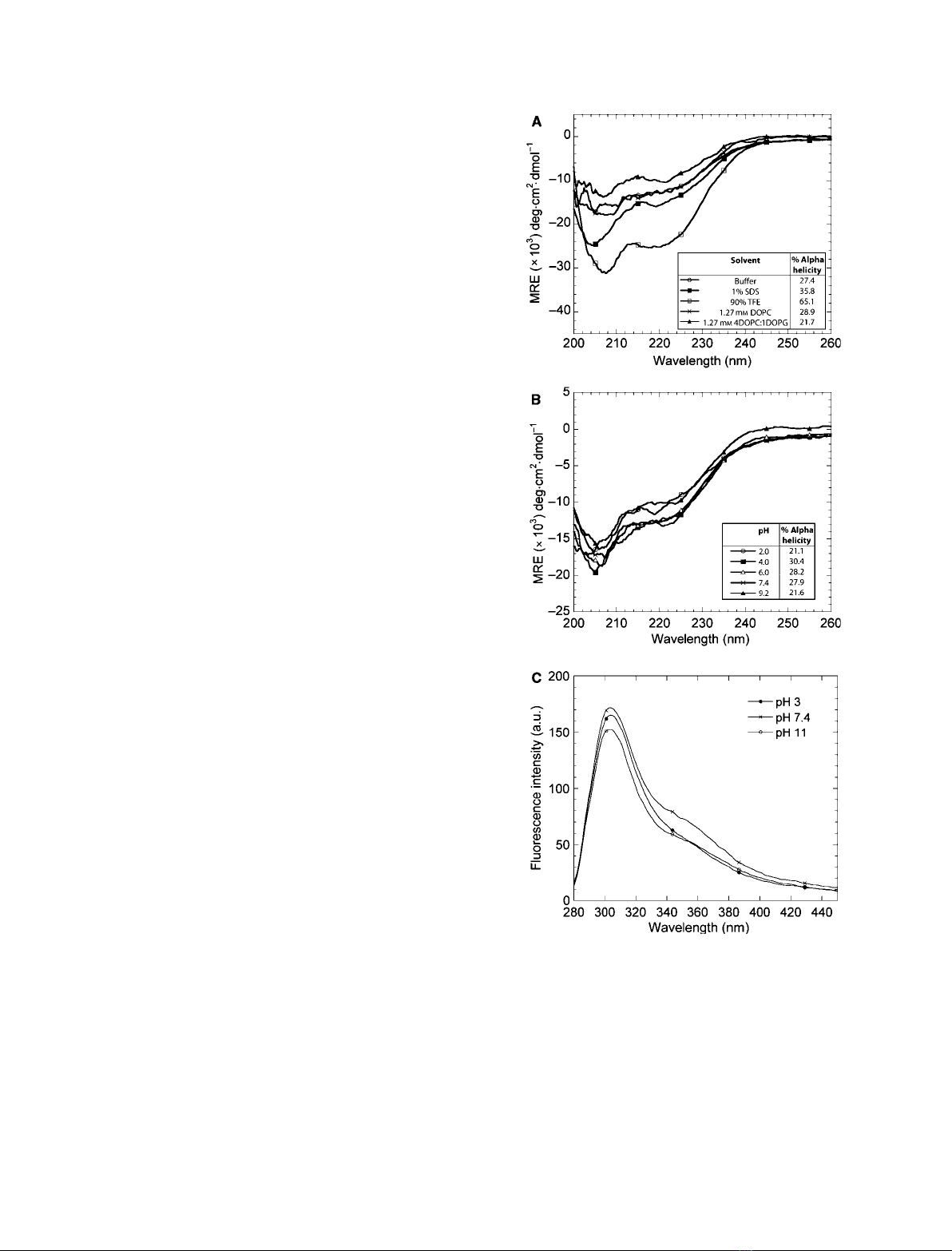
Far-UV CD spectra of PP3 were recorded under physi-
ological conditions as well as in the presence of anionic
and other organic solvents known to stabilize a-helical
structure. In all CD spectra, minima at 208 and
222 nm were observed (Fig. 1A), which indicate a-heli-
cal structure. Dissolving PP3 in micellar concentrations
of SDS (1%; 35 mM) led to a general increase in the
CD signal and thus in the degree of a-helicity. This is
consistent with the ability of micellar SDS to induce
a-helical structure [39]. The greatest signal increase
was obtained using 90% trifluoroethanol (TFE), which
is known to stabilize helical conformation as a result
of its hydrophilicity and hydrogen-bonding ability [40].
Neither 100% 1,2-dioleoylphosphatidylcholine (DOPC)
vesicles nor 1,2-dioleoylphosphatidylcholine ⁄1,2-diol-
eoylphosphatidylglycerol (DOPC ⁄DOPG) vesicles, at a
ratio of 80 : 20 (w ⁄w), induced any spectral changes,
indicating that phospholipid vesicles do not promote
further formation of a-helical structure.
For isolation of the proteose peptone fraction con-
taining PP3, high temperatures are used to precipitate
the major whey proteins a-lactalbumin and b-lacto-
globulin followed by acidification to pH 4.6 to precipi-
tate the caseins [15]. CD wavelength scans of PP3 in
aqueous buffer with pH values between 2 and 9.2 show
that the a-helical structure is very stable throughout
the tested pH range (Fig. 1B). To obtain complemen-
tary information on the tertiary structure, we turned
to fluorescence. PP3 contains no tryptophan residues,
but fluorescence emission spectra from the single tyro-
sine residue (at position 121) of PP3, acquired between
pH 3 and pH 11, indicate no shift in the emission peak
position and only small changes in fluorescence inten-
sity. This indicates that the tertiary structure is essen-
tially invariant and this is consistent with the lack of
change in CD spectra (Fig. 1C). The studies of the
biological functionalities of PP3 mainly focus on the
C-terminal part of PP3, which contains the a-helical
structure. These observations indicate that this struc-
ture is preserved during the elevated temperatures used
in the purification of the protein [15] as well as in the
acidic conditions encountered by the protein in the
gastric juice.
CD was used to study the conformational changes
that PP3 undergoes at higher temperatures and the
extent of its reversibility. Spectra recorded at different
temperatures during the scan (Fig. 2A) revealed two
isodichroic points at 205 and 235 nm, indicating
Fig. 1. The degree of a-helicity of PP3 increases in the presence of
anionic and organic solvents and the a-helical structure is essentially
invariant over the pH range 2.0–9.7. (A) CD wavelength spectra of
2.1 lMPP3 in mixtures of 10 mMNa
2
HPO
4
(pH 7.4) (denoted buffer),
1% SDS, 90% TFE, 1.27 mMcorresponding to 1 mg mL
)1
of DOPC
or 1.27 mMcorresponding to 1 mg mL
)1
of DOPC ⁄DOPG at a ratio
of 80 : 20 (w ⁄w), at 25 C. (B) CD wavelength spectra of 2.1 lMPP3
in 10 mMNa
2
HPO
4
at pH values ranging from 2 to 9.2. Percentage
a-helicity is given in the insert. (C) Steady-state tyrosine fluorescence
of the tertiary structure of PP3 at pH 3, pH 7.4 and pH 11.
Tetramerization and thermal stability of PP3 L. R. L. Pedersen et al.
338 FEBS Journal 279 (2012) 336–347 ª2011 The Authors Journal compilation ª2011 FEBS
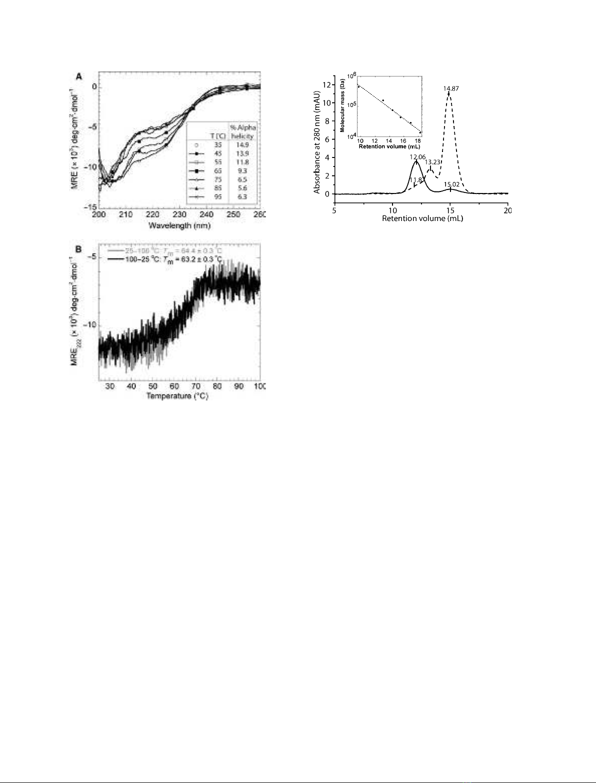
a simple two-state transition from the folded a-helical
state to the unfolded random coil state. Forward
(25 fi100 C) and backward (100 fi25 C) ther-
mal scans indicated that most of the PP3 population
was able to refold into the original conformation, as
the backward scan led to a reduction of only 4.3% in
ellipticity (Fig. 2B). Furthermore, the apparent melting
temperature was only reduced from 64.4 ± 0.3 C
(forward scan) to 63.2 ± 0.3 C (backward scan)
(Fig. 2B). CD wavelength scans of the sample were
performed at 25 C immediately before and after the
thermal scans and showed no conformational change,
confirming that PP3 essentially refolds completely
upon thermal denaturation (data not shown). These
results indicate that the a-helical structure of PP3, and
thereby the claimed positive effects conveyed by PP3
in milk, are preserved during industrial processing.
PP3 forms stable tetramers
To thoroughly analyze the multimerization or aggrega-
tion of PP3 observed in previous studies we used SEC
and AF4 combined with multi-angle light scattering
(MALS). Monomeric glycosylated PP3 has a molecular
mass of 19.4 kDa [24]; however, SEC analysis of PP3
showed a major peak eluting at 12.06 mL, correspond-
ing to a relative molecular mass of 191 kDa. A
smaller peak eluted at 15.02 mL, corresponding to a
molecular mass of 58 kDa (Fig. 3). This indicates
that PP3 preferentially assembles into multimeric struc-
tures under these conditions. These results are in
agreement with previous SEC measurements of the
PP3 multimers, where PP3 eluted at an elution volume
corresponding to a molecular mass of approxi-
mately 190 kDa [11]. Analyses of PP3 by AF4 showed
similar results, with relative molecular mass estimates
of 225.9 kDa for the high-molecular-mass peak and
23.4 kDa for the low-molecular-mass peak (Fig. 4A).
Using AF4 ⁄MALS, the molecular mass can be
determined independently from light scattering data by
extrapolation to a zero sample concentration and a
zero scattering angle using the Zimm model. This
MALS-based analysis requires the sample concentra-
tion to be determined either by the refractive index
(RI) (based on both protein and carbohydrate contri-
butions, see the Materials and methods) or by the
absorbance at 205 nm (giving only protein contribu-
tions). In separate experiments exemplified in Fig. 4B,
the molecular mass of the main PP3 peak was determined
Fig. 3. The elution profile of PP3 subjected to SEC indicates aggre-
gation of PP3 to form higher-order structures. Elution profiles of
PP3 (solid line) and BSA (dashed line) were obtained by gel-filtration
chromatography on a Superdex 200 column eluted with phosphate-
buffered saline (NaCl ⁄P
i
), pH 7.5, at a flow rate of 0.8 mLÆmin
)1
.A
relative calibration with retention volumes of a set of standard pro-
teins (14–440 kDa) is shown in the insert.
Fig. 2. PP3 refolds almost completely upon thermal denaturation.
(A) CD wavelength spectra of 2.0 lMPP3 recorded at different
temperatures. Percentage a-helicity is given in the insert. T, tem-
perature. (B) Far-UV CD spectra of a forward (25 fi100 C) and a
backward (100 fi25 C) thermal scan of 2.1 lMPP3 at 222 nm.
The melting temperature (T
m
) is indicated for each curve.
L. R. L. Pedersen et al. Tetramerization and thermal stability of PP3
FEBS Journal 279 (2012) 336–347 ª2011 The Authors Journal compilation ª2011 FEBS 339

in combination with RI data to be 79.1 ± 2.6 kDa
(mean ± standard error of three individual analyses)
using the weight-averaged (dn⁄dc)
PP3
. This value corre-
sponds to approximately four PP3 units of
19.8 ± 0.7 kDa protein assembled into a tetrameric
complex. The molecular mass of post-translationally
modified PP3 has previously been determined, by mass
spectrometric analysis, to be 19.4 ± 0.02 kDa [24],
which is in excellent agreement with the present results.
Using MALS combined with absorbance data at
205 nm (which lacks carbohydrate contributions), the
main peak was determined to be 61.1 ± 1.5 kDa, cor-
responding to four, 15.3 ± 0.4-kDa units and thus
within 3% of the theoretical molecular mass of the
PP3 polypeptide chain. The difference of 4.5 kDa
between monomer size estimates thus arises as a result
of the inability to detect glycosylations and phosphory-
lations by measuring the absorbance at 205 nm. In
fact, the molecular mass of the post-translational mod-
ifications of PP3 has previously been determined to
constitute 4.1 ± 0.02 kDa [24]. The validity of size
estimates were further confirmed by size estimation of
BSA monomers and dimers using a (dn⁄dc)
BSA
of
0.186 mLÆg
)1
[41,42] in a separate AF4 ⁄MALS experi-
ment, which revealed molecular mass values of 64.5
and 135 kDa, respectively.
In AF4 ⁄MALS, sample retention relies on the diffu-
sion coefficient (which can be related to the radius for
strictly spherical particles through the Stokes–Einstein
equation) and liquid flows inside the separation chan-
nel. Unfortunately, the 635-nm laser of our MALS
limits reliable size estimates to > 32 nm (lim-
it = k⁄20) and thus does not allow reliable estimates
of BSA and PP3 sizes and hence the evaluation of pro-
tein compactness. However, the molecular mass esti-
mates of MALS, in combination with the high
retention times (indicating a large hydrodynamic size
compared with BSA) of PP3 in AF4 ⁄MALS separa-
tion, clearly suggest that PP3 preferentially assembles
into a highly extended tetrameric complex of
79 kDa. We further analyzed the monomer ⁄tetramer
distribution as a function of protein concentration by
AF4 and found a constant ratio of 3.8 ± 0.2%
monomer and 96.2 ± 0.2% tetramer at six concentra-
tions between 0.93 mgÆmL
)1
(50 lM) and
7.72 mgÆmL
)1
(415 lM) PP3 (data not shown), sug-
gesting that the tetrameric complex is rather stable in
the absence of denaturing agents.
To monitor the unfolding and potential dissociation
of the tetramer during unfolding by GdmCl, steady-
state fluorescence anisotropy and CD were used. As
shown in Fig. 5, the anisotropy decreases above 1M
GdmCl, clearly indicating that PP3 tumbles faster in
solution under these conditions. While protein unfold-
ing is expected to increase the hydrodynamic radius of
the protein and thus decrease the tumbling rate (larger
anisotropy), the decrease in anisotropy is consistent
with dissociation of the tetrameric complex. Monitor-
ing of the GdmCl-mediated unfolding of PP3 by CD
further shows that the dissociation of the PP3 complex
is accompanied by loss of a-helix structure measured
at 220 nm and thus the PP3 complex dissociates as a
result of chemical denaturation (Fig. 5). The fluores-
cence anisotropy and the CD recordings at 50 lMPP3
show transitions with midpoints at 2.10 ± 0.06 M
GdmCl and 50% unfolding at 2.16 ± 0.04 MGdmCl,
respectively. These values are equal within error and
thus indicate that tetramer dissociation and loss of
helical structure occur in parallel.
To investigate the effect of milk fat globules or glob-
ule mimics on this structure, a simple separation-free
Fig. 4. PP3 monomers associate into highly extended and stable
tetrameric complexes. (A) Elution profile of PP3 (solid line) and BSA
(dashed line) upon separation by AF4, monitored by UV light (A
205
).
The insert shows the relative calibration in which the retention time
of BSA monomers, dimers and trimers was plotted as a function of
molecular mass. (B) AF4 ⁄MALS estimates of the molecular mass
of PP3 multimers using RI ( ) and A
205
nm ( ) data to obtain size
estimates of PP3 with and without glycosylation, respectively. The
insert shows the distribution of PP3 multimer size estimates. The
mobile phase consists of 5 mMTris ⁄HCl containing 150 mMNaCl.
Tetramerization and thermal stability of PP3 L. R. L. Pedersen et al.
340 FEBS Journal 279 (2012) 336–347 ª2011 The Authors Journal compilation ª2011 FEBS


%20--%3e%3cdefs%3e%3cstyle%3e%20.st0%20{%20fill:%20%23fff;%20}%20.st1%20{%20fill:%20%237800fa;%20}%20%3c/style%3e%3c/defs%3e%3cpath%20class='st1'%20d='M117.78,12.18H43.11c2.9,3.47,4.65,7.94,4.65,12.82,0,5.6-2.3,10.66-6.01,14.29h76.02l7.22-13.56-7.22-13.56Z'/%3e%3cg%3e%3cpath%20class='st0'%20d='M53.58,26.17h-.59v-1.46h.59v-4.96h2.83c1.78,0,2.67.94,2.67,2.82v5.76c0,1.87-.89,2.81-2.67,2.81h-2.83v-4.96ZM55.36,21.37v3.34h1.1v1.46h-1.1v3.34h1.01c.61,0,.91-.37.91-1.1v-5.93c0-.74-.3-1.1-.91-1.1h-1.01Z'/%3e%3cpath%20class='st0'%20d='M65.99,31.14h-1.8l-.31-2.07h-2.19l-.31,2.07h-1.64l1.82-11.39h2.62l1.82,11.39ZM65.28,18.04c-.25.46-.51.77-.75.94-.21.15-.47.22-.79.22-.26,0-.57-.07-.92-.22l-.38-.15c-.14-.05-.26-.07-.37-.07-.3,0-.53.18-.71.54l-.91-.68c.25-.46.51-.77.75-.94.21-.14.48-.21.79-.21.26,0,.57.07.92.21l.38.15c.14.05.26.07.37.07.3,0,.53-.18.71-.54l.91.68ZM61.91,27.52h1.73l-.87-5.76-.87,5.76Z'/%3e%3cpath%20class='st0'%20d='M74.53,26.89v1.52c0,1.91-.89,2.86-2.67,2.86s-2.67-.95-2.67-2.86v-5.93c0-1.91.89-2.86,2.67-2.86s2.67.95,2.67,2.86v1.11h-1.69v-1.22c0-.75-.31-1.12-.93-1.12s-.93.37-.93,1.12v6.15c0,.74.31,1.11.93,1.11s.93-.37.93-1.11v-1.63h1.69Z'/%3e%3cpath%20class='st0'%20d='M81.4,31.14h-1.8l-.31-2.07h-2.19l-.31,2.07h-1.64l1.82-11.39h2.62l1.82,11.39ZM75.9,19.2l1.52-1.91h1.71l1.51,1.91h-1.61l-.76-.95-.75.95h-1.61ZM77.32,27.52h1.73l-.87-5.76-.87,5.76ZM83.1,15.99l-1.76,1.91h-1.26l1.17-1.91h1.86Z'/%3e%3cpath%20class='st0'%20d='M84.86,19.75c1.78,0,2.67.94,2.67,2.82v1.48c0,1.87-.89,2.81-2.67,2.81h-.85v4.28h-1.79v-11.39h2.64ZM84.01,21.37v3.86h.85c.58,0,.87-.36.87-1.08v-1.71c0-.71-.29-1.07-.87-1.07h-.85Z'/%3e%3cpath%20class='st0'%20d='M93.51,19.75c1.78,0,2.67.94,2.67,2.82v1.48c0,1.87-.89,2.81-2.67,2.81h-.85v4.28h-1.79v-11.39h2.64ZM92.66,21.37v3.86h.85c.58,0,.87-.36.87-1.08v-1.71c0-.71-.29-1.07-.87-1.07h-.85Z'/%3e%3cpath%20class='st0'%20d='M98.8,31.14h-1.79v-11.39h1.79v4.88h2.03v-4.88h1.83v11.39h-1.83v-4.88h-2.03v4.88Z'/%3e%3cpath%20class='st0'%20d='M105.36,24.55h2.46v1.62h-2.46v3.34h3.09v1.63h-4.88v-11.39h4.88v1.63h-3.09v3.18ZM108.17,17.29l-1.76,1.91h-1.26l1.17-1.91h1.86Z'/%3e%3cpath%20class='st0'%20d='M112.2,19.75c1.78,0,2.67.94,2.67,2.82v1.48c0,1.87-.89,2.81-2.67,2.81h-.85v4.28h-1.79v-11.39h2.64ZM111.35,21.37v3.86h.85c.58,0,.87-.36.87-1.08v-1.71c0-.71-.29-1.07-.87-1.07h-.85Z'/%3e%3c/g%3e%3ccircle%20class='st1'%20cx='25'%20cy='25'%20r='20'/%3e%3cpath%20class='st0'%20d='M32.78,19.27c2.92,0,4.43,2.55,5.28,5.33l.71,2.17c.14.38-.33.75-.71.75h-5.61c.19-.33.24-.71.09-1.08l-.75-2.45c-.43-1.32-.99-2.64-1.79-3.77.75-.57,1.65-.94,2.78-.94h0ZM25,18.38c3.25,0,4.9,2.78,5.89,5.89l.76,2.45c.14.42-.33.8-.8.8h-11.69c-.42,0-.94-.38-.8-.8l.75-2.45c.99-3.11,2.64-5.89,5.89-5.89h0ZM25,11.35c1.74,0,3.11,1.37,3.11,3.11s-1.37,3.11-3.11,3.11-3.11-1.41-3.11-3.11,1.41-3.11,3.11-3.11h0ZM17.27,19.27c1.08,0,1.98.38,2.73.94-.8,1.13-1.37,2.45-1.74,3.77l-.8,2.45c-.14.38-.05.75.09,1.08h-5.56c-.42,0-.9-.38-.75-.75l.71-2.17c.9-2.78,2.41-5.33,5.33-5.33h0ZM17.27,12.91c1.51,0,2.78,1.27,2.78,2.83s-1.27,2.83-2.78,2.83-2.83-1.27-2.83-2.83,1.27-2.83,2.83-2.83h0ZM32.78,12.91c1.56,0,2.78,1.27,2.78,2.83s-1.23,2.83-2.78,2.83-2.83-1.27-2.83-2.83,1.27-2.83,2.83-2.83h0ZM27.07,28.56v.09c0,.57-.24,1.08-.61,1.46h0v.05c-.38.33-.9.57-1.46.57s-1.08-.24-1.46-.61h0c-.38-.38-.61-.9-.61-1.46v-.09h1.41v.09c0,.19.05.38.19.47v.05c.09.09.28.19.47.19s.38-.09.47-.19v-.05c.14-.09.24-.28.24-.47t-.05-.09h1.41ZM30.99,28.56v.09c0,1.65-.66,3.16-1.74,4.24-1.08,1.08-2.59,1.79-4.24,1.79s-3.16-.71-4.24-1.79l-.05-.05c-1.04-1.08-1.7-2.55-1.7-4.2v-.09h1.41v.09c0,1.27.47,2.4,1.27,3.25h.05c.85.85,1.98,1.37,3.25,1.37s2.4-.52,3.25-1.37c.85-.8,1.37-1.98,1.37-3.25v-.09h1.37ZM34.99,28.56v.09c0,2.78-1.13,5.28-2.92,7.07-1.79,1.79-4.29,2.92-7.07,2.92s-5.23-1.13-7.07-2.92c-1.79-1.79-2.92-4.29-2.92-7.07v-.09h1.41v.09c0,2.4.94,4.53,2.5,6.08,1.56,1.56,3.72,2.5,6.08,2.5s4.52-.94,6.08-2.5c1.56-1.56,2.5-3.68,2.5-6.08v-.09h1.41Z'/%3e%3c/svg%3e)