
Structure–function analysis of the filamentous actin
binding domain of the neuronal scaffolding protein
spinophilin
Herwig Schu
¨ler
1,
* and Wolfgang Peti
2
1 Max Delbru
¨ck Center for Molecular Medicine, Berlin-Buch, Germany
2 Department of Molecular Pharmacology, Physiology, and Biotechnology, Brown University, Providence, RI, USA
Dendritic spines, globular protrusions from neuronal
dendrites in the central nervous system, are the major
sites of excitatory signal transduction in dendrites.
During the past few years, it has been realized that
dendritic spines are highly dynamic structures, both
during development and in the adult nervous system.
Dendritic spine morphology changes rapidly and can
be visualized on a minutes time scale (e.g. [1,2]).
Dendritic plasticity is believed to be central for nor-
mal brain functioning [3]. The turnover of dendritic
spines is directly involved in memory formation [4],
and changes in spine plasticity caused by epileptic
Keywords
F-actin; intrinsically unstructured protein;
pointed-end capping protein; spinal
plasticity; spinophilin
Correspondence
H. Schu
¨ler, Max Delbru
¨ck Center for
Molecular Medicine, 13125 Berlin-Buch,
Germany
Fax: 0049-6221-564643
Tel: 0049-6221-568284
E-mail: herwig.schueler@med.uni-
heidelberg.de
W. Peti, Department of Molecular
Pharmacology, Physiology, and
Biotechnology, Brown University, Box G-E3,
Providence, RI 02912, USA
Fax: 001-401-8636087
Tel: 001-401-8636084
E-mail: wolfgang_peti@brown.edu
*Present address
Department of Parasitology, Heidelberg
University Medical School, Germany
(Received 21 June 2007, revised 25 October
2007, accepted 31 October 2007)
doi:10.1111/j.1742-4658.2007.06171.x
Spinophilin, a neuronal scaffolding protein, is essential for synaptic trans-
mission, and functions to target protein phosphatase-1 to distinct subcellu-
lar locations in dendritic spines. It is vital for the regulation of dendritic
spine formation and motility, and functions by regulating glutamatergic
receptors and binding to filamentous actin. To investigate its role in regu-
lating actin cytoskeletal structure, we initiated structural studies of the
actin binding domain of spinophilin. We demonstrate that the spinophilin
actin binding domain is intrinsically unstructured, and that, with increasing
C-terminal length, the domain shows augmented secondary structure con-
tent. Further characterization confirmed the previously known crosslinking
activity and uncovered a novel filamentous actin pointed-end capping
activity. Both of these functions seem to be fully contained within residues
1–154 of spinophilin.
Abbreviations
ABD, actin binding domain; ERK2, extracellular signal-regulated kinase-2; F-actin, filamentous actin; GST, glutathione S-transferase;
IUP, intrinsically unstructured protein; MBP, maltose binding protein; PKA, protein kinase-A; PP1, protein phosphatase-1; PPP1R9B, protein
phosphatase-1 regulatory subunit 9B; SAM, sterile amotif.
FEBS Journal 275 (2008) 59–68 ª2007 The Authors Journal compilation ª2007 FEBS 59
seizures may underlie cognitive deficits in epilepsy
patients [5]. Thus, a comprehensive description of the
molecular components involved in the regulation and
maintenance of dendritic spine morphology is funda-
mental to our understanding of the functions of the
central nervous system.
The molecular details that underlie the regulation of
spine morphology have advanced considerably in
recent years. As actin is the only cytoskeletal compo-
nent present in spines, actin interacting proteins are
prime candidates for the regulation of dendritic spine
plasticity [6]. Indeed, spine motility is powered by the
polymerization of actin [7,8]. In addition, actin regula-
tors, such as profilin [1,9] and rho-dependent pathways
(e.g. [10,11]), have already been shown to influence
spine morphology.
Spinophilin (Genbank ID PPP1R9B: protein phos-
phatase-1 regulatory subunit 9B), also known as neura-
bin-II, is a neuronal scaffolding protein involved in the
regulation of dendritic spine morphology [12,13]
(reviewed in [14]). Spinophilin binds and bundles actin
polymers, thereby stabilizing actin structures in the
spines [15,16]. Moreover, spinophilin can recruit rho-
family GTPases, influencing actin reorganization [17].
Spinophilin also targets protein phosphatases (pro-
tein phosphatase-1, PP1) [13,18,19] and binds to gluta-
matergic receptors [20–22]. It is currently believed
that spinophilin functions to target PP1 to gluta-
mate [a-amino-3-hydroxy-5-methyl-4-isoxazolpropio-
nate (AMPA) and N-methyl-d-aspartate (NMDA)]
receptors, and thereby modulates their activity and traf-
ficking through regulation of their phosphorylation
state [23]. Secondly, spinophilin targets PP1 to the post-
synaptic densities by providing a link to the microfila-
ment system [24].
Spinophilin shares its general domain structure and
about 65% overall sequence identity with its neuronal
isoform neurabin (Fig. 1A). Spinophilin, although
ubiquitously expressed, is predominantly found in neu-
rones, whereas neurabin is expressed almost exclusively
in neuronal cells, generally at lower levels than spino-
philin. Despite their similarity, they do not compensate
for one another [23,25,26]. Both spinophilin and neura-
bin contain N-terminal filamentous actin (F-actin)
binding, PP1 binding, PDZ and C-terminal coiled-coil
domains. In addition, neurabin, but not spinophilin,
contains a sterile amotif (SAM) domain [27] in its
A
B
C
D
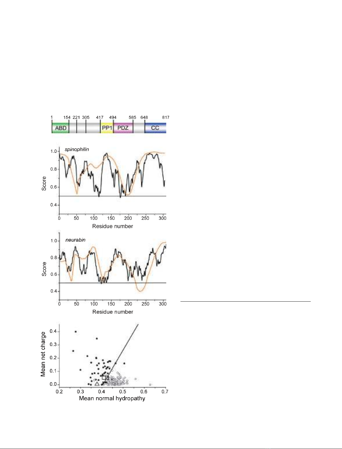
Fig. 1. N-terminal F-actin binding domains of spinophilin and neura-
bin are predicted to be disordered. (A) Schematic representation of
the Rattus norvegicus spinophilin sequence with the positions of
the construct limits used in this study and domain borders indicated
by numbers. The core actin binding domain, PP1 binding domain,
PDZ domain and C-terminal coiled-coil region are indicated. (B, C)
The sequences of human spinophilin (B) and neurabin (C) were
analysed for disorder using the programs IUPRED (black lines) [52]
and VSL2 (orange lines) [53]. Sequences scoring mostly above the
value of 0.5 (indicated) are generally regarded as intrinsically dis-
ordered. (D) Charge hydropathy plots [54] for human spinophilin
(square), neurabin (triangle) and reference sets of ordered (circles)
and disordered (dots) proteins. Both spinophilin and neurabin score
above the discriminator line, indicating intrinsic disorder. The results
of these analyses (B and D) for human and rat spinophilin were
essentially identical.
The actin binding domain of spinophilin H. Schu
¨ler and W. Peti
60 FEBS Journal 275 (2008) 59–68 ª2007 The Authors Journal compilation ª2007 FEBS

C-terminus, whereas spinophilin, but not neurabin,
may possess a dopamine receptor ⁄a-adrenergic inter-
acting domain in its N-terminus, possibly between
spinophilin residues 200 and 400 [20]. The structures
of the spinophilin and neurabin PDZ [22] and neura-
bin SAM [27] domains have been solved recently by
NMR spectroscopy.
Spinophilin interaction with F-actin is regulated by
phosphorylation of its actin binding domain (ABD) by
protein kinase-A (PKA) [28], calcium ⁄calmodulin-
dependent kinase II [29], cyclin-dependent kinase-5
and extracellular signal-regulated kinase-2 (ERK2)
[30]. PKA phosphorylates three serine residues located
in the N-terminal region of spinophilin, namely Ser94,
Ser177 and, to some extent, Ser100, whereas ERK2
phosphorylates Ser15 and Ser205. Phosphorylation of
spinophilin ABD leads to an attenuated interaction
with F-actin. Phosphorylation of these serine residues
may be reversed by different phosphatases, thus restor-
ing the F-actin binding capacity of spinophilin [30,31],
but the pathway constituents that regulate actin bind-
ing through phosphate signalling are unknown.
We have undertaken a systematic and detailed struc-
tural and functional analysis of the ABD of spinophi-
lin. We show that residues 1–154 of spinophilin are
both necessary and sufficient to mediate F-actin bind-
ing. Critically, we also show that residues 1–154 of
spinophilin and longer spinophilin ABD constructs
(residues 1–221 and 1–305 of spinophilin) are intrinsi-
cally unstructured, as tested by NMR and CD spec-
troscopy. In addition, we show that, at low molar
ratios, spinophilin ABDs bind and crosslink actin
polymers. However, at high molar ratios, they cap
F-actin polymers. Thus, we provide evidence for an
F-actin capping activity of spinophilin.
Results and Discussion
Spinophilin construct design and production
Spinophilin has previously been shown to bind to actin
polymers via its N-terminal domain [16]. Furthermore,
the spinophilin–F-actin interaction has been partially
characterized in vitro and in vivo. Here, we set out to
study spinophilin ABD and its interaction with F-actin
using an array of biophysical characterization tools to
gain insights into the mechanism of the interaction.
Proteins comprising spinophilin ABD residues 1–154,
1–221, 1–305, 154–221, 154–301 and 221–305 were pro-
duced in Escherichia coli and purified to homogeneity,
free of affinity tags used for increased solubility during
expression and purification. Thus, untagged spinophi-
lin constructs were analysed in this study, eliminating
possible interaction of actin with the hexahistidine tags
on spinophilin.
Spinophilin and neurabin ABDs are predicted
to be unstructured
We used secondary structure prediction and disorder
recognition software to analyse the sequence of spino-
philin ABD (residues 1–305). Initial analysis showed
that the sequence of spinophilin was highly biased
towards disorder-inducing amino acids (i.e. proline
and charged amino acids [32]), suggesting that it is
unstructured. Six different prediction programs were
then used to estimate the secondary structure content
of N-terminal fragments of human and rat spinophilin
and human neurabin. The results showed that only
approximately 20% of the spinophilin ABD sequence
was predicted to adopt a classified secondary structure
(Table 1), with the remainder predicted to be in ran-
dom coil. In a subsequent step, the programs iupred,
vsl2 and pondr were used to detect regions of dis-
order in the ABDs of spinophilin and neurabin. As
shown in Fig. 1, these programs also predicted a high
degree of disorder in the ABDs of spinophilin and
neurabin. On the basis of these analyses, spinophilin
and neurabin ABDs were predicted to be intrinsically
unstructured proteins (IUPs).
Spinophilin ABD is intrinsically unstructured
NMR spectroscopy is the only atomic resolution tech-
nique able to resolve the structural and dynamic char-
acteristics of IUPs. Therefore, to experimentally verify
the in silico predictions, we carried out one-dimen-
sional
1
H NMR experiments (Fig. 2A,B). The NMR
spectra of these constructs perfectly resembled the
spectra of unfolded proteins: they showed no signs of
either amide proton dispersion, which is indicative of
hydrogen bonding in secondary structure elements, or
ring current shifted methyl groups, which are caused
Table 1. Summary of secondary structure predictions for N-termi-
nal portions of human neurabin-1 (HsNEB1), human spinophilin
(HsNEB2) and rat spinophilin (RnNEB2), calculated using six differ-
ent prediction software programs.
Random coil predictions (%)
APSSP2
[46]
NORS
[47]
PORTER
[48]
PROF
[49]
PSIPRED
[50]
SPRITZ
[51]
HsNEB1 (1–308) 78.6 79.5 73.1 79.6 82.5 51.6
HsNEB2 (1–304) 79.3 89.8 74.0 89.8 81.1 60.9
RnNEB2 (1–305) 76.9 82.0 75.1 82.0 82.9 61.3
H. Schu
¨ler and W. Peti The actin binding domain of spinophilin
FEBS Journal 275 (2008) 59–68 ª2007 The Authors Journal compilation ª2007 FEBS 61
by the interaction of methyl groups with aromatic side
chains in the hydrophobic core of folded proteins. This
suggests that these recombinant spinophilin protein
constructs are intrinsically unstructured. To further
verify this result, we recorded far-UV CD spectropo-
larimetric spectra of the spinophilin ABD constructs
(Fig. 2C), which enables rapid analysis of the overall
secondary structure content of proteins. The CD spec-
tra of residues 1–154, 1–221 and 1–305 of spinophilin
were indicative of random coil structures, with a nega-
tive absorption around 202 nm. However, the CD
spectra for all three protein domain constructs showed
a negative absorption around 222 nm, indicating dif-
ferentially increasing amounts of a-helical content.
Using [h]
222 nm
, the a-helical content was calculated to
be 12%, 22% and 30% for residues 1–154, 1–221 and
1–305 of spinophilin, respectively (details in Experi-
mental procedures). Thus, both NMR and CD spec-
troscopy showed experimentally that all spinophilin
ABDs were intrinsically unstructured. However, these
unstructured proteins, similar to their folded counter-
parts, displayed different properties. The core F-ABD,
the first approximately 160 residues, seemed to be
mostly unstructured, behaving like a random coil
polymer. Additional C-terminal residues in the longer
fragments (residues 1–221 and 1–305 of spinophilin)
showed more secondary structure, as revealed by CD
spectroscopy. The percentage amino acid composition
was uniform within these three constructs, with one
exception: the number of valine residues was doubled
in the 1–221 and 1–305 sequences of spinophilin. Thus,
the increasingly structured C-terminal regions of resi-
dues 1–221 and 1–305 of spinophilin were rich in
hydrophobic valine residues. This augmented hydro-
phobic density could form the hydrophobic nucleus for
increased tertiary interactions and secondary structure
formation, probably explaining the experimental differ-
ences in the CD spectra. Finally, this was supported
by empirical observations, which indicated that resi-
dues 1–154 of spinophilin degraded more rapidly (24–
36 h) than residues 1–221 and 1–305 (5–6 days),
when stored at 4 C, indicating an easier access for
proteases to the putative random coil structure of resi-
dues 1–154 of spinophilin.
Thus, our experimental NMR and CD data clearly
demonstrated that the spinophilin ABD constructs
were largely disordered, and that their secondary struc-
ture content increased with their C-terminal length.
spinophilin1–154
spinophilin1–154
spinophilin1–221
spinophilin1–305
A
B
C
6.0 8.0 4.0 0.0
8.0 6.0 4.0 0.0
δ
δ
1H [p.p.m.]
δ
1H [p.p.m.]
222 nm
0
-20
-40
200
[Θ] (103 deg cm2/dmole)
220 240
λ (nm)
260
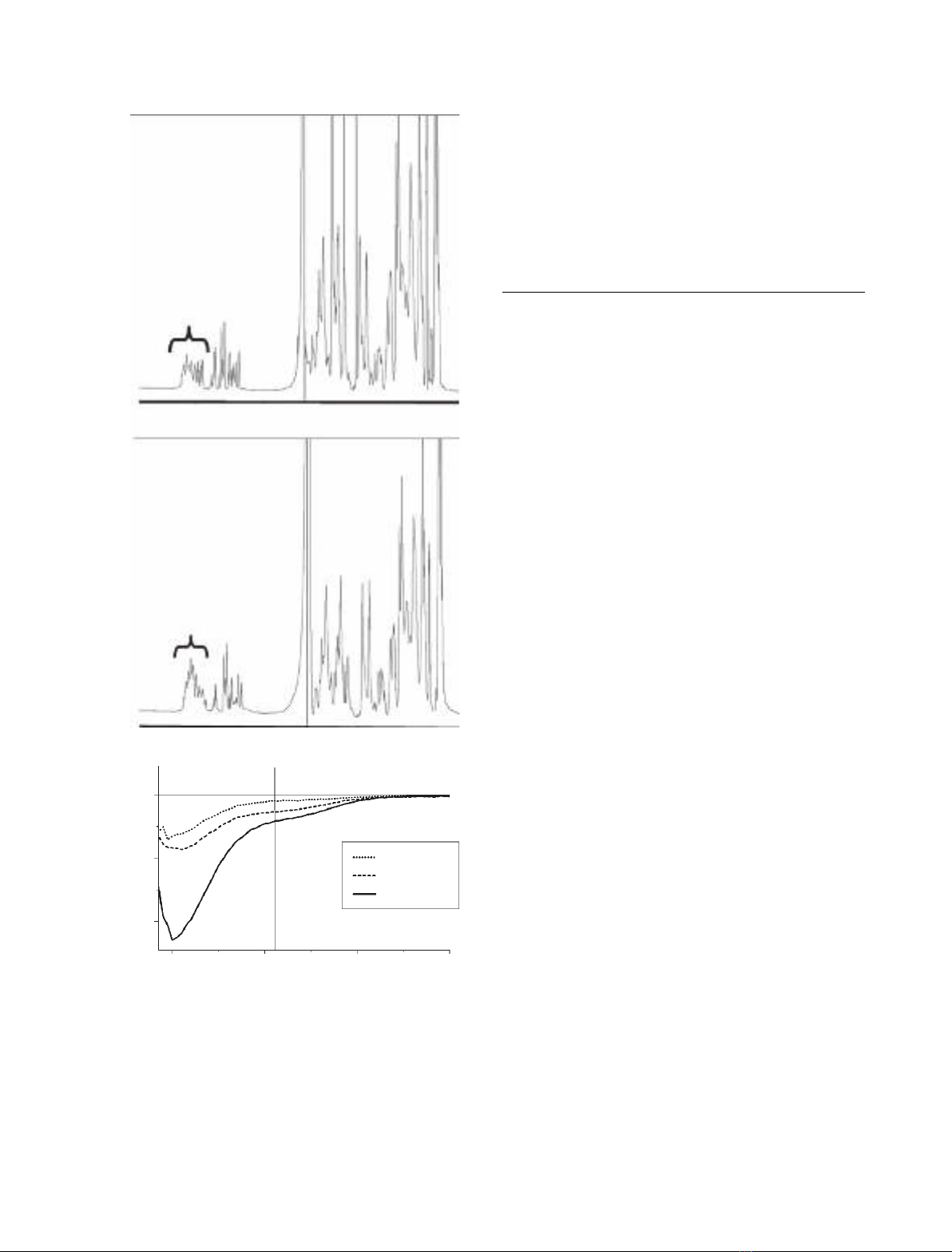
Fig. 2. Recombinant proteins containing N-terminal fragments of
rat spinophilin lack a regular secondary structure. (A, B) One-dimen-
sional
1
H NMR spectra of residues 1–154 and 1–221 of spinophilin
(spinophilin1–154 and spinophilin1–221), respectively. Parentheses
indicate the dramatically reduced H
N
chemical shift region because
of the lack of a hydrogen bonding network in IUPs. (C) Far-UV CD
spectra of spinophilin actin binding domain constructs. The molar
ellipticity differences at 222 nm are highlighted by a black bar,
clearly showing the differences in a-helical content in the three
spinophilin actin binding domain constructs.
The actin binding domain of spinophilin H. Schu
¨ler and W. Peti
62 FEBS Journal 275 (2008) 59–68 ª2007 The Authors Journal compilation ª2007 FEBS
Despite being intrinsically unstructured,
spinophilin ABD is active
It was critical to verify that spinophilin ABDs were bio-
logically active. This was accomplished using F-actin
cosedimentation assays. The spinophilin proteins were
incubated with calf brain c-actin under polymerizing
conditions and subjected to ultracentrifugation. Resi-
dues 1–154, 1–221 and 1–305 of spinophilin sedimented
with actin polymers when added at substoichiometric
amounts (4 : 1 F-actin : spinophilin construct molar
ratio; Fig. 3A). Therefore, this experiment showed
specific binding activity towards F-actin of our recom-
binant spinophilin domains, in spite of their intrinsi-
cally unstructured nature. By contrast, additional
spinophilin constructs, comprising additional fragments
of spinophilin’s ABD (residues 154–221, 221–305 and
154–305 of spinophilin), did not cosediment with
F-actin filaments (Fig. 3A). Together, these data show
that residues 1–154 of spinophilin are sufficient
to mediate the spinophilin interaction with F-actin.
Furthermore, fragments lacking residues 1–154 of
spinophilin cannot interact with actin polymers. This
contrasts with a previous study [33], where a second
actin binding site was identified in residues 154–305 of
spinophilin.
To further verify that our recombinant rat spinophi-
lin ABD constructs functioned identically to wild-type
spinophilin, we studied their activity under transient
covalent modifications. Phosphorylation at Ser94
and ⁄or Ser177, mediated by cAMP-dependent PKA,
has been shown to suppress the actin binding activity
of spinophilin from rat [28,29] (Ser177 is not conserved
in human and mouse; however, PKA phosphorylation
of mouse spinophilin Ser94 is sufficient to suppress its
association with F-actin [34]). As illustrated in Fig. 3B,
residues 1–221 of spinophilin, treated with PKA,
showed a substantially reduced capacity to cosediment
with actin polymers. This shows that our recombinant
spinophilin, like wild-type spinophilin, is responsive to
kinase regulation.
Spinophilin F-ABD is capable of F-actin
reorganization
Spinophilin has been shown to crosslink actin poly-
mers in vitro [16]. To study the effects of spinophilin
ABD on the overall morphology of F-actin, we used
fluorescence microscopy of rhodamine–phalloidin-
labelled actin polymers (Fig. 4). As expected, actin
polymers alone appeared as elongated fluorescent
filaments (Fig. 4, top panel). The addition of
residues 1–154, 1–221 or 1–305 of spinophilin
(4 : 1 F-actin : spinophilin molar ratio) strongly
induced the crosslinking of actin polymers. The result-
ing filament network resembled that obtained with
other crosslinking proteins, such as fascin [35,36], fil-
amin [37] and cortexillin [38]. In the presence of these
ABD constructs, the concentrations of fluorescent
actin polymers appeared to be higher because of the
precipitation of crosslinked actin polymer networks
onto the glass surface. In agreement with our cosedi-
mentation results, residues 154–221 and 154–305 of
spinophilin did not influence the overall morphology
of F-actin (Fig. 4).
These results show that the crosslinking of actin
polymers in vitro does not require any additional
regions outside the core ABD residues 1–154 of spino-
philin. Furthermore, although the dimerization of
spinophilin is achieved via its C-terminal coiled-coil
domain (Fig. 1A), our results demonstrated that
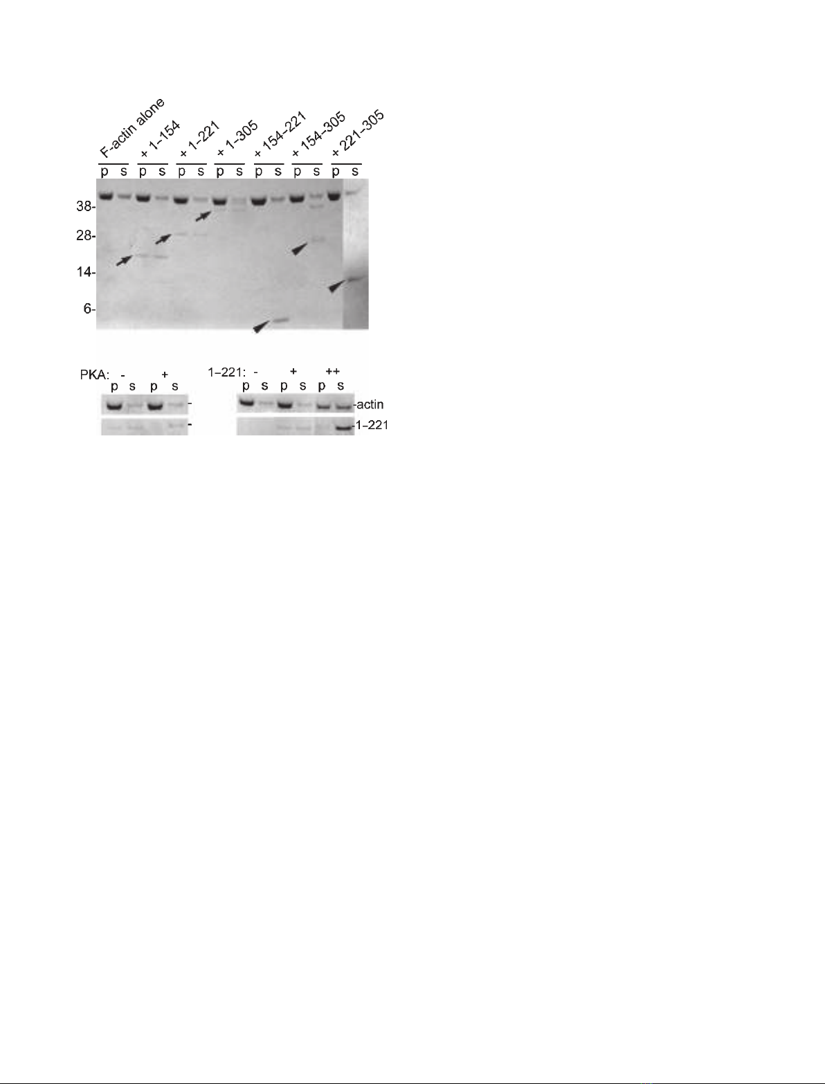
A
BC
Fig. 3. Recombinant proteins containing N-terminal fragments of
rat spinophilin are active in F-actin binding. (A) Cosedimentation
assays of 5 lMpolymers of calf brain c-actin and 2 lMspinophilin
constructs. Residues 1–154, 1–221 and 1–305 of spinophilin are
noticeably enriched in the pellet fractions on ultracentrifugation
(arrows), indicative of F-actin binding, whereas residues 154–221,
154–305 and 221–305 of spinophilin do not cosediment with
F-actin (arrowheads). (B) Cosedimentation assay of F-actin and resi-
dues 1–221 of spinophilin after incubation with PKA. The F-actin
interacting capacity of residues 1–221 of spinophilin is reduced on
PKA-mediated phosphorylation. (C) At equimolar amounts of resi-
dues 1–221 of spinophilin and F-actin, an apparent shift of actin
from the pellet to the supernatant fraction can be observed.
H. Schu
¨ler and W. Peti The actin binding domain of spinophilin
FEBS Journal 275 (2008) 59–68 ª2007 The Authors Journal compilation ª2007 FEBS 63


%20--%3e%3cdefs%3e%3cstyle%3e%20.st0%20{%20fill:%20%23fff;%20}%20.st1%20{%20fill:%20%237800fa;%20}%20%3c/style%3e%3c/defs%3e%3cpath%20class='st1'%20d='M117.78,12.18H43.11c2.9,3.47,4.65,7.94,4.65,12.82,0,5.6-2.3,10.66-6.01,14.29h76.02l7.22-13.56-7.22-13.56Z'/%3e%3cg%3e%3cpath%20class='st0'%20d='M53.58,26.17h-.59v-1.46h.59v-4.96h2.83c1.78,0,2.67.94,2.67,2.82v5.76c0,1.87-.89,2.81-2.67,2.81h-2.83v-4.96ZM55.36,21.37v3.34h1.1v1.46h-1.1v3.34h1.01c.61,0,.91-.37.91-1.1v-5.93c0-.74-.3-1.1-.91-1.1h-1.01Z'/%3e%3cpath%20class='st0'%20d='M65.99,31.14h-1.8l-.31-2.07h-2.19l-.31,2.07h-1.64l1.82-11.39h2.62l1.82,11.39ZM65.28,18.04c-.25.46-.51.77-.75.94-.21.15-.47.22-.79.22-.26,0-.57-.07-.92-.22l-.38-.15c-.14-.05-.26-.07-.37-.07-.3,0-.53.18-.71.54l-.91-.68c.25-.46.51-.77.75-.94.21-.14.48-.21.79-.21.26,0,.57.07.92.21l.38.15c.14.05.26.07.37.07.3,0,.53-.18.71-.54l.91.68ZM61.91,27.52h1.73l-.87-5.76-.87,5.76Z'/%3e%3cpath%20class='st0'%20d='M74.53,26.89v1.52c0,1.91-.89,2.86-2.67,2.86s-2.67-.95-2.67-2.86v-5.93c0-1.91.89-2.86,2.67-2.86s2.67.95,2.67,2.86v1.11h-1.69v-1.22c0-.75-.31-1.12-.93-1.12s-.93.37-.93,1.12v6.15c0,.74.31,1.11.93,1.11s.93-.37.93-1.11v-1.63h1.69Z'/%3e%3cpath%20class='st0'%20d='M81.4,31.14h-1.8l-.31-2.07h-2.19l-.31,2.07h-1.64l1.82-11.39h2.62l1.82,11.39ZM75.9,19.2l1.52-1.91h1.71l1.51,1.91h-1.61l-.76-.95-.75.95h-1.61ZM77.32,27.52h1.73l-.87-5.76-.87,5.76ZM83.1,15.99l-1.76,1.91h-1.26l1.17-1.91h1.86Z'/%3e%3cpath%20class='st0'%20d='M84.86,19.75c1.78,0,2.67.94,2.67,2.82v1.48c0,1.87-.89,2.81-2.67,2.81h-.85v4.28h-1.79v-11.39h2.64ZM84.01,21.37v3.86h.85c.58,0,.87-.36.87-1.08v-1.71c0-.71-.29-1.07-.87-1.07h-.85Z'/%3e%3cpath%20class='st0'%20d='M93.51,19.75c1.78,0,2.67.94,2.67,2.82v1.48c0,1.87-.89,2.81-2.67,2.81h-.85v4.28h-1.79v-11.39h2.64ZM92.66,21.37v3.86h.85c.58,0,.87-.36.87-1.08v-1.71c0-.71-.29-1.07-.87-1.07h-.85Z'/%3e%3cpath%20class='st0'%20d='M98.8,31.14h-1.79v-11.39h1.79v4.88h2.03v-4.88h1.83v11.39h-1.83v-4.88h-2.03v4.88Z'/%3e%3cpath%20class='st0'%20d='M105.36,24.55h2.46v1.62h-2.46v3.34h3.09v1.63h-4.88v-11.39h4.88v1.63h-3.09v3.18ZM108.17,17.29l-1.76,1.91h-1.26l1.17-1.91h1.86Z'/%3e%3cpath%20class='st0'%20d='M112.2,19.75c1.78,0,2.67.94,2.67,2.82v1.48c0,1.87-.89,2.81-2.67,2.81h-.85v4.28h-1.79v-11.39h2.64ZM111.35,21.37v3.86h.85c.58,0,.87-.36.87-1.08v-1.71c0-.71-.29-1.07-.87-1.07h-.85Z'/%3e%3c/g%3e%3ccircle%20class='st1'%20cx='25'%20cy='25'%20r='20'/%3e%3cpath%20class='st0'%20d='M32.78,19.27c2.92,0,4.43,2.55,5.28,5.33l.71,2.17c.14.38-.33.75-.71.75h-5.61c.19-.33.24-.71.09-1.08l-.75-2.45c-.43-1.32-.99-2.64-1.79-3.77.75-.57,1.65-.94,2.78-.94h0ZM25,18.38c3.25,0,4.9,2.78,5.89,5.89l.76,2.45c.14.42-.33.8-.8.8h-11.69c-.42,0-.94-.38-.8-.8l.75-2.45c.99-3.11,2.64-5.89,5.89-5.89h0ZM25,11.35c1.74,0,3.11,1.37,3.11,3.11s-1.37,3.11-3.11,3.11-3.11-1.41-3.11-3.11,1.41-3.11,3.11-3.11h0ZM17.27,19.27c1.08,0,1.98.38,2.73.94-.8,1.13-1.37,2.45-1.74,3.77l-.8,2.45c-.14.38-.05.75.09,1.08h-5.56c-.42,0-.9-.38-.75-.75l.71-2.17c.9-2.78,2.41-5.33,5.33-5.33h0ZM17.27,12.91c1.51,0,2.78,1.27,2.78,2.83s-1.27,2.83-2.78,2.83-2.83-1.27-2.83-2.83,1.27-2.83,2.83-2.83h0ZM32.78,12.91c1.56,0,2.78,1.27,2.78,2.83s-1.23,2.83-2.78,2.83-2.83-1.27-2.83-2.83,1.27-2.83,2.83-2.83h0ZM27.07,28.56v.09c0,.57-.24,1.08-.61,1.46h0v.05c-.38.33-.9.57-1.46.57s-1.08-.24-1.46-.61h0c-.38-.38-.61-.9-.61-1.46v-.09h1.41v.09c0,.19.05.38.19.47v.05c.09.09.28.19.47.19s.38-.09.47-.19v-.05c.14-.09.24-.28.24-.47t-.05-.09h1.41ZM30.99,28.56v.09c0,1.65-.66,3.16-1.74,4.24-1.08,1.08-2.59,1.79-4.24,1.79s-3.16-.71-4.24-1.79l-.05-.05c-1.04-1.08-1.7-2.55-1.7-4.2v-.09h1.41v.09c0,1.27.47,2.4,1.27,3.25h.05c.85.85,1.98,1.37,3.25,1.37s2.4-.52,3.25-1.37c.85-.8,1.37-1.98,1.37-3.25v-.09h1.37ZM34.99,28.56v.09c0,2.78-1.13,5.28-2.92,7.07-1.79,1.79-4.29,2.92-7.07,2.92s-5.23-1.13-7.07-2.92c-1.79-1.79-2.92-4.29-2.92-7.07v-.09h1.41v.09c0,2.4.94,4.53,2.5,6.08,1.56,1.56,3.72,2.5,6.08,2.5s4.52-.94,6.08-2.5c1.56-1.56,2.5-3.68,2.5-6.08v-.09h1.41Z'/%3e%3c/svg%3e)