Jin et al. Virology Journal 2010, 7:92
http://www.virologyj.com/content/7/1/92
Open Access
RESEARCH
BioMed Central
© 2010 Jin et al; licensee BioMed Central Ltd. This is an Open Access article distributed under the terms of the Creative Commons Attri-
bution License (http://creativecommons.org/licenses/by/2.0), which permits unrestricted use, distribution, and reproduction in any
medium, provided the original work is properly cited.
Research
Characterization of variants in the promoter of
BZLF1 gene of EBV in nonmalignant
EBV-associated diseases in Chinese children
Yingkang Jin
1
, Zhengde Xie*
2
, Gen Lu
2
, Shuang Yang
3
and Kunling Shen*
1
Abstract
Background: Diseases associated with Epstein-Barr virus (EBV) infections, such as infectious mononucleosis (IM), EBV-
associated hemophagocytic lymphohistiocytosis (EBV-HLH) and chronic active EBV infection (CAEBV) are not rare in
Chinese children. The association of type 1 or type 2 EBV and variants of the EBV BZLF1 promoter zone (Zp) with these
diseases is unclear.
Results: The objective of this study was to investigate the relationship between EBV genotypes (Zp variants and EBV
type 1 and 2) and the clinical phenotypes of EBV-associated diseases in Chinese children. The Zp region was directly
sequenced in 206 EBV-positive DNA samples from the blood of patients with IM, EBV-HLH, CAEBV, and healthy controls.
Type 1 or type 2 EBV was examined by PCR for EBNA2 and EBNA3C subtypes. Four polymorphic Zp variants were
identified: Zp-P, Zp-V3, Zp-P4 and Zp-V1, a new variant. The Zp-V3 variant was significantly associated with CAEBV (P ≤
0.01). The frequency of co-infection with Zp variants was higher in patients with CAEBV and EBV-HLH, compared with
IM and healthy controls, mostly as Zp-P+V3 co-infection. Type 1 EBV was predominant in all categories (81.3-95%) and
there was no significant difference in the frequency of the EBV types 1 and 2 in different categories (P > 0.05).
Conclusions: Type 1 EBV and BZLF1 Zp-P of EBV were the predominant genotypes in nonmalignant EBV associated
diseases in Chinese children and Zp-V3 variant may correlates with the developing of severe EBV infection diseases,
such as CAEBV and EBV-HLH.
Background
Epstein-Barr virus (EBV) is a member of the Lym-
phocryptovirus genus, Gammaherpesvirinae subfamily of
the Herpesviridae family of viruses. This virus is associ-
ated with a wide variety of diseases, both benign and
malignant, which ubiquitously infect humans and persist
for the lifetime of the individual. During its life cycle, EBV
has latent and productive (lytic) phases. The latent phase
maintains the virus long-term in its host and can lead to
the productive phase where virus is reactivated and pro-
duced allowing it to be transmitted. During the two
phases, EBV expresses a set of viral gene products in its
life cycle and some of these genes were proved to possess
the potential to cause changes in the interactions
between the virus and the host's immune system [1,2].
The biology and pathogenesis of EBV has been the
focus of many studies but the clinical management of the
disease is poorly understood. Whether certain EBV geno-
types are involved in the pathogenesis of specific EBV-
related diseases has been the subject of investigation in
recent years. Several viral variants can be distinguished
according to polymorphisms in EBV genes, such as EBV
nuclear antigen (EBNA) and BZLF1, a potent regulator of
the switch from latency to lytic phases encoded by the
EBV BamHI fragment Z. EBV genotypes can be catego-
rized as type 1 or type 2 on the basis of marked allelic
polymorphisms within the EBNA2, 3A, 3B, and 3C genes
[3,4]. Both EBV types have been detected in immuno-
compromised and immunocompetent hosts but type 1
EBV is predominant in Asian nasopharyngeal carcinoma
and has a greater potential to transform B lymphocytes
than EBV type 2. Type 2 EBV, on the other hand, enters
the lytic cycle more readily than type 1 EBV [5-7].
Sequence diversity of the BZLF1 gene promoter zone
* Correspondence: zhengde_xie@hotmail.com, kunlingshen@yahoo.cn
2 Department of Virology, Beijing Children's Hospital, The Capital Medical
University, Beijing 100045, China
Full list of author information is available at the end of the article

Jin et al. Virology Journal 2010, 7:92
http://www.virologyj.com/content/7/1/92
Page 2 of 7
(Zp) (from -221 to +12, with respect to the transcription
start site of BZLF1) have also been identified and variants
are differentially distributed among malignant and non-
malignant cells [8,9].
Childhood EBV infection is typically asymptomatic but
can also induce three types of non-malignant disorders,
including infectious mononucleosis (IM), EBV-associated
hemophagocytic lymphohistiocytosis (EBV-HLH) and
chronic active EBV infection (CAEBV). Certain linkages
exist between these diseases where IM, usually a benign
self-limiting disease, can develop to EBV-HLH and
CAEBV in some patients. Likewise, EBV-HLH progresses
very rapidly and becomes a life-threatening disease with-
out immunosuppressive therapy, which occurs during the
process of CAEBV sometimes or in association with ful-
minant IM[10-12]. CAEBV is characterized by chronic or
recurrent IM-like symptoms persisting over a long period
of time and has a high likelihood of developing into EBV
related malignant diseases, such as T/NK cell lympho-
mas, with a high fatality rate [13-15]. Thus, this study
aimed to investigate the association of BZLF1 Zp variants
and type 1 and type 2 EBV and to explore the relationship
between these EBV genotypes and clinical phenotypes of
EBV-associated diseases in Chinese children.
In this study, EBV DNA from blood samples of 206
patients with IM, EBV-HLH, CAEBV, and healthy con-
trols was examined by PCR for EBNA2 and EBNA3C
subtypes (EBV type 1 and type 2) and Zp variants. This
case-control study is the first investigation to explore the
association between EBV subtypes and BZLF1-Zp vari-
ants and EBV infection in the China children population.
Results
Definition of type 1 or/and type 2 EBV in patients with EBV
infection
The frequency of type 1 or type 2 EBV infection was
determined for all samples (Table 1). Collectively, type 1
EBV was present in 190 of 206 samples (92.2%) and type 2
EBV was found in 12 samples (5.8%). Among all patients,
there was no significant difference (P > 0.05) in the fre-
quency of the EBV type 1 and type 2 between categories.
The remaining four cases (1.9%) displayed co-infection
with both type 1 and type 2 EBV and were all from the
CAEBV group.
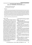
Zp variants in EBV infected children
Sequence differences identified within the major regula-
tory Zp domains (nucleotides -211 to +12) of EBV
infected individuals can be grouped into four variant
forms (Figure 1). Zp-P group sequences are identical to
the EBV prototype strain, B95.8. Zp-V3 and Zp-V4 vari-
ants have been previously described by Gutierrez et al.
[8]. Zp-V3 group sequences differ from Zp-P at three
positions: -100 (TTG), -106 (ATG), and -141 (ATG);
while Zp-4 sequences are characterized by same three
substitutions of the Zp-V3 variant in addition to a T to C
substitution at position -196. A new Zp variant was iden-
tified and named Zp-V1 and differs from Zp-P by a single
substitution at position -196 (TTC). As shown in Table 2,
The distribution of Zp subtypes involved all co-existence
variants-IM included P(n = 61), V3(n = 3), V1(n = 4),
V4(n = 6), P+V1(n = 6), P+V3(n = 2), P+V4(n = 5); EBV-
HLH included P(n = 23), V3(n = 8), V4(n = 1), P+V3(n =
12), P+V4(n = 2); CAEBV included P(n = 6), V3(n = 14),
P+V3(n = 12); controls included P(n = 29), V3 (n = 1),
V1(n = 3), V4(n = 4), P+V1(n = 1), P+V4(n = 2). We found
that Zp-P variant was the dominant genotype found in all
infection categories, except a relatively rare disease-
CAEBV, indicating that it was the primary variant of EBV
circulating in China. The Zp-V3 variant was the domi-
nant genotype in CAEBV cases (P ≤ 0.01) and relatively
high in EBV-HLH cases. The Zp-V1 variant, however,
was only found in IM and control cases, while the V4
variant was not detected in any CAEBV cases.
Co-existence of Zp variants and EBV subtypes
As shown in Table 3, the incidence of co-existence of Zp
variants (Figure 2) in HLH (30.4%) and CAEBV (37.5%)
cases was higher than for both IM (14.8%) and control
cases (7.5%). Interestingly, the Zp-P variant was present
in every co-existence case that harbored two Zp variants.
Zp-P+V1 variants were only detected in IM and control
categories, whereas Zp-P+V3 variants were predominant
Table 1: The frequency of EBV types 1 and 2 in each EBV-related disease group
Groups EBV subtypes (n/N)
Type 1Type 2Type 1 + Type 2
IM (n = 88) 94.3 (83/88) 5.7 (5/88) 0
HLH (n = 46) 93.4 (43/46) 6.6 (3/46) 0
CAEBV (n = 32) 81.3 (26/32) 6.2 (2/32) 12.5 (4/32)
Controls (n = 40) 95.0 (38/40) 5.0 (2/40) 0
Total (n = 206) 92.2 (190/206) 5.8 (12/206) 1.9(4/206)

Jin et al. Virology Journal 2010, 7:92
http://www.virologyj.com/content/7/1/92
Page 3 of 7
in CAEBV and HLH samples. The four type 1+2 EBV co-
infection cases detected in the CAEBV group all con-
tained Zp-P+V3 variants.
Discussion
This case-control study is the first investigation to
explore the association between EBV subtypes and
BZLF1-Zp variants and EBV infection in the China chil-
dren population. In this study, statistical analysis deter-
mined that differences in the distribution of Zp variants
were significant in the four patient categories. The fre-
quency of the Zp-V3 variant in the CAEBV group was
statistically higher than for other categories (P ≤ 0.01),
while Zp-P was predominant in all categories except
CAEBV. This suggests that the Zp-P variant EBV was the
most common variant found in China and that infection
by Zp-V3 is strongly correlated to CAEBV. The Zp-V3
variant is significantly associated with malignancy in
both immunocompetent and immunocompromised
patients [8,9] and the higher frequency of the Zp-V3 vari-
Figure 1 DNA sequences obtained for four EBV BZLF1 gene promoter zone (Zp) variants compared with the B95.8 prototype sequence. Po-
sitions relative to the transcription start site are indicated.

Jin et al. Virology Journal 2010, 7:92
http://www.virologyj.com/content/7/1/92
Page 4 of 7
ant in CAEBV patients observed in the current study sug-
gests that CAEBV is more likely an entity of pre-
malignancy. Similarly, the Zp-V4 variant was also identi-
fied in this study and was most associated with IM and
healthy control cases. Zp-V1 was identified as a novel
variant and was detected in 10 IM and health control
cases but not in CAEBV and EBV-HLH. The absence or
low level of ZP-V1 and Zp-V4 in CAEBV and HLH
reflects a less severe pathogenesis than for the Zp-V3
variant which may enhance the tumorigenicity of EBV.
A novel Zp variant that differed from Zp-P by one sub-
stitution at position -100 (TTG) was detect in this study
in only one patient with EBV-HLH. Due to the infrequent
isolation of this variant, we did not include this data in
correlations with disease. Previously described Zp vari-
ants, Zp-PV, Zp-V1-104, Zp-V1-105 or Zp-V1-119 [9,16]
were not detected in any patient samples. Although it
may be chance that these isolates were not detected, spe-
cific ethnic groups and geographical restrictions are likely
to contribute to the narrow distribution of variants
observed in the current study. The detection of different
new variants suggests that the accumulation of viral
mutations may contribute to the variations observed
within the host during virus persistence.
Similar to other studies that reported that type 1 EBV
was predominant in Asian nasopharyngeal carcinoma
(86.5-96%) [17,18], the current study also found that type
1 EBV was predominant in all four categories (81.3-95%).
Also in agreement to these studies, type 2 EBV infection
was rarely detected (4-13.5%). These findings suggest that
the diagnosis of EBV types 1 and 2 in patients is not likely
to be useful for predicting susceptibility to EBV-related
diseases in Chinese children. Although patients with Zp-
V4 or Zp-V1 variants were always type 1 EBV carriers,
this study did not confirm that Zp variants segregated by
EBV type due to the extremely lower frequency of type 2
EBV in the Chinese study population. Gutierrez et al. had
previously shown that the Zp-V3 variant was exclusively
associated with type 2 EBV infection; however, the cur-
rent study found that variant Zp-V3 co-existed with both
EBV types. Geographic regions, sample sizes or various
diseases are like to result in these differences.
The prevalence of co-existence EBV Zp variants within
the four categories studied ranged from 14.6 to 37.5%.
The majority of these co-existence viruses occurred in
patients with CAEBV and EBV-HLH and always was
found associated with Zp-P and not other variants. It is
likely that the majority of people are first infected with a
more prevalent variant like Zp-P, the predominant vari-
ant found in this study, but does not rule out the possibil-
ity that new point mutations are likely to be arised during
EBV replication of in its hosts from pre-existing variant.
In this way, the balance of one pre-existing virus variant
which could be controlled by its host, may be disturbed
by a specific new variant. Thus, virus replication, tro-
pism, or immune evasion in its hosts could be greatly
Table 2: The frequency of EBV BZLF1 gene promoter zone (Zp) variants in each EBV-related disease group.#
Zp-variant
%(n/N)
IM
(n = 88)
HLH
(n = 46)
CAEBV
(n = 32)
Controls
(n = 40)
P 84.1 (74/88) 80.4 (37/46) 56.3 (18/32)䉭85.0 (34/40)
V3 5.7 (5/88) 43.5 (20/46)䉭81.3 (26/32)䉭2.5 (1/40)
V4 12.5 (11/88) 6.5 (3/46) 0 15.0 (6/40)
V1 11.4 (10/88) 0 0 10.0 (4/40)
#The distribution of Zp subtypes involved all co-existence variants-IM included P(n = 61), V3(n = 3), V1(n = 4), V4(n = 6), P+V1(n = 6), P+V3(n
= 2), P+V4(n = 5); EBV-HLH included P(n = 23), V3(n = 8), V4(n = 1), P+V3(n = 12), P+V4(n = 2); CAEBV included P(n = 6), V3(n = 14), P+V3(n =
12); controls included P(n = 29), V3 (n = 1), V1(n = 3), V4(n = 4), P+V1(n = 1), P+V4(n = 2)
䉭(P ≤ 0.01) vs other groups
Table 3: The co-existence of EBV BZLF1 gene promoter zone (Zp) variants and EBV subtypes in each EBV-related disease
study group.
Co-infection
%(n/N)
IM
(n = 88)
HLH
(n = 46)
CAEBV
(n = 32)
Controls
(n = 40)
Zp-P+V1 6.8 (6/88) 0 0 2.5(1/40)
Zp-P+V4 5.7 (5/88) 4.3 (2/46) 0 5.0 (2/40)
Zp-P+V3 2.3 (2/88) 26.1(12/46) 37.5(12/32) 0
Total 14.8 (13/88) 30.4(14/46) 37.5(12/32) 7.5 (3/40)
* Four patients detected in CAEBV with both type 1 and type 2 co-infections were also presented with Zp-P and Zp-V3 co-exsistences.

Jin et al. Virology Journal 2010, 7:92
http://www.virologyj.com/content/7/1/92
Page 5 of 7
enhanced after acquiring this new variant. As the Zp-V3
variant was associated with severe diseases in this study,
the Zp-V3 type point mutations derived from Zp-P are
likely to be associated with a more invasive capacity than
Zp-V1 or Zp-V4 variants. Taken together, superinfection
by multiple strains of EBV, especially the presence of the
Zp-V3 variant, may be a contributing factor in the devel-
opment of severe EBV infections in children. Thus, these
findings may give some prospect to explore the differen-
tial distribution of Zp variants in susceptible populations
and their association with severe or even fatal EBV dis-
eases. A close dynamic follow-up on patients carrying
EBV from an early stage of infection may help us under-
stand how the host immune response allows such muta-
tions to occur.
Just how an individual acquires such mixtures of Zp
variants is unknown. This could occur by simultaneous
acquisition or by the serial accumulation from exposure
to different variant carriers. It seems implausible that
such co-infections can be co-acquired from a carrier who
was shedding multiple variants in saliva, because it is
unclear how the source can accumulate multiple infec-
tions before transmitting those orally shedding multiple
EBV variants to the next. As infection by EBV with the
Zp-P variant was a prerequisite for co-existence in this
study, it is possible that an individual is more likely to
acquire a prevalent variant, such as Zp-P, at first exposure
to the virus, and then the host immunity to this variant is
developed. However, part of hosts may fall short in resist-
ing another different variant the next time. It is more
Figure 2 DNA sequences obtained for co-exisence of EBV BZLF1 gene promoter zone (Zp) variants compared with the B95.8 prototype se-
quence. Positions relative to the transcription start site are indicated.