Zohari et al. Virology Journal 2010, 7:145
http://www.virologyj.com/content/7/1/145
Open Access
RESEARCH
© 2010 Zohari et al; licensee BioMed Central Ltd. This is an Open Access article distributed under the terms of the Creative Commons
Attribution License (http://creativecommons.org/licenses/by/2.0), which permits unrestricted use, distribution, and reproduction in
any medium, provided the original work is properly cited.
Research
Full genome comparison and characterization of
avian H10 viruses with different pathogenicity in
Mink (
Mustela vison
) reveals genetic and functional
differences in the non-structural gene
Siamak Zohari*
1,2
, Giorgi Metreveli
1
, István Kiss
2
, Sándor Belák
1,2
and Mikael Berg
1
Abstract
Background: The unique property of some avian H10 viruses, particularly the ability to cause severe disease in mink
without prior adaptation, enabled our study. Coupled with previous experimental data and genetic characterization
here we tried to investigate the possible influence of different genes on the virulence of these H10 avian influenza
viruses in mink.
Results: Phylogenetic analysis revealed a close relationship between the viruses studied. Our study also showed that
there are no genetic differences in receptor specificity or the cleavability of the haemagglutinin proteins of these
viruses regardless of whether they are of low or high pathogenicity in mink.
In poly I:C stimulated mink lung cells the NS1 protein of influenza A virus showing high pathogenicity in mink down
regulated the type I interferon promoter activity to a greater extent than the NS1 protein of the virus showing low
pathogenicity in mink.
Conclusions: Differences in pathogenicity and virulence in mink between these strains could be related to clear amino
acid differences in the non structural 1 (NS1) protein. The NS gene of mink/84 appears to have contributed to the
virulence of the virus in mink by helping the virus evade the innate immune responses.
Background
The outbreak of severe respiratory disease in mink (Mus-
tela vison) in 1984 was linked to an avian influenza virus
of subtype H10N4. At the time this was the first known
outbreak of avian influenza A virus infection in a terres-
trial mammalian species [1,2]. The only possible explana-
tion was that birds carrying the virus transmitted it via
their faeces to the mink. At the time, this was one of the
very first cases of direct transmission of avian influenza
virus to a terrestrial mammalian species [1].
Only a few months after the outbreak in Swedish mink,
some viruses of the H10N4 subtype were isolated from
domestic and wild birds in Great Britain [3]. Rather crude
full-genomic comparison by oligonucleotide (ON) map-
ping [4] and sequence analysis of the HA [5] and NP
genes [6] were conducted. The ON mapping showed a
close genomic relationship between the mink isolate (A/
Mink/Sweden/3900/84) and the concomitant avian
H10N4 viruses from fowl (A/fowl/Hampshire/378/85)
and mallard (A/mallard/Gloucestershire/374/85) respec-
tively, and a weaker genomic relationship with the H10
prototype [7] virus (A/chicken/Germany/N/49) [4].
Experimental infection of mink (Mustela vison) was ini-
tially used to link the isolated influenza virus to the clini-
cal symptoms and pathological lesions observed in the
field outbreak. In a later study, mink were infected intra-
nasally with mink/84, mallard/85, fowl/85, or chicken/49
to compare clinical symptoms, antibody response, and
possible in-contact transmission [4].
Experimental aerosol infections of mink, using mink/84
or chicken/49, were then used to compare in more detail
the pathogenesis of the two virus infections [8,9]. Follow-
* Correspondence: Siamak.zohari@sva.se
1 Swedish University of Agricultural Sciences (SLU), Department of Biomedical
Sciences and Public Health, Section of Virology, SLU, Ulls väg 2B, SE-751 89
Uppsala, Sweden
Full list of author information is available at the end of the article

Zohari et al. Virology Journal 2010, 7:145
http://www.virologyj.com/content/7/1/145
Page 2 of 11
ing intranasal infection of the mink, all three H10N4 iso-
lates, i.e. mink/84, mallard/85 and fowl/85, showed
similar clinical symptoms, causing respiratory disease,
interstitial pneumonia and specific antibody production.
All three H10N4 isolates were transmitted via contact
infection. Chicken/49 did not cause clinical disease or
contact infection, but induced antibody production and
mild lung lesions [8].
Further comparison between mink/84 and chicken/49
revealed that the infections progressed with similar pat-
terns over the first 24 hours post infection but from 48
hours post infection obvious differences were recorded.
In mink infected with chicken/49 no signs of disease were
observed, while the mink infected with mink/84 showed
severe signs of respiratory disease, with inflammatory
lesions spreading throughout the lung and viral antigen
present in substantial numbers of cells in the lung, nasal
mucosa, and trachea. The chicken/49 and mink/84 virus
have also been shown to differ in their ability to induce
interferon (IFN) production in mink lung cells [8-10].
In an effort to better understand the mechanism behind
the virulence of influenza A viruses we characterized the
complete genome of influenza A viruses that clearly
showed different pathogenicity for mink.
Results and discussion
The outcome of influenza A virus infection is influenced
both by the virus and the infected host [11,12]. The viru-
lence of an influenza virus isolate for a given host reflects
its ability to enter a host cell, replicate within the cell and
then exit and spread to new cells. Several viral gene prod-
ucts can contribute to the pathogenicity and virulence of
the influenza A virus [13,14]. Although in most instances
virulence is a multigenic trait, a single gene can also
markedly affect the pathogenicity and virulence of the
virus [15-18].
Phylogenetic and sequence analysis
We sequenced the complete genome of five H10 viruses
and analysed them along with all H10 viruses available in
the GenBank database. Phylogenetic relationships were
determined for each of the eight gene segments. The
amino acid sequences of the entire genome were analysed
to identify important amino acid residues associated with
enhanced replication and virulence in mammalian spe-
cies.
Haemagglutinin
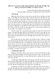
Phylogenetic analysis of the HA gene revealed that all of
the H10 viruses examined in this study belong to the Eur-
asian avian lineage of the influenza A viruses (Figure 1).
Based on the limited sequence data from the Eurasian
avian lineage of H10 influenza viruses that are available in
GenBank, a clear determination of the genetic relation-
ship among H10 viruses is very difficult. Furthermore,
the HA gene of mink/84 clustered with mallard/85, fowl/
85 and whistlingswan/88 within the Eurasian avian lin-
eage and was distinct from the HA of chicken/49, which
clusters with the early H10 Eurasian avian isolates. There
is a high degree of similarity at the amino acid level of the
haemagglutinin gene of the studied viruses. The HA
genes of mink/84 and the concomitant wild bird isolates
were 98% identical with each other and showed 95% simi-
larity to the prototype H10 virus, chicken/49. The HA
gene of H10 viruses was analysed for potential N-glycosy-
lation sites. Our analysis indicated that all the studied
H10 viruses possess five potential glycosylation sites
(positions 13, 29, 236, 406 and 447) except for fowl/85,
which displayed an additional glycosylation site at residue
123. Interestingly fowl/85 virus was originally isolated
from a flock of sick chickens with nephropathy and vis-
ceral gout [3]. Several studies indicate that the receptor
specificity of haemagglutinin plays an important role for
tissue tropism and the host range of the influenza virus
[19]. The amino acid composition of the receptor binding
pocket of the HA protein for the H10 isolates is typical of
avian influenza viruses. The H10 viruses have histidine
(H), glutamate (E) and glutamine (Q) at amino acid posi-
tions 177, 184, and 216, respectively, in H10 numbering at
the receptor-binding site [20], which favours binding of
sialic-acid α-2,3-galactose.
The haemagglutinin cleavability and the presence of
multiple basic amino acids at the HA cleavage site play a
major role in influenza H5 and H7 virus transmission and
virulence [21-23]. All five H10 isolates presented in this
study contained the amino acid sequence PQG÷RGLF at
the cleavage site in the HA molecule, indicating their low
pathogenicity. Nevertheless four of these H10 viruses
were found to be highly pathogenic in mink. It is note-
worthy that at least two H10 isolates (A/turkey/England/
384/79/H10N4 and A/mandarin duck/Singapore/8058F-
72/7/93/H10N5) that were reported previously by Wood
and co authors, would have fulfilled the definition for
highly pathogenic viruses with intravenous pathogenicity
index (IVPI) values > 1.2, without having multiple basic
amino acids at their haemagglutinin cleavage site [24].
This suggests that a factor other than the presence of
multiple basic amino acids in the cleavage site contrib-
uted to the severity of H10 viruses in mink.
Neuraminidase
Two different NA subtypes (N4 and N7) were associated
with H10 viruses in this study. Phylogenetic analysis of
the NA gene showed that all viruses belonged to the Eur-
asian avian lineage and within each NA subtype, the
viruses clustered in the same branches. The NA protein
plays an important role during the entry of the virus into
the cells and in the release of viral progeny from infected

Zohari et al. Virology Journal 2010, 7:145
http://www.virologyj.com/content/7/1/145
Page 3 of 11
Figure 1 Phylogenetic relationship between haemagglutinin genes of H10 influenza A viruses. The protein coding region tree was generated
by neighbour-joining analysis with the Tamura-Nei γ-model, using MEGA 4.0. Numbers below key nodes indicate the percentage of bootstrap values
of 2000 replicates. Isolates sequenced in this study are indicated by a red dot.

Zohari et al. Virology Journal 2010, 7:145
http://www.virologyj.com/content/7/1/145
Page 4 of 11
cells [25,26]. The active site of the NA protein consists of
15 charged amino acids that are conserved in all influenza
A viruses [27]. All of these amino acids that make up the
active site (R117, D150, R151, R224, E276, R292, R369
and Y403 in N4 numbering) and the framework site
(E119, R155, W178, S179, D/N198, I122, E227, H274,
E277, N294 and E425) of the NA are conserved in the
H10 viruses presented in this study. H10 influenza
viruses have a propensity to cause clinical symptoms in
humans; experimental and natural infections with H10N7
strains have clearly shown the zoonotic potential of some
H10 avian influenza viruses [28,29]. In the NA protein of
the analysed H10 isolates no substitutions associated
with resistance to neuraminidase inhibitor drugs (oselta-
mivir) were observed [30].
It has been suggested that the efficiency of viral replica-
tion in terrestrial domestic poultry correlates with the
length of the NA stalk and that stalk deletion has resulted
in adaptation of the virus to land-based poultry [26,31].
No deletions were found in the stalk regions of the
neuraminidase of the viruses sequenced in this study,
indicating no adaptation for growth in terrestrial domes-
tic poultry, this despite the fact that two of the studied
viruses have been isolated from sick chickens [3,7].
Internal genes
The ribonucleoprotein (RNP) complex of influenza virus
contains four proteins that are necessary for viral replica-
tion: PB1, PB2, PA and NP. Several substitutions in the
polymerase complex proteins are implicated in the viru-
lence of the influenza viruses [32,33]. Previous studies
showed that most of the host-specific markers that dis-
criminate between human and avian influenza viruses are
located in viral RNPs [34,35]. Our analysis indicates the
clear avian origin of the studied viruses with two excep-
tions; substitution E627K in PB2 has been shown to be
important for adaptation of avian viruses to replication in
mammalian hosts, and interestingly, our sequence analy-
sis showed that viruses isolated in mink and concomitant
H10 viruses carry a glutamic acid (E) at position 627
which is typically found in avian viruses, while chicken/
49-like human viruses have a lysine (K) substitution at
position 627 of PB2. This substitution has been shown to
be the main determinant of the pathogenicity of avian
influenza viruses in mammalian hosts and results in
increased replication of viruses in the upper respiratory
tract of mice and ferrets [17,36-38]. Substitution D701N
in polymerase protein PB2 has been implicated in the
adaptation of H5N1 viruses to replication and high
pathogenicity in mammalian hosts [35], this being the
same substitution as seen in mink/84.
The recently discovered PB1-F2, a 90-amino-acid pep-
tide translated from an alternative reading frame of the
PB1 gene, induces apoptosis in infected cells [39]. The
substitution N66S resulted in a more severe infection
with higher virus titres and increased production of
inflammatory cytokines in the lungs of infected mice
[40]. None of the viruses presented in this study con-
tained the N66S substitution. Similarity percentages for
the gene segments of the RNP complex varied from 88 to
95% for the PA gene to 90-100% for the PB1-F2 at the
nucleotide level.
Phylogenetic relationships were inferred for each of the
gene segments of the RNP complex. All virus genes
belong to the Eurasian avian lineage with the exception of
the PB1 gene of whistlingswan/88. With regards to the
PB1 gene, the whistlingswan/88 virus formed a sister
branch with the main American avian lineage of H10
viruses, indicating the reassortment with genes belonging
to the American avian gene pool (Figure 2).
Phylogenetic analysis showed that the M genes of the
H10 viruses presented in this study are closely related to
each other and all belong to the Eurasian avian lineage of
the influenza A viruses. Four amino acids substitutions
(L26F, V27A or T, A30T or V and S31N or R) at the M2
gene have been shown to be associated with resistance to
amantadine [41], an anti-influenza drug commonly used
in humans. Analysis of M2 protein amino acid sequences
showed that the H10 isolates are all sensitive to amanta-
dine.
Two distinct gene pools of the non structural gene
(NS), corresponding to allele A and allele B [42,43], were
present among the studied H10 viruses. The NS gene of
mink/84 clustered together with mallard/85, fowl/85 and
whistlingswan/88 in allele A within the Eurasian avian
lineage and it was clearly distinct from the NS of chicken/
49, which formed a single branch as the only Eurasian
avian H10 isolate among the allele B viruses (Figure 3).
The NS1 genes of the H10 viruses reported in this study
consisted of 890 nucleotides; there were no deletions or
insertions. Nucleotide sequence identities of the NS1
gene in allele A were 95-100%, however there was 63%
nucleotide identity and 69% amino acid identity between
mink/84 in allele A and chicken/49 in allele B.
Several studies have identified significant amino acid
motifs associated with the increased virulence of avian
influenza viruses in human (D92E) and chickens (V149A)
[37,44]. All the H10 viruses in our study contained 92D
and 149A. Obenauer and colleagues (2006) proposed that
the four C-terminal amino acid residues of the NS1 act as
a PDZ binding motif that may represent a virulence
determinant. PDZ domains are protein-interacting
domains that are involved in a variety of cell signalling
pathways. In addition, Obenauer and colleagues (2006)
showed that there were typical human, avian, equine and
swine motifs [45]. All the H10 viruses possessed the typi-
cal avian ESEV amino acid sequence at the C-terminal
end of the NS1 protein.
Zohari et al. Virology Journal 2010, 7:145
http://www.virologyj.com/content/7/1/145
Page 5 of 11
The unique property of some avian H10 viruses, partic-
ularly the ability to cause severe disease in mink without
prior adaptation, enabled our study. Coupled with previ-
ous experimental data and genetic studies we tried to
investigate the possible influence of different genes on the
virulence of these H10 avian influenza viruses in mink.
Of those amino acid residues previously described as vir-
ulence factors influencing the outcome of the avian influ-
enza virus infection in mammalian species only one was
present in the H10 viruses studied here. Although Hatta
et al. (2001) found that only a single amino acid substitu-
tion E627K of the PB2 contributes to efficient replication,
effective transmission and virulence of H5N1 influenza
virus in mammalian species [17], it seems that the exis-
tence of this mutation in PB2 of chicken/49 does not
influence the virulence of this virus in mink. There were
no differences in receptor specificity or the cleavability of
the haemagglutinin proteins between H10 viruses that
Figure 2 Phylogenetic relationship between polymerase basic protein 1 genes of H10 influenza A viruses. The protein coding region tree was
generated by neighbour-joining analysis with the Tamura-Nei γ-model, using MEGA 4.0. Numbers below key nodes indicate the percentage of boot-
strap values of 2000 replicates. Isolates sequenced in this study are indicated by a red dot.


![Vaccine và ứng dụng: Bài tiểu luận [chuẩn SEO]](https://cdn.tailieu.vn/images/document/thumbnail/2016/20160519/3008140018/135x160/652005293.jpg)