Grande et al. Journal of Inflammation 2010, 7:19
http://www.journal-inflammation.com/content/7/1/19
Open Access
REVIEW
BioMed Central
© 2010 Grande et al; licensee BioMed Central Ltd. This is an Open Access article distributed under the terms of the Creative Commons
Attribution License (http://creativecommons.org/licenses/by/2.0), which permits unrestricted use, distribution, and reproduction in
any medium, provided the original work is properly cited.
Review
Role of inflammation in túbulo-interstitial damage
associated to obstructive nephropathy
María T Grande
1,2
, Fernando Pérez-Barriocanal
1,2
and José M López-Novoa*
1,2
Abstract
Obstructive nephropathy is characterized by an inflammatory state in the kidney, that is promoted by cytokines and
growth factors produced by damaged tubular cells, infiltrated macrophages and accumulated myofibroblasts. This
inflammatory state contributes to tubular atrophy and interstitial fibrosis characteristic of obstructive nephropathy.
Accumulation of leukocytes, especially macrophages and T lymphocytes, in the renal interstitium is strongly associated
to the progression of renal injury. Proinflammatory cytokines, NF-κB activation, adhesion molecules, chemokines,
growth factors, NO and oxidative stress contribute in different ways to progressive renal damage induced by
obstructive nephropathy, as they induce leukocytes recruitment, tubular cell apoptosis and interstitial fibrosis.
Increased angiotensin II production, increased oxidative stress and high levels of proinflammatory cytokines contribute
to NF-κB activation which in turn induce the expression of adhesion molecules and chemokines responsible for
leukocyte recruitment and iNOS and cytokines overexpression, which aggravates the inflammatory response in the
damaged kidney. In this manuscript we revise the different events and regulatory mechanisms involved in
inflammation associated to obstructive nephropathy.
Introduction
Obstructive nephropathy due to congenital or acquired
urinary tract obstruction is the first primary cause of
chronic renal failure (CRF) in children, according to data
of The North American Pediatric Renal Transplant
Cooperative Study (NAPRTCS) [1]. Obstructive neph-
ropathy is also a major cause of renal failure in adults
[2,3].
The renal consequences of chronic urinary tract
obstruction are very complex, and lead to renal injury
and renal insufficiency. The experimental model of uni-
lateral ureteral obstruction (UUO) in rat and mouse has
become the standard model to understand the causes and
mechanisms of nonimmunological tubulointerstitial
fibrosis. This is because it is normotensive, nonproteinu-
ric, nonhyperlipidemic, and without any apparent
immune or toxic renal insult. The UUO consists of an
acute obstruction of one of the ureter that mimics the dif-
ferent stages of obstructive nephropathy leading to tubu-
lointerstitial fibrosis without compromising the life of the
animal, because the contralateral kidney maintains or
even increases its function due to compensatory func-
tional and anatomic hypertrophy [2,3].
The evolution of renal structural and functional
changes following urinary tract obstruction in these
models has been well described. The first changes
observed in the kidney are hemodynamic, beginning with
renal vasoconstriction mediated by increased activity of
the renin-angiotensin system and other vasoconstrictor
systems [4]. Epithelial tubular cells are damaged by the
stretch secondary to tubular distension and the increased
hydrostatic pressure into the tubules due to accumulation
of urine in the pelvis and the retrograde increase of inter-
stitial pressure. This is followed by an interstitial inflam-
matory response initially characterized by macrophage
infiltration. There is also a massive myofibroblasts accu-
mulation in the interstitium. These myofibroblasts are
formed by proliferation of resident fibroblasts, from bone
marrow-derived cells, from pericyte infiltration, as well
by epithelial-mesenchymal transformation (EMT), a
complex process by which some tubular epithelial cells
acquire mesenchymal phenotype and become activated
myofibroblasts [5,6].
Damaged tubular cells, interstitial macrophages and
myofibroblasts produce cytokines and growth factors
that promote an inflammatory state in the kidney, induce
* Correspondence: jmlnovoa@usal.es
1 Instituto "Reina Sofía" de Investigación Nefrológica, Departamento de
Fisiología y Farmacología, Universidad de Salamanca, Salamanca, Spain
Full list of author information is available at the end of the article
Grande et al. Journal of Inflammation 2010, 7:19
http://www.journal-inflammation.com/content/7/1/19
Page 2 of 14
tubular cell apoptosis and provoke the accumulation of
extracellular matrix. The end-result of severe and chronic
obstructive nephropathy is a progressive renal tubular
atrophy with loss of nephrons accompanied by interstitial
fibrosis. Thus, interstitial fibrosis is the result of these
processes in a progressive and overlapping sequence. The
evolution of renal injury in obstructive nephropathy
shares many features with other forms of interstitial renal
disease such as acute renal failure, polycystic kidney dis-
ease, aging kidney and renal transplant rejection [7-9].
The final fibrotic phase is very similar to virtually all pro-
gressive renal disorders, including glomerular disorders
and systemic diseases such as diabetes or hypertension
[4].
In this review we will analyze the role of inflammation
on renal damage associated to obstructive nephropathy,
and the cellular and molecular mechanisms involved in
the genesis of these processes. As later described, the
inflammatory process, through the release of cytokines
and growth factors, results in the accumulation of inter-
stitial macrophages which, in turn, release more cytok-
ines and growth factors that contribute directly to tubular
apoptosis and interstitial fibrosis [10,11].
Urinary obstruction induces an inflammatory state in the
kidney
In Sprague-Dawley rats subjected to chronic neonatal
UUO (from 2 to 12 days), microarray analysis revealed
that the mRNA expression of multiple immune modula-
tors, including krox24, interferon-gamma regulating fac-
tor-1 (IRF-1), monocyte chemoattractant protein-1
(MCP-1), interleukin-1β (IL-1β), CCAAT/enhancer bind-
ing protein (C/EBP), p21, c-fos, c-jun, and pJunB, were
significantly increased in obstructed compared to sham-
operated kidneys, thus suggesting that UUO induces a
pro-inflammatory environment [12]. This environment is
characterized by up-regulation of inflammatory cytok-
ines and factors that favors leukocyte infiltration. Other
cytokines with different functions are also differentially
regulated after UUO, and will contribute to the regula-
tion of inflammation and interstitial infiltration. Thus, we
will review the data available about the mechanisms
involved in this inflammatory state, including nuclear
factor κB (NF-κB) activation, increased oxidative stress,
interstitial cell infiltration, and production of proinflam-
matory cytokines and other growth factors with inflam-
matory or anti-inflammatory properties, in the renal
damage after UUO.
Thus, monocytes/macrophages, T cells, dendritic cells
and neutrophils are involved in this inflammatory state of
the kidney after UUO. Whereas interstitial macrophages
increases 4 hours after UUO and constitute the predomi-
nant infiltrating cell population in acutely obstructed kid-
neys, T cells are also evident after 24 h of obstruction
although neither B lymphocytes nor neutrophils are
observed. Moreover, interstitial macrophages increases
biphasically with an initial rapid increase during the first
24 h after UUO and the second phase following 72 h after
UUO and all reports which observed an inverse correla-
tion between interstitial macrophage number and the
degree of fibrosis was noted at the later stage of UUO
(day 14) and therefore it will be believed the possible
renoprotective role for macrophages that infiltrate in the
later phase after UUO [13].
NF-κB activation
NF-κB is a ubiquitous and well-characterized transcription
factor with a pivotal role in control of the inflammation,
among other functions. Thus, NF-κB controls the expres-
sion of genes encoding pro-inflammatory cytokines (e. g.,
IL-1, IL-2, IL-6, TNF-α, etc.), chemokines (e. g., IL-8, MIP-
1 α, MCP-1, RANTES, eotaxin, etc.), adhesion molecules
(e. g., ICAM, VCAM, E-selectin), inducible enzymes
(COX-2 and iNOS), growth factors, some of the acute
phase proteins, and immune receptors, all of which play
critical roles in controlling most inflammatory processes
[14,15]. Also the PI3K/Akt pathway, which has been
reported to be activated very early after UUO [16], results
in activation of NF-κB [17]. NF-κB also controls the
expression of EMT inducers (e.g., Snail1), and enhances
EMT of mammary epithelial cells [18,19] (Figure 1).
NF-κB is activated by several cytokines such as IL-1β,
TNF-α, by oxidative stress and by other molecules such
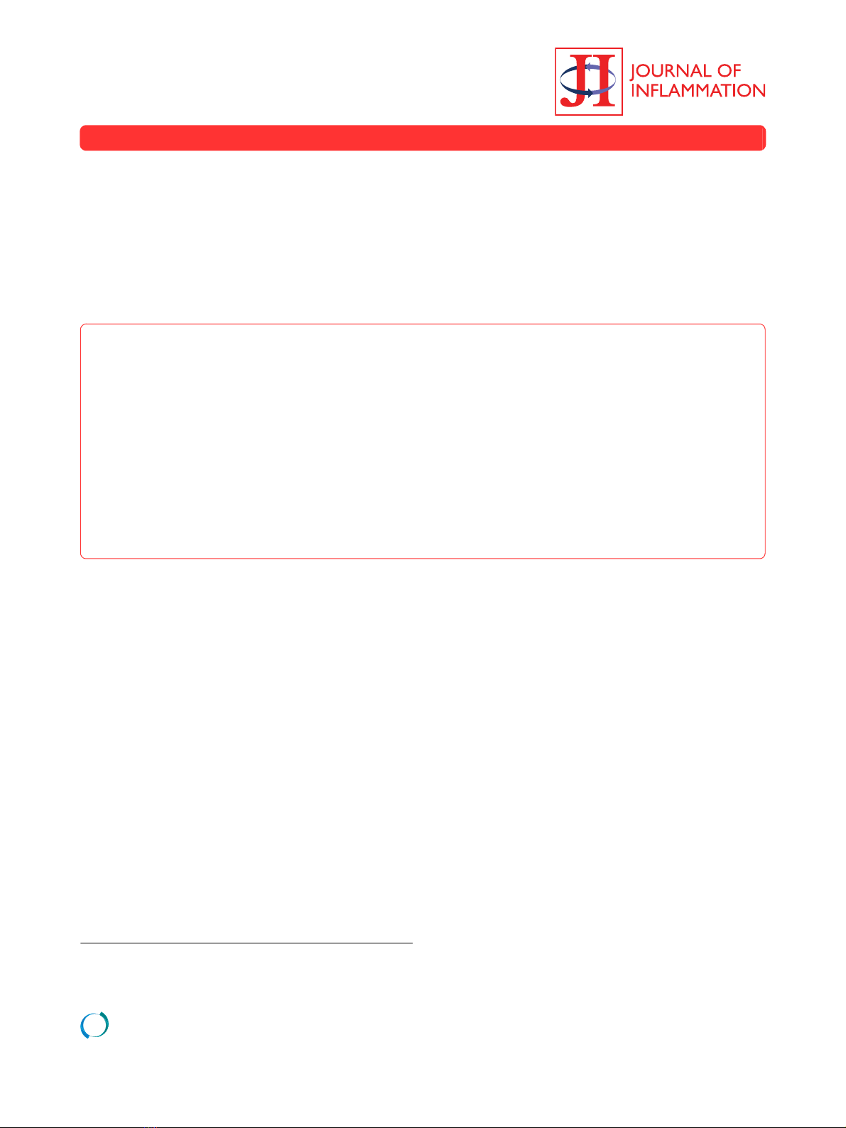
Figure 1 Schematic representation of some of the signaling inter-
mediates potentially involved in regulation of inflammatory re-
sponse after UUO. UUO induces IL-1β and TNF-α expression, leading
to NF-κB activation. UUO also induces both oxidative stress and in-
creased Angiotensin II (Ang II) levels. Ang II also activate the transcrip-
tion factor NF-κB, both directly and indirectly, by promoting oxidative
stress, which in turns activate Ang II by regulating angiotensinogen ex-
pression. TGF-β activates NF-κB through I-κB inhibition, a mechanism
shared by TNF-α. NF-κB activation concludes in IL-1β and TNF-α ex-
pression enhancing NF-κB activation. Also NF-κB controls the expres-
sion of genes encoding pro-inflammatory cytokines, adhesion
molecules and iNOS.
UUO
NF-kappaB
activation
IL-1ȕTNF-ĮROS Ang II
Angiotensinogen
HGF
TGF-ȕ
I-kappaB
Inflammation & Oxidative stress
TNF-ĮIL-1ȕMCP-1 RANTES
VCAM
ICAM
iNOS EMT
Ĺ(&0SURGXFWLRQ
Ļ(&0GHJUDGDWLRQ
TUBULOINTERSTITIAL FIBROSIS

Grande et al. Journal of Inflammation 2010, 7:19
http://www.journal-inflammation.com/content/7/1/19
Page 3 of 14
as Angiotensin II (Ang II) [20]. Obstructed kidneys pre-
sented many cells that contained activated NF-κB com-
plexes, in glomeruli, in tubulointerstitial cells and in
infiltrating cells [21]. NF-κB is activated very early follow-
ing UUO [22] and it is maintained activated during at
least 7 days after UUO [21]. Furthermore, inhibition of
NF-κB activation decreases apoptosis and interstitial
fibrosis in rats with UUO [23]. NF-κB inhibition also
diminishes monocyte infiltration and inflammation gene
overexpression after UUO [21]. The administration of a
proteasome inhibitor to maintain levels of I-κB, an
endogenous inhibitor of NF-κB, reduces renal fibrosis
and macrophage influx following UUO [24].
Renal cortical TNF-α levels increases early after UUO,
whereas TNF-α neutralization with a pegylated form of
soluble TNF receptor type 1 significantly reduced
obstruction-induced TNF-α production, as well as NF-κB
activation, IκB degradation, angiotensinogen expression,
and renal tubular cell apoptosis, thus suggesting a major
role for TNF-α in activating NF-κB via increased IκB-
alpha phosphorylation [25].
In addition, curcumin, a phenolic compound with anti-
inflammatory properties, has revealed protective action
against interstitial inflammation in obstructive nephropa-
thy by inhibition of the NF-κB-dependent pathway [26].
HGF has also been reported to inhibit renal inflamma-
tion, proinflammatory chemokine expression and renal
fibrosis in an UUO model. The anti-inflammatory effect
of HGF is mediated by disrupting nuclear factor NF-κB
signaling, as later will be described [27].
NF-κB can be also activated by oxidative stress. The
administration of antioxidant peptides to rats that suf-
fered UUO was associated to a lower activation of NF-κB,
and significantly attenuated the effects of ureteral
obstruction on all aspects of renal damage associated to
UUO [28]. Thus, oxidative stress seems to play also a
major role in the UUO-associated inflammation.
Oxidative stress
Oxidative stress has been implicated in the pathogenesis
of various forms of renal injury [29]. Oxidative stress is
also a major activator of the NF-κB and thus, an inductor
of the inflammatory state [30] (Figure 1). There are sev-
eral evidences showing that increased oxidative stress is
involved in renal inflammatory damage after UUO. Reac-
tive oxygen species are significantly increased in the
chronically obstructed kidney [31] and a positive correla-
tion was observed between the levels of free radical oxi-
dation markers in the obstructed kidney tissue and in
plasma [32]. Superoxide anion and hydrogen peroxide
production increase significantly in the obstructed kid-
ney [33]. After 5 days of obstruction, it has been reported
a slight increase on renal cortex NADPH oxidase activity
(a major source for superoxide production) whereas after
14 days of obstruction, a marked increase on NADPH
oxidase activity was observed. In addition, decreased
superoxide dismutase activity were demonstrated follow-
ing 14 days of obstruction whereas no differences were
noticed after 5 days of kidney obstruction [34].
Increased Ang II production, accumulation of activated
phagocytes in the interstitial space and elevation of
medium-weight molecules have been involved as respon-
sible for the increased oxidative stress [35] after UUO.
UUO also generate increased levels of carbonyl stress,
and subsequently advanced glycation end-products
(AGEs), and nitration adduct residues, both contributing
to the progression of renal disease in the obstructed kid-
ney [36,37]. The products of lipid peroxidation have been
also found increased in both plasma and obstructed kid-
ney after UUO [38]. Carboxymethyl-lysine, a marker for
accumulated oxidative stress, was found to be increased
in the interstitium of the obstructed kidneys [39]. Fur-
thermore, heme oxygenase-1 (HO-1) expression, a sensi-
tive indicator of cellular oxidative stress, was also found
to be induced as early as 12 hours after ureteral obstruc-
tion [39]. All these results suggest that oxidative stress is
involved in the pathogenesis of UUO. On the other hand,
levels of the antioxidant enzyme catalase and copper-zinc
superoxide dismutase, which prevent free radical dam-
age, are lower in the obstructed kidney compared with
the contralateral unobstructed kidney [33].
Antioxidant compounds, such as tocopherols reduce
the level of oxidative stress observed after UUO [38].
Moreover, the administration of isotretinoin, a retinoid
agonist, reduces renal macrophage infiltration in rats
with UUO [39]. It should be noted that an increase in cel-
lular reactive oxygen species (ROS) production stimulate
the expression of the transcription factor Snail and favors
EMT [40].
In short, oxidative stress markers levels increase in the
kidney during UUO whereas levels of enzymes that pre-
vent the oxidative damage are diminished in the
obstructed kidney. All these data suggest that oxidative
stress is increased in the obstructed kidney, and that
increased oxidative stress plays a role in inducing an
inflammatory state and in deteriorating the renal func-
tion of the obstructed kidney.
Angiotensin II
Angiotensin II (Ang II) behaves in the kidney as a proin-
flammatory mediator, as it regulates a number of genes
associated with progression of renal disease. The regula-
tion of gene expression by Ang II occurs through changes
in the activity of transcription factors within the nucleus
of target cells. In particular, several members of the NF-
κB family of transcription factors are activated by Ang II,
which in turn fuels at least two autocrine reinforcing
loops that amplify Ang II and TNF-α formation [41].

Grande et al. Journal of Inflammation 2010, 7:19
http://www.journal-inflammation.com/content/7/1/19
Page 4 of 14
Thus, it is not surprisingly the interrelation between Ang
II and proinflammatory cytokines effects in the intersti-
tial cell infiltration after UUO. Many studies have demon-
strated that obstructive nephropathy leads to activation
of the intrarenal renin-angiotensin system [4,42,43]. This
system is also activated in animal models of UUO. Ang II
has a central role in the beginning and progression of
obstructive nephropathy, both directly and indirectly, by
stimulating production of molecules that contribute to
renal injury. Following UUO, Ang II activates NF-κB, and
the subsequent increased expression of proinflammatory
genes [22]. In turn, the angiotensinogen gene is stimu-
lated by activation of NF-κB [44] (Figure 1). In relation to
the inflammatory process, Ang II type 1 receptor (AT1R)
regulates several proinflammatory genes, including
cytokines (interleukin-6 [IL-6]), chemokines (monocyte
chemoattractant protein 1 [MCP-1]), and adhesion mole-
cules (vascular cell adhesion molecule 1 [VCAM-1]) [45],
but others, as the chemokine RANTES, are regulated by
the Ang II type 2 receptor (AT2R) [46]. Some evidence
suggests that AT2R participates in the inflammatory
response in renal and vascular tissues [45-47]. In vivo and
in vitro studies have shown that Ang II activates NF-κB in
the kidney, via both AT1R and AT2R receptors [48,49].
Most studies have focused on the role of AT1R activa-
tion on kidney inflammation after UUO. For instance,
inhibition or inactivation of AT1R also reduces NF-κB
activation in the obstructed kidneys after UUO [50,51].
Also AT1R blockade, partially decreased macrophage
infiltration in the obstructed kidney [21,50,52]. Thus
AT1R activation seems to play a role in the UUO-associ-
ated inflammation. However, obstructed kidney in AT1R
KO mice showed interstitial monocyte infiltration and
NF-κB activation, and both processes were abolished by
AT2R blockade, suggesting that AT2R activation plays
also a major role in UUO-induced renal inflammation
[21]. Simultaneous blockade of both AT1R and AT2R is
able to completely prevent the inflammatory process
after UUO [21], thus giving a further proof of the role of
both receptors in the inflammatory state occurring after
UUO. It should be noted that in wild-type mice reconsti-
tuted with bone marrow cells lacking the angiotensin
AT1R gene, UUO results in more severe interstitial fibro-
sis despite fewer interstitial macrophages [53]. This effect
seems to be due to impaired phagocytic function of
AT1R-deficient macrophages [53]. This is a typical exam-
ple of the fact that manipulation of a single molecule
affecting more than one renal compartment could have
opposite effects in different compartments.
Treatment with angiotensin converting enzyme (ACE)
inhibitors greatly reduced the monocyte/macrophage
infiltration in the obstructed kidney [54] but this reduc-
tion seems to be observed only in the short-term UUO,
and 14 days after UUO ACE inhibitors did not decreased
monocyte/macrophage infiltration, maybe because in
late-stage UUO, infiltration is dependent on cytokines
formation that is independent of Ang II [55].
Ang II also stimulates the activation of the small
GTPase Rho, which in turn activates Rho-associated
coiled-coil forming protein kinase (ROCK). Furthermore,
inhibition of ROCK in mice with UUO significantly
reduces macrophage infiltration and interstitial fibrosis
[56].
Proinflammatory cytokines in urinary obstruction
TNF-α and IL-1
The prototypical pro-inflammatory cytokines, TNF-α
and interleukin-1 (IL-1), play a major role in the recruit-
ment of inflammatory cells in the obstructed kidney [57-
59]. Both TNF-α [60] and IL-1 [12,49] expression have
been found augmented after renal obstruction. TNF-
alpha production localized primarily to renal cortical
tubular cells following obstruction [61] and dendritic
cells [62]. The synthetic vitamin D analogue paricalcitol
reduced infiltration of T cells and macrophages accompa-
nied by a decreased expression of TNF-α in the
obstructed kidney [63] and TNF-α neutralization
reduced the degree of apoptotic renal tubular cell death
although it did not prevent renal apoptosis completely,
suggesting that other signaling pathways may contribute
to obstruction-induced renal cell apoptosis [60]. The IL-1
receptor antagonist (IL-1ra) administration in mice with
UUO inhibited IL-1 activity and subsequently decreased
the infiltration of macrophages, the expression of ICAM-
1 and the presence of alpha-smooth muscle actin (a
marker of myofibroblasts) [59].
Other proinflammatory cytokines
Macrophage migratory inhibitory factor (MIF) is a proin-
flammatory cytokine which regulates leukocyte activa-
tion and fibroblast proliferation but although it is
increased in the obstructed kidney after ureteral obstruc-
tion, MIF deficiency did not affect interstitial mac-
rophage and T cell accumulation induced by UUO [64],
thus suggesting that there are other factors that are also
involved.
Interstitial cell infiltration
It is now generally accepted that leukocyte infiltration
and activation of interstitial macrophages play a central
role in the renal inflammatory response to UUO [10].
The progression of renal injury in the obstructive neph-
ropathy is closely associated with accumulation of leuko-
cytes and fibroblasts in the damaged kidney. Leukocyte
infiltration, especially macrophages and T lymphocytes,
increases as early as 4 to 12 hours after ureteral obstruc-
tion and continues to increase over the course of days
thereafter [65]. There are studies suggesting that lympho-
cyte infiltration does not seem to be required for progres-

Grande et al. Journal of Inflammation 2010, 7:19
http://www.journal-inflammation.com/content/7/1/19
Page 5 of 14
sive tubulointerstitial injury since immunocompromised
mice with very low numbers of circulating lymphocytes
showed the same degree of kidney damage after UUO
[66]. However, macrophages are involved in the
obstructed pathology [65,67] and macrophage secretion
of galectin-3, a member of a large family of β-galactoside-
binding lectins, is the major mechanism for macrophage
to induce TGF-β-mediated myofibroblast activation and
extracellular matrix production [68]. Macrophages can be
functionally distinguished into two phenotypes based on
cell surface markers and cytokine profile, M1 and M2
macrophages, suggesting different roles of macrophages
in inflammation and tissue fibrosis [69]. Thus, whereas
M1 macrophages produce MMPs and induce myofibro-
blasts to produce MMPs, M2 macrophages produce large
amounts of TGF-β. It has been suggested that M1 mac-
rophages may alter the equilibrium towards degradation
during the later stages of fibrosis and play an important
anti-fibrotic role [13].
Also, mast cells seem to protect the kidney against
fibrosis by modulation of inflammatory cell infiltration
as, after UUO, obstructed kidneys from mice deficient in
mast cells showed increased fibrosis and infiltration of
ERHR3-positive macrophages and CD3-positive T cells
[70]. In a neonatal model of UUO in mice, blocking leu-
kocyte recruitment by using the CCR-1 antagonist BX471
protected against tubular apoptosis and interstitial fibro-
sis, as evidenced by reduced monocyte influx, decreased
EMT, and attenuated collagen deposition [71]. In this
model, EMT was rapidly induced within 24 hours after
UUO along with up-regulation of the transcription fac-
tors Snail1 and Snail2/Slug, preceding the induction of α-
smooth muscle actin and vimentin. In the presence of
BX471, the expression of chemokines, as well as of Snail1
and Snail2/Slug, in the obstructed kidney was completely
attenuated. This was associated with reduced mac-
rophage and T-cell infiltration, tubular apoptosis, and
interstitial fibrosis in the developing kidney. These find-
ings provide evidence that leukocytes induce EMT and
renal fibrosis after UUO [71].
The recruitment of leukocytes from the circulation is
mediated by several mechanisms including the activation
of adhesion molecules, chemoattractant cytokines and
proinflammatory and profibrotic mediators. Renal infil-
trating cells have been characterized and quantitatively
analyzed using specific blockers. For example, adminis-
tration of liposome condronate deleted F4/80-possitive
macrophages in mice and found that either F4/80+
monocytes/macrophages, F4/80+ dendritic cells, or both
cell types contribute, at least in part, to the early develop-
ment of renal fibrosis and tubular apoptosis [72]. These
dendritic cells are considered an early source of proin-
flammatory mediators after acute UUO and play a spe-
cific role in recruitment and activation of effector-
memory T-cells [62].
Adhesion molecules and leukocyte infiltration
Adhesion molecules are cell surface proteins involved in
binding with other cells or with extracellular matrix.
Adhesion molecules such as selectins, vascular cell adhe-
sion molecule 1 (VCAM-1), intercellular adhesion mole-
cule 1 (ICAM-1) and integrins plays a major role in
leukocyte infiltration in several physiological and patho-
logical conditions. We will next review their role in leuko-
cyte recruitment after UUO.
Selectins Selectins and their ligands mediate the initial
contact between circulating leukocytes and the vascular
endothelium resulting in capture and rolling of leuko-
cytes along the vessel wall [73]. There are three different
Selectins: E-selectin is expressed on endothelial cells, P-
selectin on endothelial cells and platelets, and L-selectin
on leukocytes. Whereas E-selectin expression is induced
by inflammatory cytokines, P-selectin is rapidly mobi-
lized to the surface of activated endothelium or platelets.
L-selectin is constitutively expressed on most leukocytes.
It has been reported that after ligation of the ureter,
ligands for L-selectin rapidly disappeared from tubular
epithelial cells and were relocated to the interstitium and
peritubular capillary walls, where infiltration of mono-
cytes and CD8(+) T cells subsequently occurred and
mononuclear cell infiltration was significantly inhibited
by neutralizing L-selectin, indicating the possible involve-
ment of an L-selectin-mediated pathway [74]. In mice KO
for P selectin, there is a marked decrease in macrophage
infiltration in the obstructed kidney [75]. In other study
using mice with a triple null mutation for E-, P-, and L-
selectin (EPL-/- mice), it has been reported that EPL
-/-
mice compared with wild type mice, showed markedly
lower interstitial macrophage infiltration, collagen depo-
sition and tubular apoptosis after ureteral obstruction
[76]. Furthermore, tubular apoptosis showed a significant
correlation with macrophage infiltration [76]. Sulfatide, a
sulphated glycolipid, is a L-selectin ligand in the rat kid-
ney and contributes to the interstitial monocyte infiltra-
tion following UUO [77]. Sulfation of glycolipids is
catalyzed by the enzime cerebroside sulfotransferase, and
mice with a targeted deletion of this enzyme showed a
considerable reduction in the number of monocytes/
macrophages that infiltrated the interstitium after UUO.
The number of monocytes/macrophages was also
reduced to a similar extent in L-selectin KO mice, thus
suggesting that sulfatide is a major L-selectin-binding
molecule in the kidney and that the interaction between
L-selectin and sulfatide plays a critical role in monocyte
infiltration into the kidney interstitium alter UUO [77]
ICAM and VCAM Vascular cell adhesion molecule 1
(VCAM-1) and intercellular adhesion molecule 1


![Vaccine và ứng dụng: Bài tiểu luận [chuẩn SEO]](https://cdn.tailieu.vn/images/document/thumbnail/2016/20160519/3008140018/135x160/652005293.jpg)