
The resident endoplasmic reticulum protein, BAP31, associates
with c-actin and myosin B heavy chain
Analysis by capillary liquid chromatography microelectrospray tandem MS
Axel Ducret
1
, Mai Nguyen
2
, David G. Breckenridge
2
and Gordon C. Shore
2
1
Merck Frosst Center for Therapeutic Research, Pointe-Claire-Dorval, Que
´bec, Canada;
2
Department of Biochemistry,
McIntyre Medical Sciences Building, McGill University, Montreal, Que
´bec, Canada
BAP31 is a 28-kDa integral membrane protein of the
endoplasmic reticulum whose cytosolic domain contains two
caspase recognition sites that are preferentially cleaved by
initiator caspases, such as caspase-8. Recently, we reported
that the caspase-resistant BAP31 inhibited Fas-mediated
apoptotic membrane fragmentation and the release of
cytochrome cfrom mitochondria in KB epithelial cells
(Nguyen M., Breckenridge G., Ducret A & Shore G. (2000)
Mol. Cell.Biol. 20, 6731–6740). We describe here the char-
acterization by capillary liquid chromatography microelec-
trospray tandem MS of a BAP31 immunocomplex isolated
from a HepG2 cell lysate in the absence of a death signal. We
show that BAP31 specifically associates with nonmuscle
myosin heavy chain B and nonmuscle c-actin, two compo-
nents of the cytoskeleton actomyosin complex. Collectively,
these data confirm that BAP31, in addition to its potential
role as a chaperone, may play a fundamental role in the
structural organization of the cytoplasm. Here we also show
that Fas stimulation of apoptosis releases BAP31 associa-
tions with these motor proteins, a step that may contribute to
extranuclear events, such as membrane remodelling, during
the execution phase of apoptosis.
Keywords: apoptosis; BAP31; mass spectrometry; post-
translational modifications.
Apoptosis, or programmed cell death, is a physiological
mechanism by which multicellular organisms can eliminate
in an orderly fashion unwanted or damaged cells during
development, maturation or reparation [1]. Central to the
trigger of the apoptotic pathway is the activation of a family
of cysteine proteases, the caspases, which have been shown
to (in)activate a relatively large panel of proteins involved in
essential physiological functions. Cumulatively, these pro-
teolytic events disable homeostatic and repair processes, halt
cell cycle progression, mediate structural disassembly and
morphological changes, and mark the dying cell for
engulfment and elimination.
Recently, we identified a Bcl2/Bcl-X
L
and procaspase-8
associated protein, BAP31, a 28-kDa integral membrane
protein resident in the endoplasmic reticulum (ER) of most
if not all cell types [2–5]. Sequence analysis reveals that
BAP31 can be roughly divided in two domains (Fig. 1): a
hydrophobic 15-kDa N-terminal fragment is predicted to
form three transmembrane domains in which the short
hydrophilic N terminus and a 37 amino acid loop face the
lumen of the ER. The remaining 13-kDa domain, termin-
ated by the canonical KKXX ER localization signal, is
exposed to the cytosol [4]. Functionally, BAP31 has been
suggested to represent an ER-associated chaperone as it was
first detected associated with membrane-bound immuno-
globulin in lysates of B lymphocytes [6]. Consistent with this
proposed role, BAP31 can form transient associations with
newly synthesized IgD and cellubrevin as they exit from the
ER to the Golgi apparatus [5] and it has been recently
shown to participate in the quality control of the cystic
fibrosis transmembrane conductance regulator folding [7].
In addition, BAP31 has also been suggested to be
involved in apoptotis. It is capable of selectively recruiting
the procaspase-8 isoform, procaspase-8L, as well as a
predicted adapter protein, which promotes apoptosis. These
proteins in turn contribute to the ability of BAP31 to
associate with antiapoptotic Bcl2 family proteins, which
also make direct contact with the membrane-associated
N-terminal region of BAP31 ([4,8] and references therein).
In particular, Bcl2 has been demonstrated to block the cell
death pathway induced by expression of the E1A oncogene
[4,8]. In the absence of Bcl2, however, cell death signalling
leads to the activation of procaspase-8L and the resulting
proteolytic cleavage of BAP31 at two identical caspase-8
Correspondence to A. Ducret, F. Hoffmann-La Roche Ltd,
Roche Centre of Medical Genomics, Bau93/4.40,
Grenzacherstrasse 124, CH-4070 Basel, Switzerland.
Fax: + 41 61 688 1448, Tel.: + 41 61 688 9739,
E-mail: axel.ducret@roche.com
Abbreviations: ER, endoplasmic reticulum; LC-lESI-MS/MS,
liquid chromatography microelectrospray tandem
mass spectrometry; cr, caspase-resistant.
Proteins: BAP31 (CDM_human, accession number P51572); myosin
heavy chain nonmuscle type B (MYHA_human, accession number
P35580); myosin heavy chain skeletal muscle, fetal (MYH4_human,
accession number Q9Y623); nonmuscle actin c(ACTG_human,
accession number P02571).
Enzymes: trypsin porcine (TRYP_pig. accession number P00761;
EC 3.4.21.4).
(Received 19 September 2002, revised 19 November 2002,
accepted 26 November 2002)
Eur. J. Biochem. 270, 342–349 (2003) FEBS 2003 doi:10.1046/j.1432-1033.2003.03395.x
recognition sites within its cytosolic tail, removing the
procaspase-8/Ced4 recruitment domain and generating a
p20 membrane-bound fragment of BAP31. When expressed
ectopically, p20 BAP31 causes dramatic membrane remod-
elling and is a potent inducer of cell death [4], while
cytoplasmic membrane blebbing and fragmentation and
apoptotic redistribution of actin were strongly inhibited in a
cell line containing a caspase-resistant BAP31 [9]. Interest-
ingly, the cytosolic region from Leu122 to Ala236 can be
arranged within four segments of four heptads within each
of which the frequency of hydrophobic residues at the 1 and
4 positions is 71% [2], similar to that observed in myosin
heavy chain coils [10].
In this paper, we describe the characterization of a
BAP31 immunocomplex isolated from a cell lysate in the
absence of a death signal [9]. Consistent with the above-
mentioned motif, predicting interactions between BAP31
and myosin, we show that BAP31 specifically associates
with nonmuscle myosin B heavy chain and c-actin. This
suggests an additional role for BAP31 in the ER membrane
architecture, traffic, and/or cargo movement in normal cell
physiology. Interestingly, the cleavage of BAP31 by
caspase-8 releases the tethering to these motor proteins via
BAP31, a step that may contribute to extranuclear events,
such as membrane remodelling, during the execution phase
of apoptosis [9,11]. We also show that with extended Fas
stimulation, these associations between full-length BAP31
and c-actin are lost even in the absence of BAP31 cleavage.
Materials and methods
Plasmids and cloning
cDNA encoding human BAP31 with the Flag peptide
epitope sequence inserted between the codons for amino
acids 242 and 243 was incorporated into the pcDNA3.1
expression vector as documented in [4]. BAP31–Flag was
stablyexpressedinthehumanHepG2cellline.TheKBcell
line expressing crBAP31–Flag and vectors, methodology,
and the H1299 cells used for transient transfection have
been described previously [4,8,9].
Immunoprecipitation of the preapoptotic
endoBAP31 complex
The preapoptotic endoBAP31 complex was immunopreci-
pitated as described [4]. Briefly, cells were washed in NaCl/P
i
and homogenized in 1 mL lysis medium per 10-cm culture
plate [50 m
M
Hepes pH 7.4, 150 m
M
NaCl, 1 m
M
EDTA,
0.5% (v/v) Nonidet P-40, 10 lgÆmL
)1
aprotinin, 10 lgÆmL
)1
leupeptin, and 1 m
M
phenylmethanesulfonyl fluoride]. After
centrifugation at 11 000 g, the supernatant was precleared
with 50 lL of a 1 : 1 slurry of Protein G sepharose for 1 h at
4C. The Sepharose was removed and the supernatant was
incubated with mouse M2 anti-Flag Ig (IBI-A Kodak Co.,
New Heaven, CT, USA) at 4 C for 6–8 h, at which time
20 lL of a 1 : 1 slurry of a Protein G sepharose was added.
After a 1-h incubation at 4 C, the beads were removed,
washed, and boiled in SDS electrophoresis sample buffer.
Electrophoresis and proteolytic digestion
The eluted immunocomplex was directly analysed by SDS/
PAGE using a 10% acrylamide gel (15 cm ·30 cm ·1 mm)
containing 2.6% (w/w) bis-acrylamide as a cross-linker. The
sample was run at room temperature in a Hoefer SE620 gel
apparatus for 15 h at 100 V using a 2 ·Laemmli running
buffer [50 m
M
Tris(hydroxymethyl) aminomethane, 385 m
M
glycine, 0.2% (w/v) SDS]. Protein bands were visualized by
Coomassie blue staining.
Bands of interest were excised from the gel and proteins
were digested in-gel following a published protocol [12]
modified as follows. Briefly, the acrylamide bands were
chopped into 1-mm
3
pieces that were washed for 20 min
first in 50% (v/v) acetonitrile, then in 50 m
M
ammonium
bicarbonate (unbuffered), 50% (v/v) acetonitrile, and finally
in 15 m
M
N-ethylmorpholine, 5 m
M
acetic acid, 50% (v/v)
acetonitrile. The gel pieces were then dried for 30 min in a
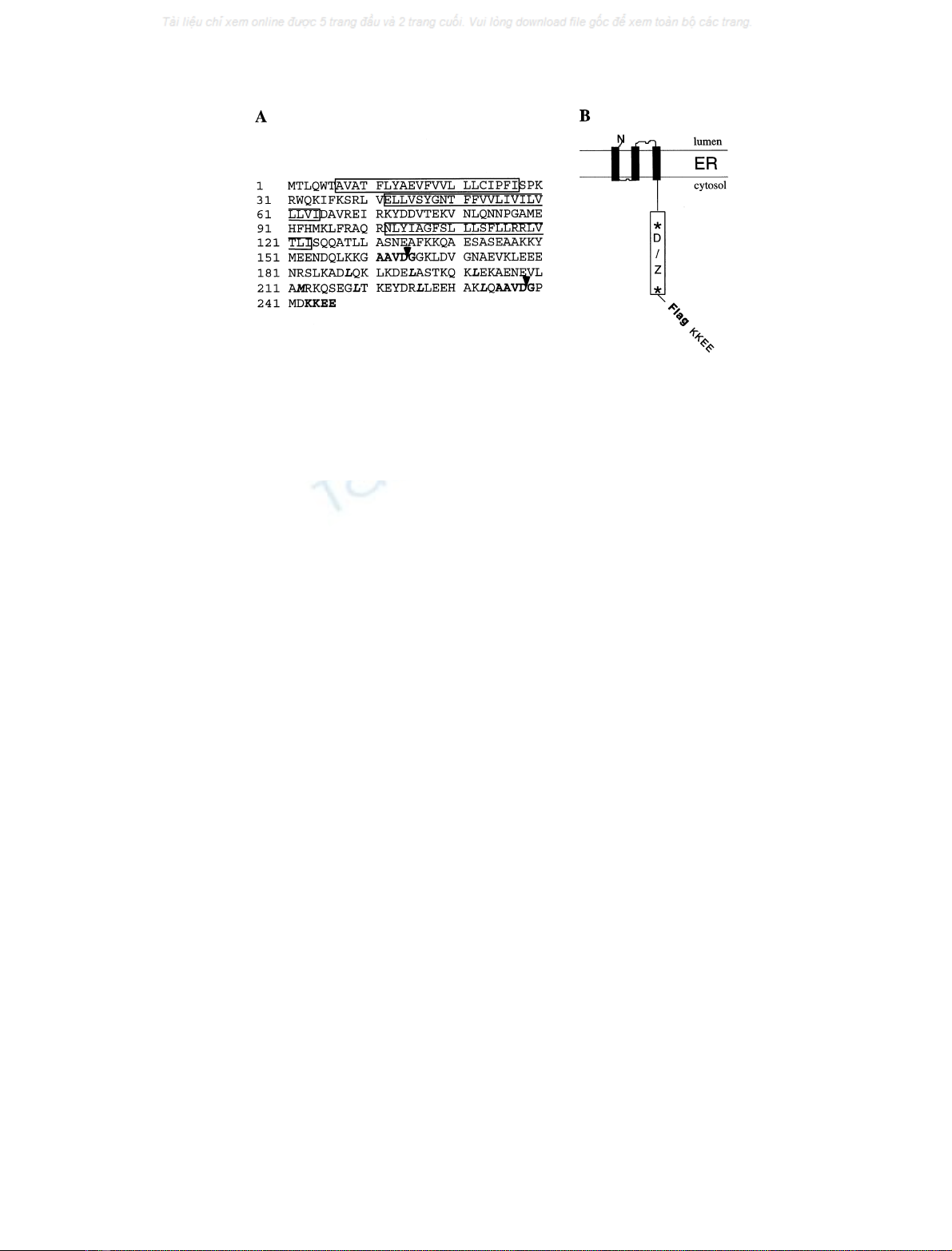
Fig. 1. Polypeptide sequence and putative arrangement of Bap31 in the ER membrane. (A) Amino acid sequence of human BAP31 (single-letter code;
the sequence data is available from GenBank under accession number X81817). The three predicted transmembrane segments are boxed and the
predicted caspase recognition sites, AAVD/G, are highlighted. Cleavage is denoted by arrows following the aspartic acid residues at positions 164
and 238. A potential leucine zipper located between the caspase recognition sites is shown in bold letters, as is the KKXX ER retention signal at the
C terminus.(B) Putative topology of BAP31 in the ER membrane. The 13-kDa cytosolic domain containing putative death effector homology (D)
and leucine zipper (Z) domains, flanked on either side by caspase-8 recognition sites (asterisks), are boxed. The Flag construct used in this work has
been inserted between amino acids 242 and 243.
FEBS 2003 BAP31 associates with c-actin and myosin B (Eur. J. Biochem. 270) 343

Speed-Vac (Savant Instruments, Hicksville, NY, USA) to
remove any remaining liquid. The dried polyacrylamide was
then rehydrated with 20–30 lL of the trypsin digest solution
[15 m
M
N-ethylmorpholine, 5 m
M
acetic acid containing
15 ngÆlL
)1
sequence-grade trypsin (Promega)] so that the
liquid was completely absorbed in the gel pieces. Digestion
was performed overnight at room temperature. Peptides were
collected by extracting the acrylamide three times for 20 min
with 40–60 lL 60% (v/v) acetonitrile, 0.5% (v/v) formic
acid. The collected fractions were combined and the peptides
weredriedinaspeed-vacandkeptat)20 C until use.
Peptide mapping by capillary liquid chromatography
microelectrospray tandem MS (LC-lESI-MS/MS)
Tryptic peptides were analysed using a self-packed capillary
column (0.1 ·120 mm, Magic-MS C
18
packing material,
Michrom BioResources Inc., Auburn CA, USA) coupled to
a Finnigan MAT TSQ7000 mass spectrometer (Thermo
Finnigan, San Jose CA, USA) using a microelectrospray
interface operated at 1.2 kV. The nanoliter flow rate
required by the capillary LC column (700 nLÆmin
)1
)was
obtained by coupling a Magic microbore HPLC system
(Michrom BioResources) with a precolumn high-pressure
flow splitter from the same supplier. Samples were recon-
stituted in 25 lL buffer A [2.5% (v/v) acetonitrile, 0.1%
(v/v) formic acid, 0.005% (v/v) heptafluorobutanoic acid],
centrifuged for 5 min at 14 000 g, and the supernatant was
injected off-line onto a C
18
precolumn cartridge
(0.5 mm ·1 mm, LC Packings Inc., San Francisco CA,
USA) at 5 lLÆmin
)1
. Peptides were eluted from the column
using a linear gradient from 10 to 60% buffer B [80% (v/v)
acetonitrile, 0.085% (v/v) formic acid, 0.005% (v/v) hepta-
fluorobutanoic acid] in 20 min.
For unambiguous identification, eluting peptides were
subjected to automated tandem MS by collision-induced
dissociation essentially as described by Ducret et al. [13] with
some minor modifications. Briefly, peptides were subjected
to tandem MS if the ion current for a particular species
exceeded a relative intensity of 200 000 counts. After
analysis, the mass of the investigated species was recorded
into a user table that prevented the re-analysis of the same
ion until its intensity had decreased under a user-defined
threshold. This modification was essential to analyse
complex mixtures when several peptides were usually
coeluting in a chromatographic peak.
Database searching
Uninterpreted tandem mass spectra were correlated to
protein databases using the program
SEQUEST
version C1
[14,15] essentially as described by Ducret et al. [13]. For
identification purposes, all tandem mass spectra were
matched against a subset of the NCBI GenBank protein
database (http://www.ncbi.nlm.nih.gov; nonredundant pro-
tein database release July 2, 2001) filtered with the word
human(resulting in a database of 67 000 entries).
Automated
SEQUEST
identifications were performed using
default parameters with the peptide and fragment tolerance
set to 1.5 Da and 1.0 Da, respectively, and with methionine
dynamically searched for the commonly found methionine
sulfoxide derivative (+16 Da). Further characterization
was achieved by manually excluding tandem mass spectra of
poor quality, by restricting the database to the proteins that
were identified in a first pass approach, and by changing the
SEQUEST
parameter file to account for post-translational
modifications as indicated in the text.
Results
Immunoprecipitation of the preapoptotic endoBAP31
complex and preliminary characterization by SDS/PAGE
In a recent work [9], we reported on the characterization of a
caspase-resistant (cr) BAP31 that inhibited Fas-mediated
apoptotic membrane blebbing and fragmentation in KB
epithelial cells. crBAP31–Flag, whose caspase recognition
aspartate residues were mutated to alanine residues (Fig. 1),
only modestly slowed down the time-course for activation
of caspases, as assayed by the processing of procaspases 8
and 3, by the measurement of total DEVDase activity, and
by the cleavage of the caspase targets poly(ADP-ribosyl)
polymerase and endogenous BAP31. In contrast, cytoplas-
mic membrane blebbing and fragmentation and apoptotic
redistribution of actin were strongly inhibited, cell mor-
phology was retained near normal, and the irreversible loss
of cell growth potential following removal of the Fas
stimulus was delayed. In its unmutated form, BAP31 is a
preferred substrate for caspases 8 and 1 whose cleavage
product generates a p20 fragment that remains integrated in
the ER membrane (Fig. 1). When expressed ectopically, the
p20 fragment is a potent inducer of cell death. These results
argue that the cytosolic domain of BAP31 is important for
regulating cytoplasmic apoptotic events associated with
membrane fragmentation.
To examine proteins that might be potentially associated
with BAP31, BAP31–Flag was inserted and stably
expressed in the human HepG2 cell line. The preapoptotic
endoBAP31 complex was immunoprecipitated using the
anti-Flag Ig and the immunopurified proteins were analysed
by SDS/PAGE. The immunocomplex was found to contain
both the BAP31–Flag and the endogenous BAP31 proteins,
migrating at apparent masses of 33 kDa and 28 kDa,
respectively, and two additional protein bands at apparent
masses of 42 and 190 kDa [9]. For sequence analysis, the
BAP31 immunoprecipitation protocol was scaled up and
the immunocomplex obtained was analysed by preparative
SDS/PAGE using a 10% separating gel (Fig. 2).
Peptide fragmentation mapping by LC-lESI-MS/MS
Specifically recruited proteins (at 190, 42 and 28 kDa) were
excised from the gel, destained, and in-gel digested for
protein identification. Peptides obtained by the proteolytic
in-gel digestion were analysed by LC-lESI-MS/MS.
Detailed analysis of the 28-kDa protein confirmed its
identity as the endogenous BAP31 protein while the 42 and
the 190 kDa bands were identified as nonmuscle c-actin and
myosin heavy chain nonmuscle type B, respectively.
Unambiguous identification was made difficult by the large
number of described protein variants in the human data-
base. Concomitantly, several good quality MS/MS spectra
were not correlated to the database by
SEQUEST
, indicating
the potential presence of post-translational modifications
344 A. Ducret et al. (Eur. J. Biochem. 270)FEBS 2003
and/or additional protein variants. We therefore re-analysed
the data with a smaller database, containing only human
actin or human myosin entries, and MS/MS spectrum that
failed to be confidently identified in the first pass analysis
were manually interpreted (Table 1).
Of the initial 51 tandem mass specta obtained by LC-MS/
MS analysis of the 190 kDa band 45 were selected for a
second pass identification using a protein database filtered
for the words humanand myosin(144 entries). In total, 41
MS/MS spectra were assigned to myosin heavy chain
nonmuscle type B tryptic peptides, covering 33% of the
total amino acid sequence (Fig. 3A). Two MS/MS spectra
were found to contain peptides that deviated from the
published amino acid sequence. One of them, at positions
217–232, differed from the published amino acid sequence
by a Ser227Ala mutation while nine of the 14 amino acids of
the second peptide at positions 130–143 were exchanged.
The mutated sequence was almost identical to a corres-
ponding peptide in the skeletal muscle myosin heavy chain-2
sequence (MYHC-IIB; GeneBank accession number
Q9Y623). In both cases, the predicted peptide and its
mutated counterpart were present in an approximately
equimolar amounts, indicating either that two distinct
myosin species (namely, myosin heavy chain nonmuscle
type B and skeletal muscle myosin heavy chain-2) were
coprecipitated or that the nonmuscle myosin expressed in
the HepG2 cells was expressed in two (or several) allelic
forms. The former hypothesis, however, is unlikely as these
twomyosinspeciesshareonly40% identity. Therefore,
several peptides specific for each myosin variant should
have been identified during the LC-MS/MS analysis.
Finally, four MS/MS spectra could not be unambiguously
assigned to a given peptide sequence. All spectra were of
medium quality and the absence of characteristic immo-
nium ions specific for a terminal lysine or arginine might
indicate that those peptides were not generated by a tryptic
cleavage. As a result, they were not analysed further.
Similarly, 39 spectra of the initial 42 tandem mass spectra
obtained by LC-MS/MS analysis of the 42-kDa band were
selected for a second pass identification using a protein
database filtered for the words humanand actin(280
entries). In total, 38 MS/MS spectra could be assigned to
c-actin tryptic peptides, covering 69% of the total amino acid
sequence (Fig. 3B). In particular, the presence of a methyl-
histidine reported in the literature at position 72 was con-
firmed in our analysis. Of particular interest were five MS/
MS spectra of good spectral quality that could not be initially
matched to any specific actin sequence. Manual interpret-
ation of the fragmentation patterns (Fig. 4) indicated that all
five analysed species were derived from a heterogeneous
N-terminal peptide. Fig. 4A shows the tandem mass spec-
trum of the N-terminal peptide as reported in the sequence
database: the N-terminal methionine residue has been
Table 1. Overview of the tandem MS analysis of myosin and actin by LC-MS/MS.
Analysis of MS/MS spectra (n) Database entries (n) Identification
28-kDa band 15 MS/MS Human (67051 entries) 5 MS/MS: BAP31 human
10 MS/MS: no identification
42-kDa band 42 MS/MS Human (67051 entries) 30 MS/MS b-orc-actin
39 MS/MS Human&actin(280 entries) 36 MS/MS c-actin
2 MS/MS: c-actin variants
1 MS/MS: no identification
180-kDa band 51 MS/MS Human(67051 entries) 35 MS/MS: myosin heavy chain non-muscle B
16 MS/MS: no identification
45 MS/MS Human&myosin(144 entries) 39 MS/MS: myosin heavy chain non-muscle B
2 MS/MS: myosin variants
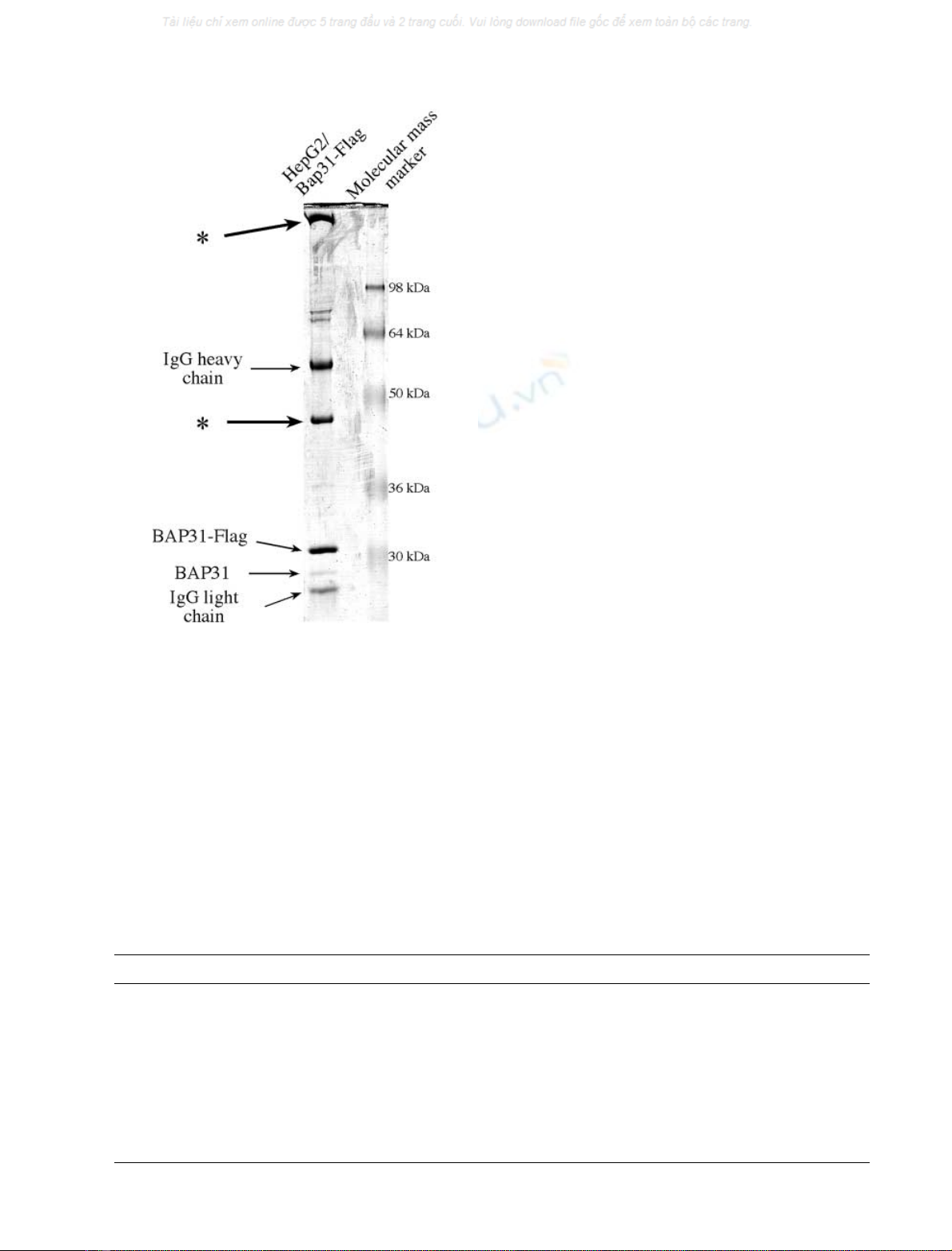
Fig. 2. SDS/PAGE analysis of the immunoprecipitated pre-apoptotic
BAP31–Flag complex in transfected HepG2 cells. Pre-apoptotic HepG2
cells, stably expressing the BAP31–Flag construct, were lysed and
BAP31 was immunoprecipitated with the anti-Flag M2 Ig. The
immunocomplex was subjected to SDS/PAGE analysis and visualized
by Coomassie blue staining. The two bands of interest, at apparent
molecular masses of 42 and 175 kDa, are labelled with stars.
FEBS 2003 BAP31 associates with c-actin and myosin B (Eur. J. Biochem. 270) 345
removed and the first glutamic acid residue has been
acetylated. In addition, Cys16 was alkylated by an acryl-
amide monomer, a common experimental artefact when
un-alkylated proteins are purified by an SDS/PAGE step
[16]. A very rich fragmentation pattern was essential to
confirm the putative peptide sequence in its entirety. Further,
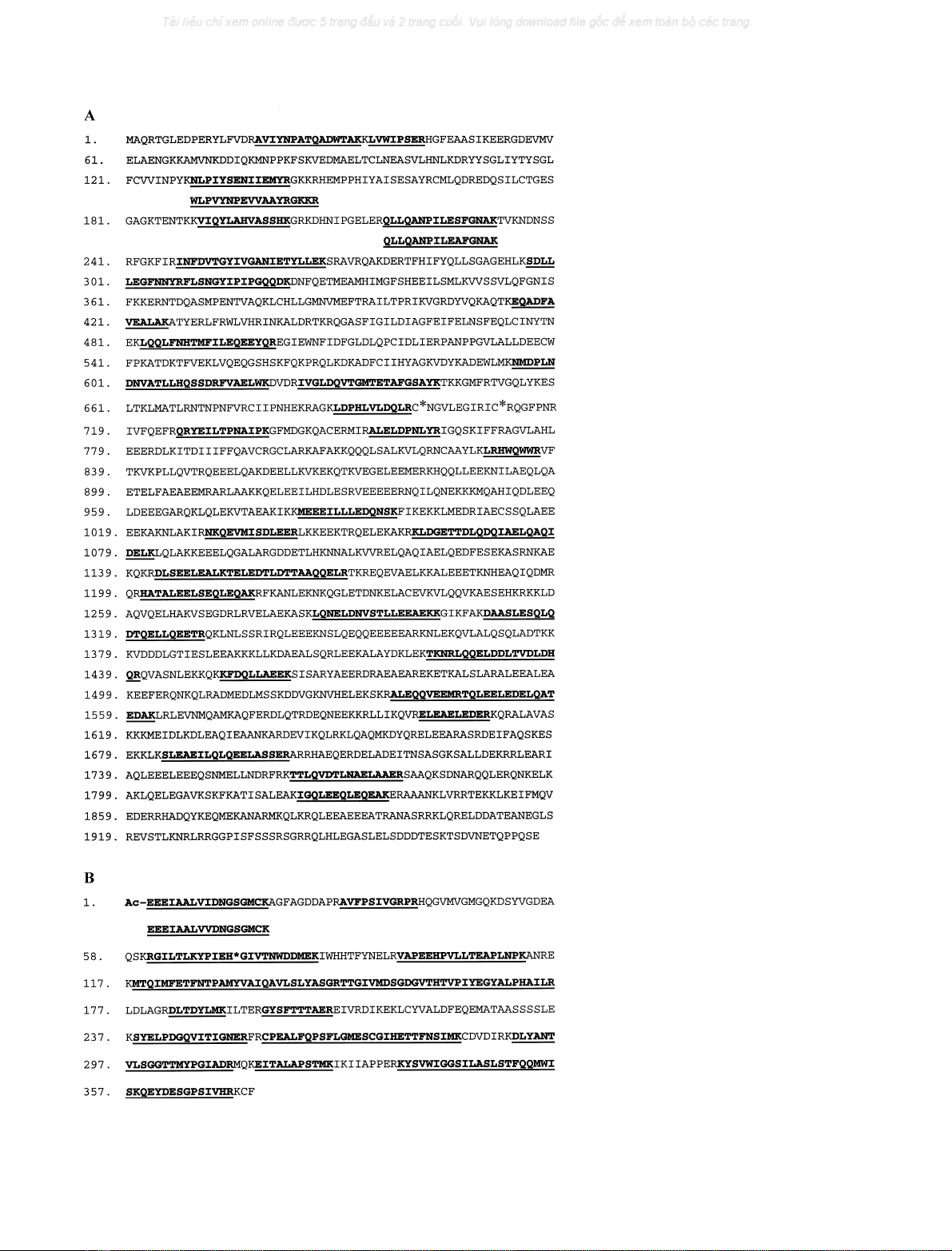
Fig. 3. Detailed amino acid sequence analysis
of (A) the myosin heavy chain nonmuscle type B
(GeneBank accession P35580) and of (B) non-
muscle c-actin (GeneBank accession P02571)
byLC-lESI-MS/MS. All amino acids are in
single-letter code. The peptides unequivocally
identified by spectral matching of the tandem
mass spectra with the sequence database by
SEQUEST
areunderlined.(A)Thetwopotential
alkylated cysteine residues (SH1/SH2 sites) at
positions 701 and 711 are each marked with a
star. Two peptides were found to deviate from
the predicted sequence (at position 130–147
and position 217–232). (B) The methyl-histi-
dine at position 72 is marked with a star. The
two N-terminal peptide variants described in
this work are indicated. See text for more
details.
346 A. Ducret et al. (Eur. J. Biochem. 270)FEBS 2003


%20--%3e%3cdefs%3e%3cstyle%3e%20.st0%20{%20fill:%20%23fff;%20}%20.st1%20{%20fill:%20%237800fa;%20}%20%3c/style%3e%3c/defs%3e%3cpath%20class='st1'%20d='M117.78,12.18H43.11c2.9,3.47,4.65,7.94,4.65,12.82,0,5.6-2.3,10.66-6.01,14.29h76.02l7.22-13.56-7.22-13.56Z'/%3e%3cg%3e%3cpath%20class='st0'%20d='M53.58,26.17h-.59v-1.46h.59v-4.96h2.83c1.78,0,2.67.94,2.67,2.82v5.76c0,1.87-.89,2.81-2.67,2.81h-2.83v-4.96ZM55.36,21.37v3.34h1.1v1.46h-1.1v3.34h1.01c.61,0,.91-.37.91-1.1v-5.93c0-.74-.3-1.1-.91-1.1h-1.01Z'/%3e%3cpath%20class='st0'%20d='M65.99,31.14h-1.8l-.31-2.07h-2.19l-.31,2.07h-1.64l1.82-11.39h2.62l1.82,11.39ZM65.28,18.04c-.25.46-.51.77-.75.94-.21.15-.47.22-.79.22-.26,0-.57-.07-.92-.22l-.38-.15c-.14-.05-.26-.07-.37-.07-.3,0-.53.18-.71.54l-.91-.68c.25-.46.51-.77.75-.94.21-.14.48-.21.79-.21.26,0,.57.07.92.21l.38.15c.14.05.26.07.37.07.3,0,.53-.18.71-.54l.91.68ZM61.91,27.52h1.73l-.87-5.76-.87,5.76Z'/%3e%3cpath%20class='st0'%20d='M74.53,26.89v1.52c0,1.91-.89,2.86-2.67,2.86s-2.67-.95-2.67-2.86v-5.93c0-1.91.89-2.86,2.67-2.86s2.67.95,2.67,2.86v1.11h-1.69v-1.22c0-.75-.31-1.12-.93-1.12s-.93.37-.93,1.12v6.15c0,.74.31,1.11.93,1.11s.93-.37.93-1.11v-1.63h1.69Z'/%3e%3cpath%20class='st0'%20d='M81.4,31.14h-1.8l-.31-2.07h-2.19l-.31,2.07h-1.64l1.82-11.39h2.62l1.82,11.39ZM75.9,19.2l1.52-1.91h1.71l1.51,1.91h-1.61l-.76-.95-.75.95h-1.61ZM77.32,27.52h1.73l-.87-5.76-.87,5.76ZM83.1,15.99l-1.76,1.91h-1.26l1.17-1.91h1.86Z'/%3e%3cpath%20class='st0'%20d='M84.86,19.75c1.78,0,2.67.94,2.67,2.82v1.48c0,1.87-.89,2.81-2.67,2.81h-.85v4.28h-1.79v-11.39h2.64ZM84.01,21.37v3.86h.85c.58,0,.87-.36.87-1.08v-1.71c0-.71-.29-1.07-.87-1.07h-.85Z'/%3e%3cpath%20class='st0'%20d='M93.51,19.75c1.78,0,2.67.94,2.67,2.82v1.48c0,1.87-.89,2.81-2.67,2.81h-.85v4.28h-1.79v-11.39h2.64ZM92.66,21.37v3.86h.85c.58,0,.87-.36.87-1.08v-1.71c0-.71-.29-1.07-.87-1.07h-.85Z'/%3e%3cpath%20class='st0'%20d='M98.8,31.14h-1.79v-11.39h1.79v4.88h2.03v-4.88h1.83v11.39h-1.83v-4.88h-2.03v4.88Z'/%3e%3cpath%20class='st0'%20d='M105.36,24.55h2.46v1.62h-2.46v3.34h3.09v1.63h-4.88v-11.39h4.88v1.63h-3.09v3.18ZM108.17,17.29l-1.76,1.91h-1.26l1.17-1.91h1.86Z'/%3e%3cpath%20class='st0'%20d='M112.2,19.75c1.78,0,2.67.94,2.67,2.82v1.48c0,1.87-.89,2.81-2.67,2.81h-.85v4.28h-1.79v-11.39h2.64ZM111.35,21.37v3.86h.85c.58,0,.87-.36.87-1.08v-1.71c0-.71-.29-1.07-.87-1.07h-.85Z'/%3e%3c/g%3e%3ccircle%20class='st1'%20cx='25'%20cy='25'%20r='20'/%3e%3cpath%20class='st0'%20d='M32.78,19.27c2.92,0,4.43,2.55,5.28,5.33l.71,2.17c.14.38-.33.75-.71.75h-5.61c.19-.33.24-.71.09-1.08l-.75-2.45c-.43-1.32-.99-2.64-1.79-3.77.75-.57,1.65-.94,2.78-.94h0ZM25,18.38c3.25,0,4.9,2.78,5.89,5.89l.76,2.45c.14.42-.33.8-.8.8h-11.69c-.42,0-.94-.38-.8-.8l.75-2.45c.99-3.11,2.64-5.89,5.89-5.89h0ZM25,11.35c1.74,0,3.11,1.37,3.11,3.11s-1.37,3.11-3.11,3.11-3.11-1.41-3.11-3.11,1.41-3.11,3.11-3.11h0ZM17.27,19.27c1.08,0,1.98.38,2.73.94-.8,1.13-1.37,2.45-1.74,3.77l-.8,2.45c-.14.38-.05.75.09,1.08h-5.56c-.42,0-.9-.38-.75-.75l.71-2.17c.9-2.78,2.41-5.33,5.33-5.33h0ZM17.27,12.91c1.51,0,2.78,1.27,2.78,2.83s-1.27,2.83-2.78,2.83-2.83-1.27-2.83-2.83,1.27-2.83,2.83-2.83h0ZM32.78,12.91c1.56,0,2.78,1.27,2.78,2.83s-1.23,2.83-2.78,2.83-2.83-1.27-2.83-2.83,1.27-2.83,2.83-2.83h0ZM27.07,28.56v.09c0,.57-.24,1.08-.61,1.46h0v.05c-.38.33-.9.57-1.46.57s-1.08-.24-1.46-.61h0c-.38-.38-.61-.9-.61-1.46v-.09h1.41v.09c0,.19.05.38.19.47v.05c.09.09.28.19.47.19s.38-.09.47-.19v-.05c.14-.09.24-.28.24-.47t-.05-.09h1.41ZM30.99,28.56v.09c0,1.65-.66,3.16-1.74,4.24-1.08,1.08-2.59,1.79-4.24,1.79s-3.16-.71-4.24-1.79l-.05-.05c-1.04-1.08-1.7-2.55-1.7-4.2v-.09h1.41v.09c0,1.27.47,2.4,1.27,3.25h.05c.85.85,1.98,1.37,3.25,1.37s2.4-.52,3.25-1.37c.85-.8,1.37-1.98,1.37-3.25v-.09h1.37ZM34.99,28.56v.09c0,2.78-1.13,5.28-2.92,7.07-1.79,1.79-4.29,2.92-7.07,2.92s-5.23-1.13-7.07-2.92c-1.79-1.79-2.92-4.29-2.92-7.07v-.09h1.41v.09c0,2.4.94,4.53,2.5,6.08,1.56,1.56,3.72,2.5,6.08,2.5s4.52-.94,6.08-2.5c1.56-1.56,2.5-3.68,2.5-6.08v-.09h1.41Z'/%3e%3c/svg%3e)