
Different roles of functional residues in the hydrophobic
binding site of two sweet orange tau glutathione
S-transferases
Angela R. Lo Piero, Valeria Mercurio, Ivana Puglisi and Goffredo Petrone
Dipartimento di Scienze Agronomiche, Agrochimiche e delle Produzioni Animali (DACPA), Universita
`di Catania, Italy
Introduction
The glutathione S-transferases (GSTs; EC 2.5.1.18) are
members of a multifunctional superfamily of enzymes
catalyzing the conjugation of glutathione (GSH) to the
electrophilic groups of hydrophobic and usually
cytotoxic molecules of either endogenous or exogenous
origin [1–4]. GSTs are widely distributed in nature
from humans to bacteria [5–7]. In plants, the GSH
addition reaction is coupled to the vacuolar compart-
mentation of the GSH conjugates because of the lack
of an effective excretion pathway, which is active in
Keywords
glutathione S-transferase; H-site;
site-directed mutagenesis; sweet orange;
tau class glutathione S-transferase
Correspondence
A. R. Lo Piero, Dipartimento di Scienze
Agronomiche, Agrochimiche e delle
Produzioni Animali (DACPA), Universita
`di
Catania, Via S. Sofia 98, 95123, Catania,
Italy
Fax: +39 95 7141581
Tel: +39 95 7580238
E-mail: rlopiero@unict.it
(Received 27 July 2009, revised 7 October
2009, accepted 5 November 2009)
doi:10.1111/j.1742-4658.2009.07481.x
Glutathione S-transferases (GSTs) catalyze the conjugation of glutathione
to hydrophobic compounds, contributing to the metabolism of toxic
chemicals. In this study, we show that two naturally occurring tau GSTs
(GSTUs) exhibit distinctive kinetic parameters towards 1-chloro-2,4-dini-
trobenzene (CDNB), although they differ only in three amino acids
(Arg89, Glu117 and Ile172 in GSTU1 are replaced by Pro89, Lys117 and
Val172 in GSTU2). In order to understand the effects of the single mis-
matched residues, several mutant GSTs were generated through site-direc-
ted mutagenesis. The analysis of the kinetic parameters of the mutants
led to the conclusion that Glu117 provides a critical contribution to the
maintenance of a high-affinity CDNB-binding site. However, the substitu-
tion E117K gives rise to mutants showing increased k
cat
values for
CDNB, suggesting that Lys117 might positively influence the formation
of the transition state during catalysis. No changes in the K
m
values
towards glutathione were found between the naturally occurring GSTs
and mutants, except for the mutant caused by the substitution R89P in
GSTU1, which showed a sharp increase in K
m
. Moreover, the analysis of
enzyme reactivation after denaturation showed that this R89P substitution
leads to a two-fold enhancement of the refolded enzyme yield, suggesting
that the insertion of proline might induce critical structural modifications.
In contrast, the substitution P89R in GSTU2 does not modify the reacti-
vation yield and does not impair the affinity of the mutant for glutathi-
one, suggesting that all three residues investigated in this work are
fundamental in the creation of enzymes characterized by unique biochem-
ical properties.
Abbreviations
4-NPB, 4-nitrophenethyl bromide; CDNB, 1-chloro-2,4-dinitrobenzene; ECA, ethacrynic acid; GmGSTU-4-4, Glycine max tau glutathione
S-transferase-4-4; GSH, glutathione; G-site, glutathione-binding site; GST, glutathione S-transferase; GSTU, tau glutathione S-transferase;
H-site, hydrophobic binding site; NBD-Cl, 7-chloro-4-nitrobenzo-2-oxa-1,3-diazole.
FEBS Journal 277 (2010) 255–262 ª2009 The Authors Journal compilation ª2009 FEBS 255

animals [8,9]. GSTs, in addition to transfer of GSH to
toxic compounds, act as GSH-dependent peroxidase,
isomerase and oxidoreductases, playing pivotal roles in
plant cell protection, as reviewed by Moons [10]. Plant
GSTs are abundantly expressed, and show major tran-
scriptional regulation. It has been reported that GST
transcript levels can markedly increase in response to
a wide variety of stressful conditions, such as herbi-
cides [1,11], chilling [11,12], hypoxic stress [13], dehy-
dration [14], wounding [15], and pathogen attack
[16,17]. Most GSTs are active as dimers, either homo-
dimers or heterodimers of subunits ranging from 23 to
30 kDa in size. Subunits of all known GST structures
exhibit a two-domain fold, the N-terminal domain and
C-terminal domain, including the highly conserved
GSH-binding site (G-site) and the more divergent
cosubstrate-binding domain or hydrophobic binding
site (H-site) [7,18]. The G-site includes both a-helices
and b-strands as secondary structure elements. The
topological arrangement of these elements is usually
bababba, similar to the thioredoxin fold of other
GSH-binding or cysteine-binding proteins. The H-site
is entirely helical, with a variable number of a-helices,
depending on the specific enzymes [19]. Mechanisti-
cally, the catalysis of the nucleophilic aromatic substi-
tution reactions comprises the substrate binding to the
enzyme’s active site, ionization of GSH to form the
highly reactive thiolate anion, and nucleophilic attack
by the thiolate at the substrate electrophilic center.
A hydroxyl group, provided in most plant GSTs by a
serine, is considered to be required for the correct ori-
entation and stabilization of the deprotonated thiolate
anion in the active site of the enzyme [20]. On the basis
of protein sequence similarity, active site residue and
gene organization plant GSTs are grouped in four
main classes (phi, tau, zeta, and theta) [18,21,22]. The
majority of the plant GSTs belongs to the tau (GSTU)
and phi classes, which are plant-specific. Among the
plant GSTs, GSTUs are the most numerous, and
members of this class overlap in their function of
enhancing crop stress tolerance. Despite the important
roles of the GSTUs, extensive analysis of critical resi-
dues in both domain sites is needed, although the
highly conserved nature of the G-site [23] and the
recent crystallographic characterization of two GSTUs
[24–26] have allowed researchers to formulate some
general considerations about the N-terminal domains
of the enzymes. Moreover, the existence of a conserved
electron-sharing network that helps the glutamyl c-car-
boxylate of GSH to function as a catalytic base,
accepting the proton from the thiol group to form an
anionic GSH, has been reported [27]. This network is
characterized by an electrostatic interaction between
negatively and positively charged amino acids stabi-
lized by an array of hydrogen bonds, and appears to
be a functionally conserved motif in all GST classes
[27]. In the Glycine max GSTU-4-4 (GmGSTU4-4)–
GSH complex, the strictly conserved residues Arg18,
Glu66, Ser67 and Asp103 appear to form the proposed
electron-sharing network [26]. Studies of the catalytic
properties of Pinus tabulaeformis GSTU1 by site-direc-
ted mutagenesis, which demonstrate the crucial role of
both Ser67 and Glu66 in GSH binding, corroborate
these findings [28]. In contrast, the H-sites of GSTs
exhibit a low degree of sequence identity, and hence
unique structures that reflect different functions in vivo
[18]. In a previous study, we isolated from sweet
orange leaves two distinct GSTU genes sharing 98.6%
homology at the nucleotide levels and containing a
651 bp ORF. The encoded proteins differ in only three
amino acids: the triad Arg89, Glu117 and Ile172 found
in the isoform GSTU1 is replaced by the triad Pro89,
Lys117 and Val172 in the GSTU2 isoform, all of the
mismatches being located in the H-sites of the enzymes
[11]. As long as the enzymes were expressed in vitro
and purified, they exhibited different specific activity
with 1-chloro-2,4-dinitrobenzene (CDNB) as substrate,
GSTU1 showing a value three-fold lower than that
observed for GSTU2 [11]. In the present work, site-
directed mutagenesis was used to evaluate the effects
of the single mismatched residues on the kinetic prop-
erties of isoforms GSTU1 and GSTU2, and also to
address questions regarding the functional roles of
these H-site residues. The results have both academic
relevance and practical importance, as they will form
the basis for the design of new engineered GSTs show-
ing altered substrate specificity and enhanced activity
towards xenobiotics.
Results and Discussion
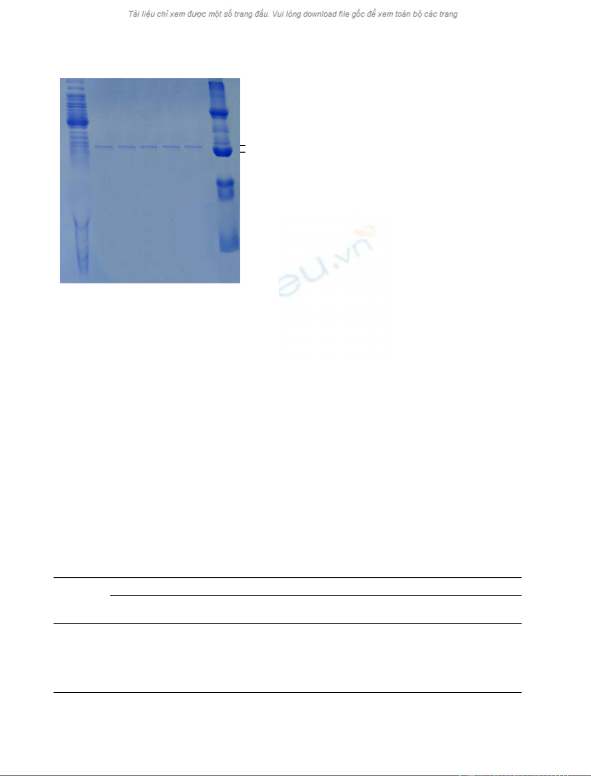
Wild-type (GSTU1 and GSTU2) and mutant GSTs
were expressed and purified as described in Experimen-
tal procedures. After purification, all recombinant pro-
teins showed a single band in SDS ⁄PAGE, and an
identical molecular mass of about 26.0 kDa, corre-
sponding to the calculated molecular mass of the
recombinant sweet orange GST subunits (Fig. 1).
CDNB is generally considered to be the classic GST
substrate, because most GST isoenzymes display high
activity towards it. It has been shown that the sweet
orange GST isoforms exhibit quite different specific
activities with CDNB as substrate, GSTU2 being
three-fold more active than GSTU1 [11]. Conse-
quently, steady-state kinetic characterization of both
isoforms with respect to CDNB was performed prior
Functional role of the GST H-site residues A. R. Lo Piero et al.
256 FEBS Journal 277 (2010) 255–262 ª2009 The Authors Journal compilation ª2009 FEBS
to any other investigation concerning their catalytic
mechanism. The values of kinetic parameters obtained
by non-linear regression analysis are listed in Table 1.
The results showed that GSTU1 had a lower apparent
K
m
for CDNB (0.75 mm) than that exhibited by
GSTU2 (1 mm), this finding being consistent with a
higher affinity of GSTU1 for this substrate. The K
m
values of GSTU1 and GSTU2 for CDNB are in the
range of those of plant GSTs, whose values vary from
0.12 to 4.43 mm[24,29,30]. As expected on the basis of
the complete identity of the G-site sequences, the same
K
m
for GSH was registered for both isoforms
(0.5 mm), which is in general agreement with other
published K
m
values for GSH [31]. However, in the
case of both substrates, GSTU2 showed higher cata-
lytic efficiency (k
cat
⁄K
m
) as well as higher k
cat
values
(Table 1). The alignment of the H-site sequences of 20
GSTUs from different plant sources showed that
Pro89 and the Lys117 found in GSTU2 are strictly
conserved residues, whereas, at the 172 position, the
isoleucine is well conserved but valine or threonine can
also be found (Fig. S1). Therefore, the naturally occur-
ring GSTU1 (Arg89, Glu117, and Ile172) probably has
unique features, and might perform a different func-
tional role in vivo than GSTU2. This assumption is
also supported by the analysis of gene expression,
which showed that the GSTU1 gene is induced by cad-
mium sulfate, CDNB, cyhalothrin, and cold stress,
whereas GSTU2 is constitutively expressed [11]. Given
that these naturally occurring GSTUs provided a good
opportunity to understand how specific amino acids
might contribute to the enzyme’s biochemical behav-
ior, several mutant GSTs, hereafter referred to by their
distinctive amino acid triad, were generated by site-
directed mutagenesis. In particular, the RKI and RKV
mutants were obtained by individually replacing the
Glu117 of GSTU1 (REI) with Lys117, and the Pro89
of GSTU2 (PKV) with Arg89, respectively. Moreover,
the PEI and PKI mutants were obtained by replacing
Arg89 of GSTU1 (REI) with Pro89, and Glu117 of
the PEI mutant with Lys117, respectively. Steady-state
kinetic analysis of purified RKI and RKV mutants
showed sharp increases of approximately six-fold and
five-fold in the K
m
value for CDNB as compared with
GSTU1 (REI) (Table 1). Consequently, the results sug-
gest that Lys117-containing enzymes, either wild type
or mutant, do not easily accommodate CDNB in their
active site, and also highlight the critical contribution
of Glu117 to CDNB recognition. Accordingly, the PEI
mutant, in which the Glu117 is restored as compared
with the PKI mutant, shows a K
m
value for CDNB
similar to that of GSTU1 (REI) (Table 1). However,
the k
cat
values for both GSTU2 (PKV) and the E117K
36 kDa
29 kDa
1234576
Fig. 1. SDS ⁄PAGE of the purified wild-type and mutant GSTs. Lane
1: unpurified E. coli extract. Lane 2: purified wild-type GSTU1 (REI).
Lane 3: purified wild-type GSTU2 (PKV). Lane 4: purified PEI
mutant. Lane 5: RKV mutant. Lane 6: purified PKI mutant. Lane 7:
molecular mass marker. The mutant enzymes are referred to by
their distinctive triad of amino acids located, respectively, at posi-
tions 89, 117, and 172.
Table 1. Steady-state kinetic constants of wild-type and mutant GSTs. The kinetic parameters were calculated by using nonlinear regression
analysis with the HYPER32 program. Each value represents the mean ± standard deviation of three replicates.
GST
CDNB GSH
K
m
(mM)
V
max
(lMÆmin
)1
)
k
cat
(s
)1
·10
3
)
k
cat
⁄K
m
(M
)1
Æs
)1
)
K
m
(mM)
V
max
(lMÆmin
)1
)
k
cat
(s
)1
·10
3
)
k
cat
⁄K
m
(M
)1
Æs
)1
)
GSTU1 (REI) 0.75 ± 0.03 2.0 ± 0.10 23.8 ± 1 31.7 ± 1.45 0.5 ± 0.02 1.0 ± 0.1 13.8 ± 0.24 27.7 ± 1.15
GSTU2 (PKV) 1.0 ± 0.07 4.0 ± 0.06 108.1 ± 1 108.1 ± 5.4 0.5 ± 0.03 4.0 ± 0.15 76.9 ± 0.4 153.8 ± 6.94
RKI 5.0 ± 0.2 9.0 ± 0.5 257.1 ± 2 51.4 ± 2.0 0.6 ± 0.03 5.0 ± 0.20 147.0 ± 0.9 245.0 ± 4.26
RKV 4.0 ± 0.4 8.0 ± 0.25 119.4 ± 1 29.8 ± 0.6 0.35 ± 0.02 3.3 ± 0.10 129.4 ± 0.5 369.7 ± 21.12
PKI 4.0 ± 0.4 4.5 ± 0.15 160.7 ± 1.6 40.0 ± 1.5 0.7 ± 0.04 4.0 ± 0.15 64.0 ± 0.63 91.4 ± 2.16
PEI 0.85 ± 0.05 3.0 ± 0.07 28.8 ± 0.07 33.9 ± 2.4 4.0 ± 0.02 7.0 ± 0.3 70.7 ± 0.7 17.6 ± 0.24
A. R. Lo Piero et al. Functional role of the GST H-site residues
FEBS Journal 277 (2010) 255–262 ª2009 The Authors Journal compilation ª2009 FEBS 257

mutants (RKI, RKV, and PKI) were increased as com-
pared with those of the Glu117-containing enzymes
(REI and PEI) (Table 1). This finding suggests a nega-
tive influence of Glu117 on the catalytic event. It has
been shown that the addition of GSH to CDNB
occurs via an addition–elimination sequence involving
a short-lived r-complex intermediate (Meisenheimer
complex) as transition state [32]. The structure of the
transition state [32] indicates that the enzyme might
provide electrophilic assistance interactions to the
developing charge of the r-complex o-nitro group [33].
The comparison of the crystal structure of the rat M1-
1 GST in complex with the transition state analog
and with the product reveals two completely different
binding modes for the intermediate and the product,
suggesting that a specific motion is associated with
the collapse of the intermediate into the product
[34]. More recently, Axarli et al. [26] have also shown
that the catalytic reaction of GmGSTU4-4 is barely
sensitive to the nature of the leaving group, as the sub-
stitution of the chlorine atom with the more electro-
negative fluorine in the CDNB molecule did not affect
the k
cat
values. Altogether, these results are consistent
with the idea that the rate-limiting step of the CDNB–
GSH catalytic reaction is the physical event of product
release, probably involving structural motions or con-
formational changes of the ternary complex. In this
context, we propose that the substitution in the orange
GSTU1 of a nucleophilic residue by an electrophilic
one (E117K) could function in strongly favoring the
formation of the transition state during GSH addition
to CDNB by providing the required electrophilic assis-
tance to the developing transition state.
As regards the analysis of kinetic parameters regard-
ing the natural in vivo substrate GSH, the mutants
containing Lys117 showed apparent K
m
values similar
to those reported for the wild-type GSTs (Table 1).
However, the RK and PK enzymes, both wild type
and mutants, showed a strong increase in the k
cat
⁄K
m
values with respect to the Glu117-containing enzymes
(REI and PEI) (Table 1), indicating that the substitu-
tion E117K is crucial to the formation of enzymes with
higher catalytic efficiency. Interestingly, the PEI
mutant showed an eight-fold increase in the K
m
value
for GSH as compared with GSTU1 (REI) (substitution
R89P) (Table 1), suggesting that such mutation of the
H-site might exert its effects upon the G-site kinetic
properties. Recently, Axarli et al. [25] reported the
crystal structure of the GSTU4-4 from soybean, which
shares 71% sequence similarity with the orange
GSTUs [11]. The consistency in the fold of GSTs and
the availability of the structure of GmGSTU4-4
prompted us to construct molecular homology models
for Citrus sinensis GSTU1 (REI) and the PEI mutant,
as well as for Citrus sinensis GSTU2 (PKV) and the
RKV mutant, by submitting the amino acid sequences
to swiss-model [35]. Then, the alignments of the 3D
structural homology models were performed between
the wild-type GSTs and their respective mutants at
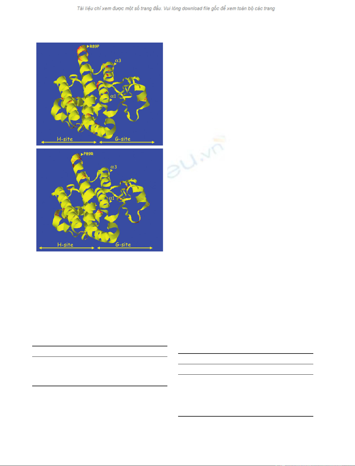
the 89 position (REI–PEI, and PKV–RKV) by the
web-based program matras [36]. The analysis of the
superimposed REI–PEI models revealed relevant con-
formational differences between the enzyme structures,
mainly involving a-helix1 (from Ser13 to Gly27) and
a-helix3 (from Ser67 to Asp80), which represent the
catalytic ‘core’ of the G-site [26] (numbering of helices
is consistent with that of other GSTs) (Fig. 2A). In
contrast, minimal structural perturbations of the afore-
mentioned a-helix1 and a-helix3 were observed in the
case of the superimposed PKV–RKV models, whose
major nonoverlapping regions are, instead, localized in
the H-site of the enzymes (Fig. 2B). These findings are
in agreement with the different values of apparent K
m
for GSH observed between GSTU1 (REI) and the PEI
mutant, and overall indicate that the substitution
R89P in GSTU1 might modify the architecture of the
G-site, thus negatively influencing the enzyme’s affinity
for GSH. Furthermore, Table 2 shows the recovered
activity following denaturation and refolding of wild-
type GSTUs and the PEI and RKV mutants. The reac-
tivation yield of the PEI mutant was two-fold higher
than that observed in GSTU1 (REI) (Table 2), thus
supporting our hypothesis that Pro89 might have a
structural role in the PEI mutant. However, the recov-
ered activities of both GSTU2 (PKV) and the RKV
mutant were similar, suggesting that the substitution
P89R in GSTU2 (PKV) does not affect the refolding
process. Consequently, the putative structural role
assigned to the Pro89 of the PEI mutant cannot be
attributed to the Pro89 of GSTU2 (PKV), as the
RKV mutant, arising from the P89R substitution in
GSTU2 (PKV), has both similar K
m
values (Table 1)
and similar reactivation yields after denaturation
(Table 2) as GSTU2 (PKV). Therefore, the results
lead to the conclusion that all three amino acids inves-
tigated in this work take part in the creation of
enzymes showing unique structures and, consequently,
functions.
The substrate specificity of the sweet orange GSTs
was also investigated in order to identify catalytic
activities that may be related to their biological func-
tion. To this end, some substrates in addition to
CDNB were examined, including 7-chloro-4-nitro-
benzo-2-oxa-1,3-diazole (NBD-Cl), with which mam-
malian a-class GSTs show high activity [37], the alkyl
halide 4-nitrophenethyl bromide (4-NPB), related to
Functional role of the GST H-site residues A. R. Lo Piero et al.
258 FEBS Journal 277 (2010) 255–262 ª2009 The Authors Journal compilation ª2009 FEBS
the role of GSTs in detoxification processes, and ethac-
rynic acid (ECA), a phenylacetic derivative that con-
tains an electrophilic group, similar to the cytotoxic
a,b-alkenals, which may be formed during oxidative
stress [29] (Table 3). Overall, CDNB is the preferred
substrate of both naturally occurring (REI and PKV)
and mutant GSTs, with the RKV and PKV enzymes
showing the highest specific activity. Relatively lower
activity was detected with the mammalian GST sub-
strate NBD-Cl (Table 3). Interestingly, the mutant
enzyme RKV is able to conjugate 4-NPB to GSH,
whereas wild types, as well as other mutant GSTs, are
not, thus suggesting the crucial role of Val172, but not
of Ile172, in creating the active site architecture that is
suitable to accommodate the aforesaid substrate. This
finding is of particular interest, as alkyl halides have
toxicological interest in view of their occurrence as
environmental pollutants [38]. Therefore, the RKV
mutant, owing to the distinguishing ability to conju-
gate 4-NPB to GSH (Table 3) and to the extremely
high catalytic efficiency towards GSH (Table 1), exhib-
its great potential for the development of enzymes with
novel properties, e.g. the ability to reclaim strongly
contaminated environments through nucleophilic sub-
stitution reactions involving either aryl halide or alkyl
halide xenobiotics. None of the enzymes is active with
ECA, suggesting that wild-type and mutant GSTs
might not be directly involved in the removal of harm-
ful oxidative stress byproducts (Table 3). The data
showing that recombinant orange GSTs do not exhibit
in vitro glutathione peroxidase activity (not shown)
support this last hypothesis.
Experimental procedures
Molecular cloning of sweet orange GSTU1 and
GSTU2
Cloning of sweet orange GSTU1 and GSTU2 genes and
their transfer into the expression vector pEXP1–DEST
(Invitrogen, Carlsbad, CA, USA) was as described by Lo
Table 2. Recovered activity after denaturation and refolding of
wild-type and mutant GSTs. The recovered GST activities were
measured in standard conditions after dilution of the denaturating
agent to an ineffective concentration. Each value represents the
mean ± standard deviation of three replicates.
GST Recovered activity after denaturation (%)
GSTU1 (REI) 25 ± 0.5
PEI 49 ± 1
GSTU2 (PKV) 36 ± 0.8
(RKV) 37 ± 0.7
A
B
Fig. 2. Representation of the 3D homology models of the wild-type
GSTs and their respective mutants at position 89. (A) Superposition
of wild-type GSTU1 (REI) and the PEI mutant (R89P). (B) Superposi-
tion of the wild-type GSTU2 (PKV) and the RKV mutant (P89R).
Wild-type GSTs are shown in yellow, and mutants are shown in
red. The nonoverlapping regions between the superimposed 3D
models appear as red areas.
Table 3. Specific activity of the wild-type and mutant GSTs
towards different substrates. The GST assay was performed in
standard conditions in the presence of 1 mMof different sub-
strates. Each value represents the mean ± standard deviation of
three replicates. ND, not detectable.
Activity [nmol (minÆmg
)1
)]
GST CDNB 4-NPB NBD-Cl ECA
GSTU1 (REI) 45.7 ± 1.2 ND 26.1 ± 0.8 ND
PEI 56.3 ± 1.4 ND 23.1 ± 0.2 ND
RKI 66.8 ± 1.8 ND 53.6 ± 0.9 ND
RKV 81.1 ± 1.7 148 ± 1.5 17.7 ± 0.2 ND
PKI 75 ± 1.5 ND 38.3 ± 0.6 ND
GDSTU2 (PKV) 79.2 ± 1.6 ND 27.2 ± 0.3 ND
A. R. Lo Piero et al. Functional role of the GST H-site residues
FEBS Journal 277 (2010) 255–262 ª2009 The Authors Journal compilation ª2009 FEBS 259


![Đề thi trắc nghiệm giữa kì môn Hóa đại cương 2: [Kèm đáp án/ Tổng hợp đề thi/ Mới nhất]](https://cdn.tailieu.vn/images/document/thumbnail/2026/20260514/quangbinhmc2006@gmail.com/135x160/56141778723328.jpg)

![Tài liệu Hóa học đại cương Trường Đại học Công nghệ Giao thông Vận tải [PDF]](https://cdn.tailieu.vn/images/document/thumbnail/2026/20260511/vispacex_27/135x160/7711778501472.jpg)

%20--%3e%3cdefs%3e%3cstyle%3e%20.st0%20{%20fill:%20%23fff;%20}%20.st1%20{%20fill:%20%237800fa;%20}%20%3c/style%3e%3c/defs%3e%3cpath%20class='st1'%20d='M117.78,12.18H43.11c2.9,3.47,4.65,7.94,4.65,12.82,0,5.6-2.3,10.66-6.01,14.29h76.02l7.22-13.56-7.22-13.56Z'/%3e%3cg%3e%3cpath%20class='st0'%20d='M53.58,26.17h-.59v-1.46h.59v-4.96h2.83c1.78,0,2.67.94,2.67,2.82v5.76c0,1.87-.89,2.81-2.67,2.81h-2.83v-4.96ZM55.36,21.37v3.34h1.1v1.46h-1.1v3.34h1.01c.61,0,.91-.37.91-1.1v-5.93c0-.74-.3-1.1-.91-1.1h-1.01Z'/%3e%3cpath%20class='st0'%20d='M65.99,31.14h-1.8l-.31-2.07h-2.19l-.31,2.07h-1.64l1.82-11.39h2.62l1.82,11.39ZM65.28,18.04c-.25.46-.51.77-.75.94-.21.15-.47.22-.79.22-.26,0-.57-.07-.92-.22l-.38-.15c-.14-.05-.26-.07-.37-.07-.3,0-.53.18-.71.54l-.91-.68c.25-.46.51-.77.75-.94.21-.14.48-.21.79-.21.26,0,.57.07.92.21l.38.15c.14.05.26.07.37.07.3,0,.53-.18.71-.54l.91.68ZM61.91,27.52h1.73l-.87-5.76-.87,5.76Z'/%3e%3cpath%20class='st0'%20d='M74.53,26.89v1.52c0,1.91-.89,2.86-2.67,2.86s-2.67-.95-2.67-2.86v-5.93c0-1.91.89-2.86,2.67-2.86s2.67.95,2.67,2.86v1.11h-1.69v-1.22c0-.75-.31-1.12-.93-1.12s-.93.37-.93,1.12v6.15c0,.74.31,1.11.93,1.11s.93-.37.93-1.11v-1.63h1.69Z'/%3e%3cpath%20class='st0'%20d='M81.4,31.14h-1.8l-.31-2.07h-2.19l-.31,2.07h-1.64l1.82-11.39h2.62l1.82,11.39ZM75.9,19.2l1.52-1.91h1.71l1.51,1.91h-1.61l-.76-.95-.75.95h-1.61ZM77.32,27.52h1.73l-.87-5.76-.87,5.76ZM83.1,15.99l-1.76,1.91h-1.26l1.17-1.91h1.86Z'/%3e%3cpath%20class='st0'%20d='M84.86,19.75c1.78,0,2.67.94,2.67,2.82v1.48c0,1.87-.89,2.81-2.67,2.81h-.85v4.28h-1.79v-11.39h2.64ZM84.01,21.37v3.86h.85c.58,0,.87-.36.87-1.08v-1.71c0-.71-.29-1.07-.87-1.07h-.85Z'/%3e%3cpath%20class='st0'%20d='M93.51,19.75c1.78,0,2.67.94,2.67,2.82v1.48c0,1.87-.89,2.81-2.67,2.81h-.85v4.28h-1.79v-11.39h2.64ZM92.66,21.37v3.86h.85c.58,0,.87-.36.87-1.08v-1.71c0-.71-.29-1.07-.87-1.07h-.85Z'/%3e%3cpath%20class='st0'%20d='M98.8,31.14h-1.79v-11.39h1.79v4.88h2.03v-4.88h1.83v11.39h-1.83v-4.88h-2.03v4.88Z'/%3e%3cpath%20class='st0'%20d='M105.36,24.55h2.46v1.62h-2.46v3.34h3.09v1.63h-4.88v-11.39h4.88v1.63h-3.09v3.18ZM108.17,17.29l-1.76,1.91h-1.26l1.17-1.91h1.86Z'/%3e%3cpath%20class='st0'%20d='M112.2,19.75c1.78,0,2.67.94,2.67,2.82v1.48c0,1.87-.89,2.81-2.67,2.81h-.85v4.28h-1.79v-11.39h2.64ZM111.35,21.37v3.86h.85c.58,0,.87-.36.87-1.08v-1.71c0-.71-.29-1.07-.87-1.07h-.85Z'/%3e%3c/g%3e%3ccircle%20class='st1'%20cx='25'%20cy='25'%20r='20'/%3e%3cpath%20class='st0'%20d='M32.78,19.27c2.92,0,4.43,2.55,5.28,5.33l.71,2.17c.14.38-.33.75-.71.75h-5.61c.19-.33.24-.71.09-1.08l-.75-2.45c-.43-1.32-.99-2.64-1.79-3.77.75-.57,1.65-.94,2.78-.94h0ZM25,18.38c3.25,0,4.9,2.78,5.89,5.89l.76,2.45c.14.42-.33.8-.8.8h-11.69c-.42,0-.94-.38-.8-.8l.75-2.45c.99-3.11,2.64-5.89,5.89-5.89h0ZM25,11.35c1.74,0,3.11,1.37,3.11,3.11s-1.37,3.11-3.11,3.11-3.11-1.41-3.11-3.11,1.41-3.11,3.11-3.11h0ZM17.27,19.27c1.08,0,1.98.38,2.73.94-.8,1.13-1.37,2.45-1.74,3.77l-.8,2.45c-.14.38-.05.75.09,1.08h-5.56c-.42,0-.9-.38-.75-.75l.71-2.17c.9-2.78,2.41-5.33,5.33-5.33h0ZM17.27,12.91c1.51,0,2.78,1.27,2.78,2.83s-1.27,2.83-2.78,2.83-2.83-1.27-2.83-2.83,1.27-2.83,2.83-2.83h0ZM32.78,12.91c1.56,0,2.78,1.27,2.78,2.83s-1.23,2.83-2.78,2.83-2.83-1.27-2.83-2.83,1.27-2.83,2.83-2.83h0ZM27.07,28.56v.09c0,.57-.24,1.08-.61,1.46h0v.05c-.38.33-.9.57-1.46.57s-1.08-.24-1.46-.61h0c-.38-.38-.61-.9-.61-1.46v-.09h1.41v.09c0,.19.05.38.19.47v.05c.09.09.28.19.47.19s.38-.09.47-.19v-.05c.14-.09.24-.28.24-.47t-.05-.09h1.41ZM30.99,28.56v.09c0,1.65-.66,3.16-1.74,4.24-1.08,1.08-2.59,1.79-4.24,1.79s-3.16-.71-4.24-1.79l-.05-.05c-1.04-1.08-1.7-2.55-1.7-4.2v-.09h1.41v.09c0,1.27.47,2.4,1.27,3.25h.05c.85.85,1.98,1.37,3.25,1.37s2.4-.52,3.25-1.37c.85-.8,1.37-1.98,1.37-3.25v-.09h1.37ZM34.99,28.56v.09c0,2.78-1.13,5.28-2.92,7.07-1.79,1.79-4.29,2.92-7.07,2.92s-5.23-1.13-7.07-2.92c-1.79-1.79-2.92-4.29-2.92-7.07v-.09h1.41v.09c0,2.4.94,4.53,2.5,6.08,1.56,1.56,3.72,2.5,6.08,2.5s4.52-.94,6.08-2.5c1.56-1.56,2.5-3.68,2.5-6.08v-.09h1.41Z'/%3e%3c/svg%3e)