
Probing the role of glutamic acid 139 of
Anabaena
ferredoxin-NADP
+
reductase in the interaction with substrates
Merche Faro
1
, Susana Frago
1
, Tomas Mayoral
2
, Juan A. Hermoso
2
, Julia Sanz-Aparicio
2
,
Carlos Go
´mez-Moreno
1
and Milagros Medina
1
1
Departamento de Bioquı´mica y Biologı´a Molecular y Celular, Facultad de Ciencias, Universidad de Zaragoza, Spain;
2
Grupo de
Cristalografı´a Macromolecular y Biologı´a Estructural, Instituto Quı´mica-Fı´sica Rocasolano, C.S.I.C. Serrano 119, Madrid, Spain
The role of the negative charge of the E139 side-chain of
Anabaena Ferredoxin-NADP
+
reductase (FNR) in steering
appropriate docking with its substrates ferredoxin, flavo-
doxin and NADP
+
/H, that leads to efficient electron
transfer (ET) is analysed by characterization of several E139
FNR mutants. Replacement of E139 affects the interaction
with the different FNR substrates in very different ways.
Thus, while E139 does not appear to be involved in the
processes of binding and ET between FNR and NADP
+
/H,
the nature and the conformation of the residue at position
139 of Anabaena FNR modulates the precise enzyme
interaction with the protein carriers ferredoxin (Fd) and
flavodoxin (Fld). Introduction of the shorter aspartic acid
side-chain at position 139 produces an enzyme that interacts
more weakly with both ET proteins. Moreover, the removal
of the charge, as in the E139Q mutant, or the charge-reversal
mutation, as in E139K FNR, apparently enhances
additional interaction modes of the enzyme with Fd, and
reduces the possible orientations with Fld to more produc-
tive and stronger ones. Hence, removal of the negative
charge at position 139 of Anabaena FNR produces a dele-
terious effect in its ET reactions with Fd whereas it appears
to enhance the ET processes with Fld. Significantly, a large
structural variation is observed for the E139 side-chain
conformer in different FNR structures, including the E139K
mutant. In this case, a positive potential region replaces a
negative one in the wild-type enzyme. Our observations
further confirm the contribution of both attractive and
repulsive interactions in achieving the optimal orientation
for efficient ET between FNR and its protein carriers.
Keywords: catalytic mechanism; electron transfer; ferre-
doxin-NADP
+
reductase; protein–protein interaction.
During the photosynthetic light-driven reactions solar
energy is converted into chemical energy and stored in the
cell in the form of ATP and NADPH reducing equivalents.
Ferredoxin-NADP
+
reductase (FNR, EC 1.18.1.2) is an
FAD containing flavoenzyme that catalyses the electron
transfer (ET) from each of two molecules of the one electron
carrier ferredoxin (Fd), and uses them to convert NADP
+
into NADPH via hydride (H
–
) transfer from the N5 of the
FAD isoalloxazine ring to the NADP
+
nicotinamide ring,
according to the reaction:
2Fdrd þNADP þþHþ !
FNR 2Fdox þNADPH
In cyanobacteria and certain algae when the organism is
grown under iron deficient conditions flavodoxin (Fld) is
synthesized instead of Fd and replaces it in the ET from
photosystem I to FNR [1,2]. Three-dimensional structures
of free FNRs from different organisms have been reported
[3–6], as well of those of nonproductive complexes with
NADP
+
[3,7]. FNR has also been shown to be a prototype
for a large family of flavin-dependent oxidoreductases that
function as transducers between nicotinamide dinucleotides
(two-electron carriers) and various one-electron carrier
proteins [4,5,8]. Moreover, recently, the structures of biolo-
gically relevant FNR
ox
:Fd
ox
complexes, in Anabaena and
maize, have been solved [9,10], whereas no structures con-
cerning the FNR interaction with Fld have been reported.
In Anabaena FNR it has been shown that electrostatic
interactions contribute to the stabilization of a 1 : 1
complex with either Fd or Fld [11–13]. Thus, it is proposed
that both ET proteins occupy the same region for the
interaction with the reductase, although each individual
residue on FNR does not appear to participate to the same
extent in the different processes with Fd and Fld [14]. A
wide range of results is consistent with a plus–minus
electrostatic interaction in which FNR contributes with
basic residues, while the ET protein contributes with acidic
ones, to the stabilization of the complex [13–18]. Neverthe-
less, in the FNR : Fd complex it has been proven that these
are not the only forces involved in the ET interaction and a
crucial role has been established for some hydrophobic
residues in optimal binding and orientation for efficient ET
[19,20]. The crystal structure of the Anabaena FNR : Fd
Correspondence to M. Medina, Departamento de Bioquı
´mica y
Biologı
´a Molecular y Celular, Facultad de Ciencias, Universidad de
Zaragoza, 50009-Zaragoza, Spain.
Fax: + 34 976762123, Tel.: + 34 976762476,
E-mail: mmedina@posta.unizar.es
Abbreviations: FNR, ferredoxin-NADP
+
reductase; FNR
ox
,
FNR in the oxidized state; FNR
rd
, FNR in the reduced state;
FNR
sq
, FNR in the semiquinone state; Fd, ferredoxin; Fd
ox
,Fdinthe
oxidized state; Fd
rd
, Fd in the reduced state; Fld, flavodoxin; Fld
ox
,
Fld in the oxidized state; Fld
rd
, Fld in the reduced state; ET, electron
transfer; DCPIP, 2,6-dichloroindophenol.
Enzymes: ferredoxin-NADP
+
reductase (FNR; EC 1.18.1.2).
(Received 6 June 2002, revised 13 August 2002,
accepted 21 August 2002)
Eur. J. Biochem. 269, 4938–4947 (2002) FEBS 2002 doi:10.1046/j.1432-1033.2002.03194.x

complex is consistent with both the electrostatic nature of
the interaction as well as the critical contribution of
hydrophobic interactions to the binding specificity [9].
Moreover, the structure of FNR suggested that not only
positive charges, but also some negative ones, might play an
important role at the Fd interaction surface. Thus, site-
directed mutagenesis studies indicated that the carboxylate
group of E301 in FNR plays a critical role in the redox
processes between the isoalloxazine moiety of FAD and Fd
or Fld [21], probably by stabilizing the flavin semiquinone
intermediate while transferring protons from the external
medium to the FNR isoalloxazine N5 atom through S80
[3,5,21]. E301A FNR showed important altered properties
with regard to wild-type FNR, which were ascribed to
structural differences in the microenvironment of the
isoalloxazine ring [21,22]. Moreover, the structure of
E301A FNR also showed interesting conformational chan-
ges in the side-chain of another glutamic acid residue, E139,
that in the mutant points towards the FAD cofactor in the
active centre cavity and is stabilized by a network of
hydrogen bonds that connects it to the flavin ring through
the S80 side-chain [22]. Such observation also suggested that
in E301A FNR the side-chain of E139 might influence the
properties of the flavin, assuming some of the functions
carried out by E301 in the wild-type enzyme [22]. In this
context, a special reactivity of the side-chain of E139 had
already been shown [23]. Therefore, since in Anabaena
FNR, E301 and E139 are the only negatively charged side-
chains exposed around the putative ET protein-binding site,
it is worthwhile to analyse the function of the glutamic acid
residue at position 139. A previous characterization of the
reduction of several E139 FNR mutants by Fd suggested
the formation of less productive complexes induced by
nonconservative replacements at E139, which were respon-
sible for the impairment in accepting electrons from Fd at
low ionic strength (l) [24]. In the present study, further
characterization of E139D, E139K and E139Q FNR forms
has been carried out in order to elucidate the role of E139
not only in the protein interaction and ET with Fd, but also
with the other two FNR substrates, Fld and NADP
+
.
Kinetic data will be used together with the three-dimen-
sional structure of E139K FNR to reveal the function of this
versatile glutamic acid residue in the interaction of FNR
with its substrates.
MATERIALS AND METHODS
Biological material
Wild-type, E139K, E139Q and E139D forms from
Anabaena PCC 7119 FNR were produced as described
previously [24]. UV–visible absorption spectroscopy and
SDS/PAGE were used as purity criteria.
Steady-state kinetic analysis
The FNR diaphorase, assayed with 2,6-dichloroindophenol
(DCPIP) as electron acceptor, and the FNR NADPH-
dependent cytochrome creductase, using either Fd or Fld as
protein electron carrier, activities were determined for all of
the FNR mutants in 50 m
M
Tris/HCl pH 8.0 at 25 ± 1 C
as described [21,25]. Ionic strength was adjusted by adding
aliquots of a 5
M
NaCl to each standard reaction mixture.
Stopped-flow kinetic measurements
Fast ET processes between the different FNR forms,
either in the oxidized or reduced states, and its substrates
(Fd, Fld and NADPH), were studied by stopped-flow
methodology under anaerobic conditions using an
Applied Photophysics SX17.MV spectrophotometer inter-
faced with an Acorn 5000 computer using the
SX
18.
MV
software from Applied Photophysics [21]. The observed
rate constants (k
obs
) were calculated by fitting the data to
a mono- or bi-exponential process. Samples were made
anaerobic by successive evacuation and flushing with O
2
-
free Air, before being introduced into the stopped-flow
syringes. Equimolecular concentrations of FNR and each
of its substrates were used. Final concentrations were kept
in the range 10–15 l
M
. Since the protocol for anaerobic
sample production does not allow an exact control of
protein concentration, only a qualitative analysis of the
amplitudes ascribed to the different processes was per-
formed. Appropriate wavelengths to follow the reaction
were chosen for each process taking into account the
extinction coefficient changes of both reactants resulting
from the processes of oxidation and reduction. Measure-
ments were carried out in 50 m
M
Tris/HCl, pH 8.0 at
13 ± 1 C. Each kinetic trace was the mean of 4–10
independent measurements. Errors in the determination of
k
obs
values were ± 10%.
Crystal growth, data collection and structure
refinement
Crystals of E139K FNR were grown by the hanging drop
method. The 5-lL droplets consisted of 2 lL0.75m
M
protein in 10 m
M
Tris/HCl pH 8.0, 1 lLb-octylglucoside
at 5% (w/v), 18% polyethylene glycol 6000, 20 m
M
ammonium sulphate and 0.1
M
Mes/NaOH pH 5.5.
The droplets were equilibrated against 1 mL reservoir
solution at 20 C. Crystals grew to a maximum size of
0.7 ·0.4 ·0.4 mm in the presence of phase separation
caused by the detergent. X-ray data for the E139K FNR
were collected at 100 K on a Mar Research (Norderstedt,
Germany) IP area detector using a graphite monochro-
matic CuKaradiation generated by an Enraf-Nonius
(Delft, the Netherlands) rotating anode generator up to
2.5 A
˚resolution. The crystal belongs to the P6
5
hexagonal
space group with unit cell dimensions a ¼b¼87.03 A
˚
and c¼96.37 A
˚.TheVmis3.3A
˚
3
/Da with one FNR
molecule in the asymmetric unit and 63% solvent content.
Data were processed and reduced with
MOSFLM
and
SCALA
from the
CCP
4 package [26]. The E139K structure
was solved by molecular replacement using the program
AMORE
[27] on the basis of the 1.8-A
˚resolution native
FNR model [3], without FAD cofactor, SO
42–
anion and
water molecules (Table 1). An unambiguous single solu-
tion for the rotation and translation functions was
obtained, which was refined by the fast rigid-body
refinement program
FITTING
. The model was subjected
to alternate cycles of conjugate gradient refinement with
the program
X
-
PLOR
[28] and manual model building with
the software package
O
[29]. Finally, 202 water molecules
were added. The coordinates and structure factors for the
E139K FNR mutant have been deposited in the Protein
Data Bank (accession number 1GR1).
FEBS 2002 Role of Glu139 in FNR substrate interaction (Eur. J. Biochem. 269) 4939

RESULTS
Steady-state kinetic parameters of the different FNR
forms
The steady-state kinetic parameters of the different FNR
mutants at E139 were determined for two reactions
catalysed in vitro by FNR by fitting the experimental data
to the Michaelis–Menten equation.
Diaphorase activity. The analysis of the kinetic param-
eters of E139K, E139Q and E139D FNR variants
determined when using the DCPIP-diaphorase assay
yielded values in the same range as those obtained for
the wild-type FNR (Table 2). Thus, at the ionic strength
range assayed, all of the mutants had KNADPH
mand k
cat
values that were within a factor of 2 of those of the wild-
type enzyme. Increasing the salt concentration produced
larger KNADPH
mvalues for all the FNR forms (between 3-
and 5-fold from l¼28 m
M
to l¼200 m
M
), as expected
due to the electrostatic nature of the interaction between
FNR and NADP
+
[3,30,31]. When analysing the k
cat
values, the largest effect was found for E139K FNR at
l¼28 m
M
(50 m
M
Tris/HCl pH 8.0) that is 72% that of
wild-type FNR. Moreover, while the k
cat
values for E139Q
and E139D FNRs diminish with increasing ionic strength
similar to those of the wild-type enzyme, the k
cat
value for
the charge reversal mutant, E139K FNR, is salt concen-
tration independent. Thus, when studying the catalytic
efficiency for these mutants in the diaphorase reaction, all
of them yield values very close to those of the wild-type
enzyme at the different ionic strengths assayed (within a
factor of 1.5). Moreover, in all cases an important decrease in
the efficiency of the assay was observed upon increasing the
ionic strength, which, as indicated above, is due mainly to the
increases observed in the KNADPH
mvalues.
NADPH-dependent cytochrome c reductase activity. The
effects observed by replacement of E139 in FNR were
larger when analysing cytochrome creductase activity
(Table 3), where, apart from the interaction and ET
Table 1. Data collection and refinement statistics.
Data collection
Temperature (K) 100
X-ray source Rotating anode
Space group P6
5
Cell a,b,c (A
˚) 87.03; 87.03; 96.37
Resolution Range (A
˚) 27.3–2.5
N. of unique refections 13944
Completeness of data (%)
All data 97.1
Outer shell 99.9
R
syma
(%) 16.7
Refinement statistics
Sigma cutoff 0
Resolution Range (A
˚) 10–2.5
Nof protein atoms 2338
Nof heterogen atoms 58
Nof solvent atoms 203
R
factor b
18%
Free R
factor
25%
RMS deviation
Bond lengths (A
˚) 0.008
Bond angles (A
˚) 0.882
Ramachandran outliers None
a
R
sym
¼S
hkl
S
i
|I
i
–ÆIæ|/S
hkl
S
i
ÆIæ
b
R
factor
¼S||F
o
|–|F
c
||/S|F
o
|
Table 3. Kinetic parameters for wild-type and mutated FNR variants as obtained in the NADPH-dependent cytochrome creductase assay at different
ionic strengths using either Fd or Fld as electron carrier protein.
Ionic
strength
(m
M
)
Wild-type FNR E139D FNR E139K FNR E139Q FNR
k
cat
(s
)1
)
K
m
(l
M
)
k
cat
/K
m
(l
M
)1
Æs
)1
)
k
cat
(s
)1
)
K
m
(l
M
)
k
cat
/K
m
(l
M
)1
Æs
)1
)
k
cat
(s
)1
)
K
m
(l
M
)
k
cat
/K
m
(l
M
)1
Æs
)1
)
k
cat
(s
)1
)
K
m
(l
M
)
k
cat
/K
m
(l
M
)1
Æs
)1
)
Ferredoxin
28 225 ± 3 23 ± 1 9.7 ± 0.2 280 ± 18 100 ± 13 2.8 ± 0.7 176 ± 5 4.3 ± 1.5 41 ± 9 117 ± 1 0.27 ± 0.01 433 ± 19
100 209 ± 9 20 ± 3 10.4 ± 2.1 192 ± 6 23 ± 2 8.4 ± 0.8 155 ± 10 2.5 ± 0.4 62 ± 11 58 ± 10 0.5 ± 0.1 116 ± 15
200 135 ± 5 21 ± 2 6.4 ± 0.9 148 ± 6 40 ± 4 3.8 ± 0.6 120 ± 8 3.5 ± 0.2 34 ± 8 70 ± 10 0.9 ± 0.4 78 ± 8
Flavodoxin
28 24 ± 1 33 ± 5 0.7 ± 0.1 38 ± 10 99 ± 6 0.4 ± 0.2 17 ± 1 10 ± 2 1.7 ± 0.1 25 ± 1 2.4 ± 0.1 10.4 ± 0.6
100 19 ± 1 60 ± 9 0.3 ± 0.5 – – – 26 ± 2 28 ± 1 0.9 ± 0.1 25 ± 1 10.9 ± 0.6 2.3 ± 0.2
200 14 ± 1 127 ± 23 0.11 ± 0.04 – – – 28 ± 4 49 ± 9 0.6 ± 0.1 20 ± 1 26.6 ± 3.1 0.7 ± 0.1
Table 2. Kinetic parameters for wild-type and mutated FNR variants as obtained in the diaphorase assay at different ionic strengths.
Ionic
strength
(m
M
)
Wild-type FNR E139D FNR E139Q FNR E139K FNR
k
cat
(s
)1
)
KNADPH
m
(l
M
)
k
cat
/K
m
(l
M
)1
Æs
)1
)
k
cat
(s
)1
)
KNADPH
m
(l
M
)
k
cat
/K
m
(l
M
)1
Æs
)1
)
k
cat
(s
)1
)
KNADPH
m
(l
M
)
k
cat
/K
m
(l
M
)1
Æs
)1
)
k
cat
(s
)1
)
KNADPH
m
(l
M
)
k
cat
/K
m
(l
M
)1
Æs
)1
)
28 81 ± 3 6.0 ± 0.6 13.5 ± 0.5 89 ± 3 4.7 ± 0.2 19.1 ± 1.2 88 ± 5 3.4 ± 0.2 26.1 ± 3.0 59 ± 1 5.8 ± 0.3 10.2 ± 0.6
100 66 ± 3 7.6 ± 0.3 8.6 ± 0.1 85 ± 2 13.7 ± 1.2 6.3 ± 0.4 76 ± 8 11.6 ± 0.3 6.6 ± 0.6 60 ± 1 9.7 ± 0.2 6.2 ± 0.5
200 54 ± 3 17.8 ± 0.8 3.0 ± 0.3 58 ± 4 29.7 ± 5.9 2.1 ± 0.5 60 ± 4 23.7 ± 2.2 2.5 ± 0.4 63 ± 1 33.4 ± 0.8 1.9 ± 0.1
4940 M. Faro et al. (Eur. J. Biochem. 269)FEBS 2002
between FNR and NADPH, complex formation and ET
between the FNR and the electron carrier protein is
required.
Thus, nonconservative replacement of E139 produced
large decreases in the K
m
values when using Fd as protein
carrier (KFd
m) from FNR to cytochrome c. Thus, under the
standard conditions (l¼28 m
M
), E139K and E139Q FNR
variants show KFd
mvalues 85- and 5-fold, respectively, lower
than that found for the wild-type enzyme. This effect is
observed at all ionic strengths assayed and suggests that the
presence of a negatively charged residue at this position is in
some way involved in weakening the interaction between
FNR and Fd. In line with this, the conservative replacement
of E139 by aspartic acid produces an increase in the KFd
m
value (more than 4-fold). Moreover, while E139K and
E139Q FNRs had k
cat
values that were 52% and 78%,
respectively, of that observed for wild-type enzyme, when
assayed under the same conditions (l¼28 m
M
), E139D
FNR reaches k
cat
values slightly higher (124%) than that of
wild-type enzyme. The dependence of k
cat
on increasing
ionic strength was the same in all of the FNR forms,
showing a decrease in the k
cat
as the salt concentration was
increased.
When the FNR NADPH-dependent cytochrome c
reductase activity was assayed using Fld as protein carrier,
the corresponding kinetic parameters were also altered by
E139 replacement. However, the magnitudes of the
observed changes were smaller than those observed when
using Fd. Thus, at the standard conditions (l¼28 m
M
),
E139K and E139Q FNRs also show K
m
values for Fld
(KFld
m) considerably smaller (13- and 3-fold, respectively),
than that for wild-type FNR, whereas their corresponding
k
cat
values are similar to that of wild-type. With regard to
the ionic strength dependence, the KFld
mis more sensitive to
salt concentration than KFd
m, leading to KFld
mvalues at
l¼200 m
M
at least 4-fold larger than those obtained at
l¼28 m
M
, for all mutated and wild-type FNRs. Again, in
contrast with the nonconservative replacements, the substi-
tution of E139 by aspartic acid causes a large increase in the
KFld
mvalue (3-fold with regard to the wild-type) and results
in a k
cat
value 1.6-fold larger than that obtained for wild-
type enzyme. Moreover, linear concentration dependencies
were observed for E139D at l¼100 m
M
and l¼200 m
M
in the Fld concentration range studied (up to 150 l
M
)
making it impossible to calculate the corresponding kinetic
parameters.
As a direct consequence of the changes observed for the
K
m
values, either with Fd or Fld, the corresponding catalytic
efficiencies (k
cat
/K
m
) are, when compared with the wild-type
values, higher for E139K and E139Q FNRs, and slightly
smaller for the E139D mutant.
Fast kinetic stopped-flow analysis of the reaction
of the different FNR variants with their substrates
Stopped-flow methodology allows further analysis of the
time course of association and ET between FNR, either in
the oxidized or reduced states, and its substrates (Fd, Fld
and NADPH) [21].
Reactions of FNR with NADP
+
/NADPH. Reduction of
the Anabaena FNR variants by NADPH and reoxidation of
the reduced enzyme by NADP
+
were followed by the FNR
flavin spectral changes produced at 458 nm. Wild-type
FNR reacted rapidly with NADPH, producing a decrease
in absorption that was best fit by two processes that have
been attributed to the production of the charge-transfer
complex [FNR
ox
:NADPH](k
obs
>500Æs
)1
) followed by
the H
–
transfer from NADPH to FAD (k
obs
> 140Æs
)1
),
resulting in the equilibrium mixture of both charge-transfer
complexes, [FNR
ox
:NADPH]and[FNR
rd
:NADP
+
]
[21,32]. The time courses observed for the reduction of the
different FNR E139 mutants by NADPH show kinetic
profiles that are similar to that of the wild-type enzyme
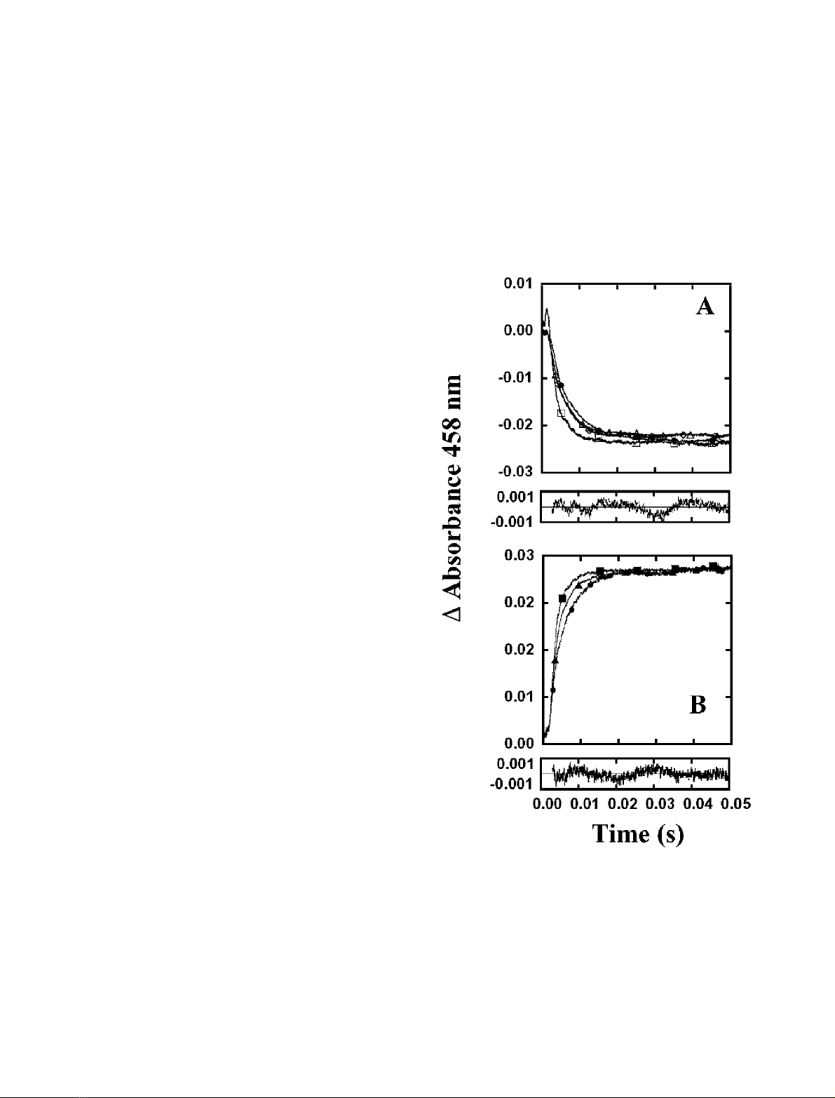
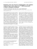
Fig. 1. Time course of the anaerobic reactions of FNR forms with its
NADP
+
/H cofactor as measured by stopped-flow. Reactions were
carried out in 50 m
M
Tris/HCl pH 8.0, at 13 C and followed at
458 nm. Equimolar concentrations of both reactants were used in the
range 10–15 l
M
.(A)ReactionofFNR
ox
with NADPH. Also shown is
the residual for the fit of the transient corresponding to E139D to a
biexponential equation. h, Wild-type FNR; e, E139D FNR; n,
E139Q FNR; d, E139K FNR. (B) Reaction of FNR
rd
with NADP
+
.
Also shown the residual for the fit of the transient corresponding to
E139K to a monoexponential process. j, E139Q FNR; m, E139D
FNR; d, E139K FNR.
FEBS 2002 Role of Glu139 in FNR substrate interaction (Eur. J. Biochem. 269) 4941

(Fig. 1A), and fitting of the kinetic traces shows only slightly
slower k
obs
values for the process ascribed to the formation
of the initial charge-transfer complex with regard to that of
the wild-type (Table 4). The kinetics of reoxidation of the
wild-type enzyme by NADP
+
produces an increase in
absorbance at 458 nm that is best fit to a single exponential
process having a rate constant > 550Æs
)1
. This reaction has
been attributed to ET within the complex, i.e.
[FNR
rd
:NADP
+
]fi[FNR
ox
: NADPH] [21,32]. When
analysing this reaction for the different E139 FNR mutants,
a fast increase in absorption was also observed, which takes
place on the same time scale and with equivalent amplitudes
as those observed for the wild-type enzyme reaction
(Fig. 1B). Moreover, the observed kinetic traces were all
best fit to mono exponential processes with k
obs
values
between 40% (for E139D and E139K) and 64% (for
E139Q) of that found for the wild-type enzyme (Table 4).
Therefore, although it is not possible to quantify exactly the
magnitude of the impairment due to the E139 replacement,
considering that equivalent amplitudes are detected for the
wild-type and the mutants’ processes, it appears that only
subtle changes have occurred for the overall interaction
process between FNR and its coenzyme in both redox states.
Reactions of FNR with Fd. Reactions between FNR and
Fd were followed at 507 nm; this wavelength is an
isosbestic point for FNR
ox
and FNR
sq
and, although it
is not an isosbestic point for FNR
sq
and FNR
rd
,the
absorbance change associated with the FNR
sq
fiFNR
rd
transition is negligible when compared with that due to the
redox state change of Fd at this wavelength. When
following the ET process between Fd
rd
and FNR
ox
no
reaction was detected in the cases of the wild-type or the
E139D FNRs. Previous transient kinetic studies predict
k
obs
values for both wild-type and E139D FNRs to be
> 1000Æs
)1
for the ET between FNR
ox
and Fd
rd
to
produce FNR
sq
and Fd
ox
[24], and thus under our
stopped-flow experimental conditions the reaction should
occur within the instrument’s dead time. Moreover,
previous stopped-flow experiments performed with wild-
type FNR and a 3-fold excess of Fd
rd
[21] showed evidence
of a fast reaction (k
obs
> 250Æs
)1
) which was ascribed to
the reoxidation of a second molecule of Fd
rd
by the
FNR
sq
, expected to have been rapidly formed within the
stopped-flow experimental dead time. However, because
under our present experimental conditions FNR and Fd
are mixed in equimolecular amounts, there is no Fd
rd
in
excess and this second-sequential reaction is not likely
to occur. Moreover, according to the thermodynamic
driving force of the reaction, the reoxidation of Fd
rd
(E¼)384 mV) by FNR (E¼)323 mV) [33] is expected
to take place completely and no Fd
rd
would be in
equilibrium with the rapidly formed products Fd
ox
and
FNR
sq
. For the reaction between Fd
rd
and E139Q FNR,
we were able to observe only the final traces of the Fd
reoxidation to which corresponds a k
obs
>550Æs
)1
indica-
ting that this process has been affected to some degree
although we are not able to quantify it. No reaction was
detected also for the ET from Fd to E139K FNR.
However, taking into account the large impairment
reported for the E139K mutant in accepting electrons
from Fd at low ionic strength [24], the lack of observable
reaction in this particular case must be attributed to the
fact that the reaction does not take place at all under our
stopped-flow conditions. In order to confirm this hypothe-
sis, and to rule out the possibility of the reaction taking
place within the instrument’s dead time, it was followed at
higher salt concentration. A process observed at
l¼133 m
M
and having a k
obs
>370Æs
)1
(Fig. 2A) was
ascribed to the reduction of the mutant by Fd
rd
. This final
ionic strength was chosen so that the expected process
could be detected after taking into account the k
obs
values
reported previously for the ET from Fd
rd
and E139K
FNR
ox
when the reaction was measured by laser flash
photolysis (Fig. 3 [24]).
When the reverse reaction, i.e. ET from FNR
rd
to Fd
ox
was studied, different behaviours were also observed for
the E139 FNR mutants (Fig. 2B). Reduction of Fd by
wild-type FNR, although mostly limited by the instru-
ment’s dead time, yielded a decay at 507 nm which
corresponded to a k
obs
>500Æs
)1
. E139D FNR reacts in a
manner indistinguishable from wild-type, whereas E139Q
FNR shows a k
obs
of 140Æs
)1
, demonstrating that neutral-
ization of the negative charge at position 139 produces a
sizeable impairment on the enzyme ET to Fd. Again,
E139K FNR was, by far, the most impaired in its ET to
Table 4. Fast kinetic parameters for the reactions of wild-type and mutated FNR forms with its substrates as studied by stopped-flow methodology. ND,
no data available.
FNR variant
k
obs
(s
)1
) for the mixing of FNR
ox
with k
obs
(s
)1
) for the mixing of FNR
rd
with
NADPH
a
Fd
rdb
Fld
rdc
NADP
+a
Fd
oxb
Fld
oxc
Wild-type > 500
e
ND
d
ND
d
> 550
e
> 500
e
2.5
> 140
e
0.5
E139D > 350
e
ND
d
ND
d
250 > 500
a
4
> 140
e
0.7
E139Q > 350
e
> 550
e
ND
d
348 140 3
> 140
e
0.6
E139K > 330
e
ND
f
ND
d
220 180
e
17
> 130
e
> 370
g
13 2.2
(l¼133 m
M
)
a
Reaction followed at 458 nm.
b
Reaction followed at 507 nm.
c
Reaction followed at 600 nm.
d
Reaction occurred within the dead time of
the instrument.
e
Most of the reaction took place within the instrument’s dead time.
f
No reaction was detected.
g
Ionic strength was
adjusted to 133 m
M
by adding NaCl from a 5
M
stock solution.
4942 M. Faro et al. (Eur. J. Biochem. 269)FEBS 2002


%20--%3e%3cdefs%3e%3cstyle%3e%20.st0%20{%20fill:%20%23fff;%20}%20.st1%20{%20fill:%20%237800fa;%20}%20%3c/style%3e%3c/defs%3e%3cpath%20class='st1'%20d='M117.78,12.18H43.11c2.9,3.47,4.65,7.94,4.65,12.82,0,5.6-2.3,10.66-6.01,14.29h76.02l7.22-13.56-7.22-13.56Z'/%3e%3cg%3e%3cpath%20class='st0'%20d='M53.58,26.17h-.59v-1.46h.59v-4.96h2.83c1.78,0,2.67.94,2.67,2.82v5.76c0,1.87-.89,2.81-2.67,2.81h-2.83v-4.96ZM55.36,21.37v3.34h1.1v1.46h-1.1v3.34h1.01c.61,0,.91-.37.91-1.1v-5.93c0-.74-.3-1.1-.91-1.1h-1.01Z'/%3e%3cpath%20class='st0'%20d='M65.99,31.14h-1.8l-.31-2.07h-2.19l-.31,2.07h-1.64l1.82-11.39h2.62l1.82,11.39ZM65.28,18.04c-.25.46-.51.77-.75.94-.21.15-.47.22-.79.22-.26,0-.57-.07-.92-.22l-.38-.15c-.14-.05-.26-.07-.37-.07-.3,0-.53.18-.71.54l-.91-.68c.25-.46.51-.77.75-.94.21-.14.48-.21.79-.21.26,0,.57.07.92.21l.38.15c.14.05.26.07.37.07.3,0,.53-.18.71-.54l.91.68ZM61.91,27.52h1.73l-.87-5.76-.87,5.76Z'/%3e%3cpath%20class='st0'%20d='M74.53,26.89v1.52c0,1.91-.89,2.86-2.67,2.86s-2.67-.95-2.67-2.86v-5.93c0-1.91.89-2.86,2.67-2.86s2.67.95,2.67,2.86v1.11h-1.69v-1.22c0-.75-.31-1.12-.93-1.12s-.93.37-.93,1.12v6.15c0,.74.31,1.11.93,1.11s.93-.37.93-1.11v-1.63h1.69Z'/%3e%3cpath%20class='st0'%20d='M81.4,31.14h-1.8l-.31-2.07h-2.19l-.31,2.07h-1.64l1.82-11.39h2.62l1.82,11.39ZM75.9,19.2l1.52-1.91h1.71l1.51,1.91h-1.61l-.76-.95-.75.95h-1.61ZM77.32,27.52h1.73l-.87-5.76-.87,5.76ZM83.1,15.99l-1.76,1.91h-1.26l1.17-1.91h1.86Z'/%3e%3cpath%20class='st0'%20d='M84.86,19.75c1.78,0,2.67.94,2.67,2.82v1.48c0,1.87-.89,2.81-2.67,2.81h-.85v4.28h-1.79v-11.39h2.64ZM84.01,21.37v3.86h.85c.58,0,.87-.36.87-1.08v-1.71c0-.71-.29-1.07-.87-1.07h-.85Z'/%3e%3cpath%20class='st0'%20d='M93.51,19.75c1.78,0,2.67.94,2.67,2.82v1.48c0,1.87-.89,2.81-2.67,2.81h-.85v4.28h-1.79v-11.39h2.64ZM92.66,21.37v3.86h.85c.58,0,.87-.36.87-1.08v-1.71c0-.71-.29-1.07-.87-1.07h-.85Z'/%3e%3cpath%20class='st0'%20d='M98.8,31.14h-1.79v-11.39h1.79v4.88h2.03v-4.88h1.83v11.39h-1.83v-4.88h-2.03v4.88Z'/%3e%3cpath%20class='st0'%20d='M105.36,24.55h2.46v1.62h-2.46v3.34h3.09v1.63h-4.88v-11.39h4.88v1.63h-3.09v3.18ZM108.17,17.29l-1.76,1.91h-1.26l1.17-1.91h1.86Z'/%3e%3cpath%20class='st0'%20d='M112.2,19.75c1.78,0,2.67.94,2.67,2.82v1.48c0,1.87-.89,2.81-2.67,2.81h-.85v4.28h-1.79v-11.39h2.64ZM111.35,21.37v3.86h.85c.58,0,.87-.36.87-1.08v-1.71c0-.71-.29-1.07-.87-1.07h-.85Z'/%3e%3c/g%3e%3ccircle%20class='st1'%20cx='25'%20cy='25'%20r='20'/%3e%3cpath%20class='st0'%20d='M32.78,19.27c2.92,0,4.43,2.55,5.28,5.33l.71,2.17c.14.38-.33.75-.71.75h-5.61c.19-.33.24-.71.09-1.08l-.75-2.45c-.43-1.32-.99-2.64-1.79-3.77.75-.57,1.65-.94,2.78-.94h0ZM25,18.38c3.25,0,4.9,2.78,5.89,5.89l.76,2.45c.14.42-.33.8-.8.8h-11.69c-.42,0-.94-.38-.8-.8l.75-2.45c.99-3.11,2.64-5.89,5.89-5.89h0ZM25,11.35c1.74,0,3.11,1.37,3.11,3.11s-1.37,3.11-3.11,3.11-3.11-1.41-3.11-3.11,1.41-3.11,3.11-3.11h0ZM17.27,19.27c1.08,0,1.98.38,2.73.94-.8,1.13-1.37,2.45-1.74,3.77l-.8,2.45c-.14.38-.05.75.09,1.08h-5.56c-.42,0-.9-.38-.75-.75l.71-2.17c.9-2.78,2.41-5.33,5.33-5.33h0ZM17.27,12.91c1.51,0,2.78,1.27,2.78,2.83s-1.27,2.83-2.78,2.83-2.83-1.27-2.83-2.83,1.27-2.83,2.83-2.83h0ZM32.78,12.91c1.56,0,2.78,1.27,2.78,2.83s-1.23,2.83-2.78,2.83-2.83-1.27-2.83-2.83,1.27-2.83,2.83-2.83h0ZM27.07,28.56v.09c0,.57-.24,1.08-.61,1.46h0v.05c-.38.33-.9.57-1.46.57s-1.08-.24-1.46-.61h0c-.38-.38-.61-.9-.61-1.46v-.09h1.41v.09c0,.19.05.38.19.47v.05c.09.09.28.19.47.19s.38-.09.47-.19v-.05c.14-.09.24-.28.24-.47t-.05-.09h1.41ZM30.99,28.56v.09c0,1.65-.66,3.16-1.74,4.24-1.08,1.08-2.59,1.79-4.24,1.79s-3.16-.71-4.24-1.79l-.05-.05c-1.04-1.08-1.7-2.55-1.7-4.2v-.09h1.41v.09c0,1.27.47,2.4,1.27,3.25h.05c.85.85,1.98,1.37,3.25,1.37s2.4-.52,3.25-1.37c.85-.8,1.37-1.98,1.37-3.25v-.09h1.37ZM34.99,28.56v.09c0,2.78-1.13,5.28-2.92,7.07-1.79,1.79-4.29,2.92-7.07,2.92s-5.23-1.13-7.07-2.92c-1.79-1.79-2.92-4.29-2.92-7.07v-.09h1.41v.09c0,2.4.94,4.53,2.5,6.08,1.56,1.56,3.72,2.5,6.08,2.5s4.52-.94,6.08-2.5c1.56-1.56,2.5-3.68,2.5-6.08v-.09h1.41Z'/%3e%3c/svg%3e)