RESEARC H Open Access
MiR-106b promotes cell proliferation via
targeting RB in laryngeal carcinoma
Kemin Cai
†
, Yu Wang
†
and Xueli Bao
*
Abstract
MiR-106b is frequently up-regulated in various types of human cancer including laryngeal carcinoma. However the
underlying mechanism of miR-106b involved in laryngeal carcinoma remains elusive. Here we showed that
reduction of miR-106b induced cell cycle G0/G1 arrest by targeting tumor suppressor RB in human laryngeal
carcinoma cells. Further, Introducing RB cDNA without 3’UTR abrogated miR-106b-induced cell proliferation. Finally,
there was an inverse relationship between RB and miR-106b expression in laryngeal carcinoma tissues. To our
knowledge, these data indicate for the first time that miR-106b directly regulate cell cycle by targeting RB in
laryngeal carcinoma and that miR-106b could be potential therapeutic approaches for laryngeal carcinoma.
Keywords: laryngeal carcinoma, miR-106b, RB, cell proliferation
Background
Laryngeal carcinoma is a common head and neck malig-
nancy with high incidence as it accounts for approxi-
mately 2.4% of new malignancies worldwide every year
[1,2]. Despite recent advances in cancer treatment, the
prognosis for patients with laryngeal carcinoma espe-
cially at advanced stage remains poor. Therefore, it is
essential to investigate the mechanism involved in the
development and progression of laryngeal carcinoma.
MicroRNAs (miRNAs) are a new class of small, non-
coding RNAs and regulate gene expression by binding
to the 3’-untranslated regions (3’UTRs) of specific
mRNAs. miRNAs could function as oncogenic miRNAs
or tumor suppressor miRNAs, playing crucial roles in
the development and progression of carcer [3,4]. Recent
studies have indicated that frequent deregulation of
miRNA in laryngeal carcinoma [5,6]. Let-7a was signifi-
cantly downregulated both in human laryngeal squa-
mous cancer tissues and Hep-2 cells, and functions as a
potential tumor suppressor in human laryngeal cancer
[5]. Hui et al investigated the significance of miRNA in
patients with locally advanced head and neck squamous
cell carcinoma and identified that thirty-eight miRNAs
were significantly differentially expressed between
malignant versus normal tissues [6]. Of note, upregula-
tion of miR-106b, miR-423, miR-20a, and miR-16 as
well as downregulation of miR-10a were newly observed.
In present work, we determined the function of miR-
106b involved in laryngeal carcinoma. Reduction of miR-
106b by antisense oligonucleotides inhibited cell prolifera-
tion and induced cell cycle G0/G1 arrest in laryngeal carci-
noma cells. Moreover, RB was a direct target of miR-106b
by luciferase reporter assay. Introduction of RB cDNA
without 3’UTR abrogated miR-106b-induced cell prolifera-
tion. Finally, there was an inverse correlation of expression
of miR-106b and RB in laryngeal carcinoma tissues.
Materials and methods
Clinical sample collection
Twenty laryngeal carcinoma tissues used in this study
were obtained from Taizhou People’s Hospital in China.
Specimens were snap-frozen in liquid nitrogen, incuding
10 laryngeal carcinomas with stage I and II, and 10 lar-
yngeal carcinomas with stage III and IV. The collection
and use of the patient samples were reviewed and
approved by Institutional Ethics Committees, and writ-
ten informed consent from all patients was appropriately
obtained.
Cell culture and transfection
Hep-2 and TU212 cells were purchased from Chinese
Academy of Sciences Cell Bank. Cells were maintained
* Correspondence: entcaikemin7173@126.com
†Contributed equally
Department of Otorhinolaryngology Head and Neck Surgery, Taizhou
People’s Hospital, Taizhou 225300, P.R. China
Cai et al.Journal of Experimental & Clinical Cancer Research 2011, 30:73
http://www.jeccr.com/content/30/1/73
© 2011 Cai et al; licensee BioMed Central Ltd. This is an Open Access article distributed under the terms of the Creative Commons
Attribution License (http://creativecommons.org/licenses/by/2.0), which permits unrestricted use, distribution, and reproduction in
any medium, provided the original work is properly cited.

in DMEM medium supplemented with 10% fetal bovine
serum. Cells were transfected using Lipofectamine 2000
(Invitrogen, USA) at the time of 50-60% confluent. 48 h
after transfection, cells were harvested for further
studies.
Plasmids and oligonucleotides
For expression plasmid construct, wild-type RB cDNA
sequence without 3’UTR was selected and cloned into
Pgenesil-1 vector. 2’-O-methyl (OMe)-oligonucleotides
were chemically synthesized and purified by Gene-
Pharma Co., Ltd. (Shanghai, China). The amount of oli-
gonucleotides transfected was 50 nmol/L. Sequences as
follows: miR-106b, 5’- UAAAGUGCUGACAGUGCA-
GAU-3’; anti-miR-106b (As-miR-106b), 5’-AUCUGCA-
CUGUCAGCACUUUA-3’; scrambled miRNA (negative
control), 5’-UUGUACUACACAAAAGUACUG-3’.
Real time PCR
Trizol reagent was used to isolate total RNA from cells
48 h after transfection. The RT-real-time PCR was car-
ried out with the miRNA detection kit (Ambion, USA).
Amplification reaction protocol was performed for 40
cycles consisting 95°C for 3 min, 95°C for 15 sec, 60°C
for 30 sec. Both RT and PCR primer were purchased
from Ambion. 5S RNA was used for normalization.
Relative quantification was conducted using amplifica-
tion efficiencies derived from cDNA standard curves
and obtained relative gene expression. Relative gene
expression was calculated via a 2
ΔΔCt
method.
MTT assay
Cells were plated at 10
4
cells per well in 96-well plates
with six replicate wells. After transfection as described
previously, 20 μlofMTT(5g/L,Sigma,USA)was
added into each well at each day of consecutive 4 days
after treatment and the cells were incubated for addi-
tional 4 h, the supernatant was then discarded. 200 μlof
DMSO was added to each well to dissolve the precipi-
tate. Optical density (OD) was measured at wave length
of 550 nm. The data are presented as the mean ± SD,
which are derived from triplicate samples of at least
three independent experiments.
Cell cycle analysis
Cells were washed with PBS, fixed with 70% ethanol for
at least 1 h. After extensive washing, the cells were sus-
pended in HBSS (Hank’s Balanced Salt Solution) con-
taining 50 μg/mL PI and 50 μg/ml RNase A and
incubated for 1 h at room temperature, and analyzed by
FACScan (Becton Dickinson, USA). Cell cycle analysis
was analyzed by ModFit software. Experiments were
performed in triplicate. Results were presented as % of
cell in a particular phase.
Western blot analysis
Equal amounts of protein per lane were separated by 8%
SDS-polyacrylamide gel and transferred to PVDF mem-
brane. The membrane was blocked in 5% skim milk for
1 h and then incubated with a specific antibody for 2 h.
The antibodies used in this study were: antibodies to RB
(Santa Cruz, USA). The antibody against b-actin (Santa
Cruz, USA) was used as control. The specific protein
was detected by using a SuperSignal protein detection
kit (Pierce, USA). The band density of specific proteins
was quantified after normalization with the density of b-
actin.
Luciferase reporter assay
The human RB 3’UTR (bases 813-959) were amplified
and cloned into the XbaI site of the pGL3-control vec-
tor (Promega, USA), downstream of the luciferase gene,
to generate the plasmids pGL3-WT-RB-3’UTR. pGL3-
MUT-RB-3’UTR plasmids were generated from pGL3-
WT-RB-3’UTR by deleting the binding site (bases 883-
889) for miR-106b “GCACUUU”. For the luciferase
reporter assay, cells were cultured in 96-well plates,
transfected with the plasmids and As-miR-106b using
Lipofectamine 2000. 48 h after transfection, luciferase
activity was measured using the Dual Luciferase Repor-
ter Assay System (Promega). Firefly luciferase activity
was normalized to renilla luciferase activity for each
transfected well.
Statistical analysis
Statistics was determined by ANOVA, or t test using
SPSS11.0. Statistical significance is determined as P <
0.05.
Results
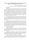
MiR-106b expression in laryngeal carcinomas
To explore miR-106b expression in laryngeal carcino-
mas, we examined 20 human laryngeal carcinoma speci-
mens with different clinical stages using Real time PCR.
As shown in Figure 1, the levels of miR-106b increased
markedly in laryngeal carcinomas with stage III and IV
in comparison to those with stage I and II (P < 0.01).
And we also found high miR-106b expression in Hep-2
and TU212 laryngeal carcinoma cells (Figure 1).
MiR-106b inhibition suppresses cell proliferation and
induces G0/G1 arrest
As-miR-106b and miR-106b mimic oligonucleotides
were employed to change miR-106b expression in Hep-
2 and TU212 cells to evaluate the significance of miR-
106b in laryngeal carcinoma. In both two cells, miR-
106b expression significantly decreased in As-miR-106b
group and increased in miR-106b group 48 h after
transfection (Figure 2A). MTT assay data showed that a
Cai et al.Journal of Experimental & Clinical Cancer Research 2011, 30:73
http://www.jeccr.com/content/30/1/73
Page 2 of 6
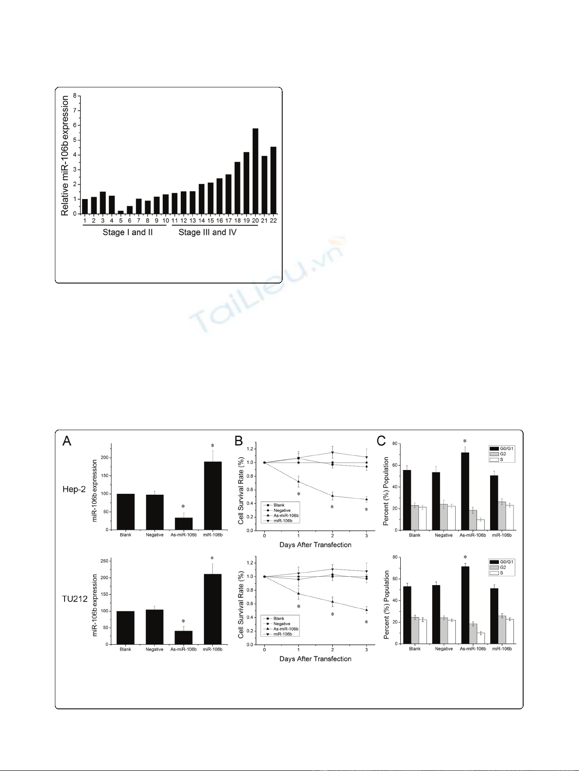
statistically significant cell proliferation inhibition was
found in As-miR-106b group of Hep-2 cells, compared
with control groups respectively. Similar trend was
observed in TU212 cells (Figure 2B). There was no dif-
ference between blank control group and negative con-
trol group in the whole experiment. Next we analyzed
the cell cycle distribution by FACS. As-miR-106b treated
cells represented significant ascends in G0/G1 phase
in comparison to untreated Hep-2 and TU212 cells
(Figure2C).However,wedidnotobserveasignificant
difference in the rate of growth inhibition between miR-
106b group and blank control group; although a slightly
increasing trend of cell survival rate and G0/G1 phase
was seen in Hep-2 and TU212 cells. These results raise
the possibility that there exists a threshold value for
miR-106b up-regulation. Taken together, reduction of
miR-106b can induce cells arrest at G0/G1 phases,
thereby inhibiting cell proliferation in laryngeal carci-
noma cells.
RB is a direct target of miR-106b
To further explore the molecular mechanism of
As-miR-106b induced cell cycle in laryngeal carcinoma
cells, bioinformatics analysis of miR-106b potential tar-
get genes was performed through the databases Target-
Scan http://www.targetscan.org and PicTar http://www.
pictar.bio.nyu.edu, We found that tumor suppressor RB
associated with cell cycle contained the highly conserved
putative miR-106b binding sites (Figure 3A). To deter-
mine whether RB is directly regulated by miR-106b,
Western blot analysis and Luciferase reporter assay were
employed. Western blot analysis showed that a notable
induction of RB expression was detected after
knockdown of miR-106b in Hep-2 and TU212 cells
(Figure 3B). Further, we created pGL3-WT-RB-3’UTR,
and pGL3-MUT-RB-3’UTR plasmids. Reporter assay
revealed that inhibition of miR-106b triggered a marked
Figure 1 Expression of miR-106b in laryngeal carcinoma.
Expression levels of miR-106b in laryngeal carcinoma tissues and
cell lines (21: Hep-2 cells, 22: TU212 cells) were measured by Real
time PCR and quantified as described in methods.
Figure 2 Reduction of miR-106b suppressed laryngeal carcinoma cell proliferation. (A) Expression levels of miR-106b in laryngeal
carcinoma cells 48 h after As-miR-106b and miR-106b treatment. (B) MTT assay displayed that cells treated with As-miR-106b proliferated at a
significantly lower rate than control groups after transfection. (C) After 48 h treatment, cells were harvested and performed by cell cycle assay.
Data are expressed as the mean ± SD of 3 independent experiments. * P < 0.05 compared with control group.
Cai et al.Journal of Experimental & Clinical Cancer Research 2011, 30:73
http://www.jeccr.com/content/30/1/73
Page 3 of 6
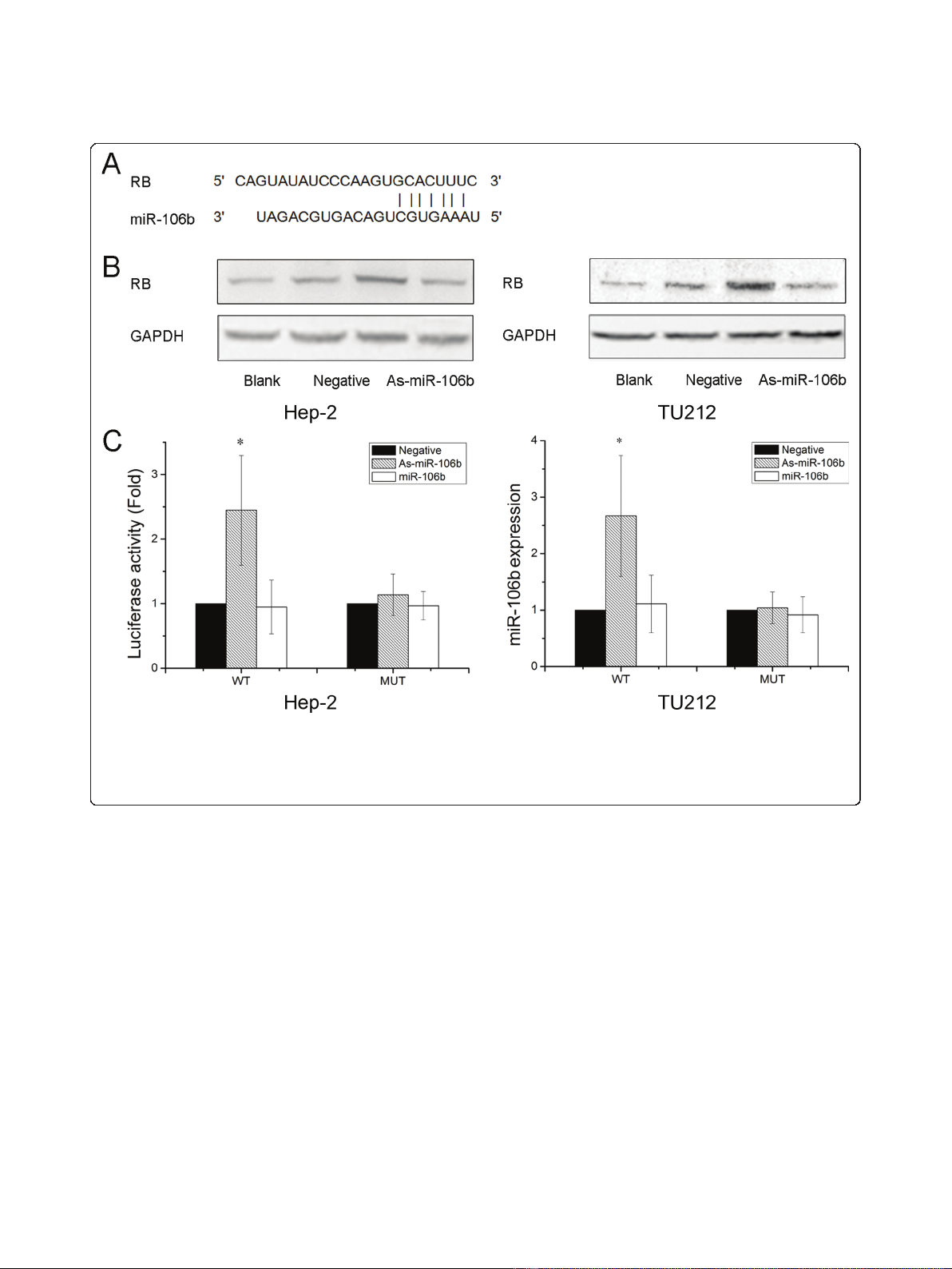
increase of luciferase activity of pGL3-WT-RB-3’UTR
plasmid both in Hep-2 and TU212 cells, without change
in luciferase activity of pGL3-MUT-RB-3’UTR (Figure
3C). These data indicate that RB is a direct target of
miR-106b in laryngeal carcinoma.
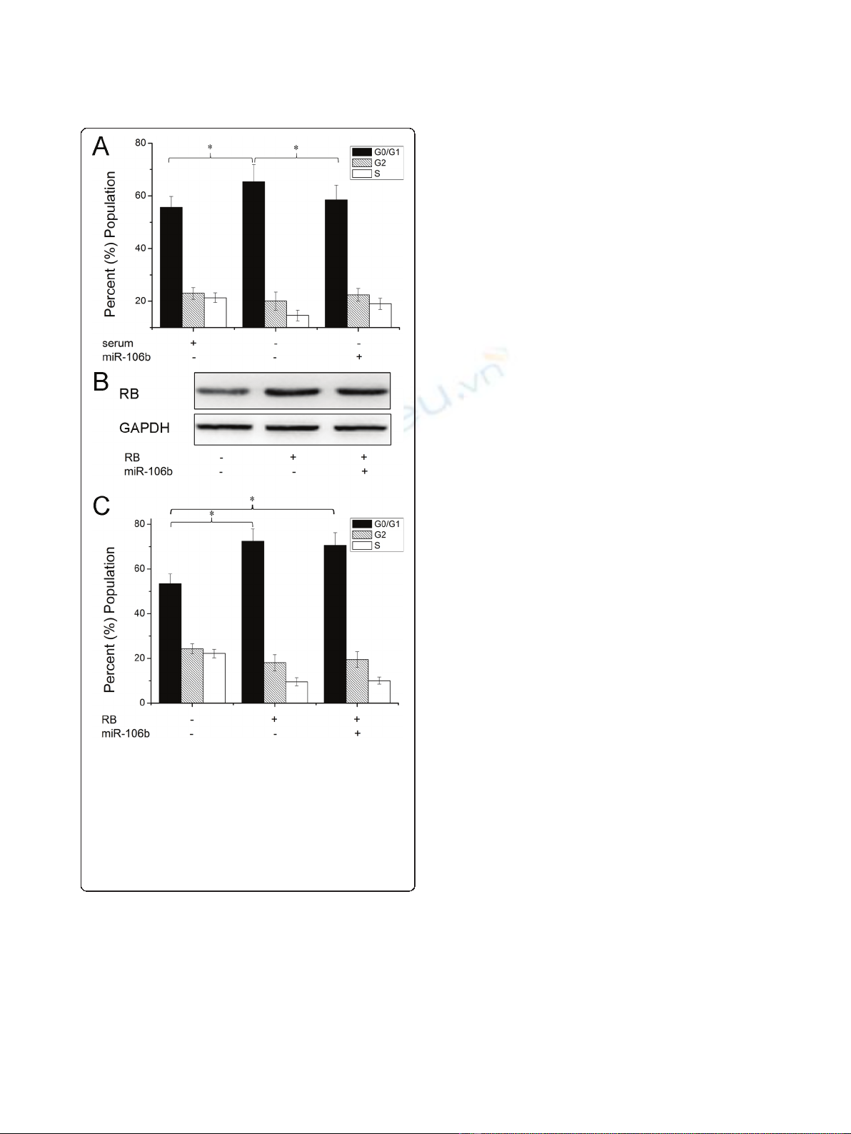
Core role of RB in miR-106b-mediated cell proliferation
Having demonstrated RB as a direct target of miR-106b,
we next examined the importance of RB in miR-106b-
mediated cell proliferation. The cell cycle distribution
analysis showed that upregulation of miR-106b signifi-
cantly reduced cell cycle G0/G1 phase arrest induced by
serum starvation (Figure 4A). Then we transfected Rb
without 3’UTR into Hep-2 cells. Western blot assay
showed that transfection with RB without 3’UTR over-
rided RB expression targeted by miR-106b (Figure 4B).
As shown in Figure 4C, the cells transfected RB
significantly induced G0/G1 phase arrest. However,
when we transfected with RB without 3’UTR and miR-
106b, expression of RB largely abrogated the effect of
miR-106b on cell cycle distribution. These findings sug-
gest that RB is a major target of miR-106b involved in
laryngeal carcinoma cell proliferation.
Inverse correlation of expression of miR-106b and RB in
laryngeal carcinoma tissues
We further explored the correlation of between miR-
106b and RB expression in laryngeal carcinomas. We
tested RB expression in these 20 human laryngeal carci-
noma specimens and found RB expression was down-
regulated in laryngeal carcinomas with stage III and IV
in comparison to those with stage I and II (Figure 5A).
Further, Pearson correlation showed that a significant
negative correlation existed between miR-106b and RB
Figure 3 RB was identified as target genes of miR-106b. (A) A schematic representation showing the putative target site for miR-106b in RB
mRNA 3’UTR. (B) Cells were transfected with As-miR-106b and miR-106b, and the expression of RB was analyzed by Western blot. The expression
of b-actin was used as a loading control. (C) Luciferase constructs were transfected into cells transduced with As-miR-106b and miR-106b.
Luciferase activity was determined 48 h after transfection. The ratio of normalized sensor to control luciferase activity is shown. Data are
expressed as the mean ± SD of 3 independent experiments. * P < 0.05 compared with control group.
Cai et al.Journal of Experimental & Clinical Cancer Research 2011, 30:73
http://www.jeccr.com/content/30/1/73
Page 4 of 6
expression in laryngeal carcinoma tissues (R = 0.673, P
< 0.005) (Figure 5B).
Discussion
Recent evidences indicate that miR-106b has partici-
pated in development and progression of human
tumors, such as hepatocellular cancer, prostate cancer,
gastric cancers and renal cell carcinoma [7-10]. In this
study, repression of miR-106b resulted in cell prolifera-
tion inhibition and cell cycle G0/G1 arrest in laryngeal
carcinoma cells. Further, As-miR-106b regulated RB
expression via targeting 3’UTR of RB. Finally, expression
of RB abolished cell proliferation of miR-106b.
MiR-106b, located at Chr 7, is one member of miR-
106b-25 cluster. Several genes have been evidenced to
be the targets of miR-106b, such as p21/CDKN1A and
TGF-btype II receptor (TbR II). Ivanovska et al
reported that miR-106b gain of function promotes cell
cycle progression, whereas loss of function reverses this
phenotype. And p21/CDKN1A is a direct target of miR-
106b and that its silencing plays a key role in miR-106b-
induced cell cycle phenotypes[11].Inthepathogenesis
of Alzheimer’s diseases, miR-106b regulated TbRII
expression via binding 3’UTR of the TbRIImRNA,
thereby leads to impairment in TGF-bsignaling [12].
Here, we evidenced that RB was a novel direct and func-
tional target of miR-106b involved in cell proliferation of
laryngeal carcinoma cells. Reduction of miR-106b regu-
lated RB expression via targeting 3’UTR of RB, and
expression of RB largely abrogated miR-106b-induced
cell proliferation in laryngeal carcinoma cells. And miR-
106b increased with the increasing stages of laryngeal
carcinoma tissues, and inversely correlated with RB
expression.
The RB-pathway, consisting of inhibitors and activa-
tors of cyclin-dependent kinases, the retinoblastoma
tumor suppressor (RB), the E2F-family of transcription
factors and cyclin-dependent protein kinases, plays criti-
cal roles in the regulation of cell cycle progression and
cell death [13,14]. Components of this pathway, particu-
larly RB, p16Ink4a, and cyclin D1, are frequently altered
in human cancers to promote deregulated cellular pro-
liferation [15,16]. Recently, a comprehensive analysis of
the genome and transcriptome has shown that the RB-
pathway is altered in 78% of the primary glioblastoma
tumor samples [17]. In our study, RB was lower expres-
sion in laryngeal carcinomas with stage III and IV in
comparison to those with stage I and II, in line with the
previous study [18]. And upregulation of RB controls
G1/S transition in the cell cycle. Up to now, the
approaches that specifically target the RB-pathway have
been used in preclinical models, but not yet in the clini-
cal setting [19,20]. However, the RB-pathway is still a
promising target in cancer intervention and further
investigations are needed.
In conclusion, we have showed that miR-106b is one
of oncogenic miRNAs in laryngeal carcinomas and RB is
a novel and critical target of miR-106b. These results
suggest that miR-106b might be useful as a potential
therapeutic target for laryngeal carcinoma and more in
depth analysis is required.
Figure 4 Expression of RB abrogates miR-106b -induced cell
proliferation. (A) Cells were transfected with miR-106b and then
treated with serum starvation and cell proliferation was performed
by cell cycle analysis. (B) Cells were transfected with pcDNA-RB
(without the 3’UTR) and miR-106b, RB protein level was detected by
Western blot assay. b-actin protein was regarded as endogenous
normalizer. (C) Cells were transfected with pcDNA-RB (without the
3’UTR) and miR-106b, cell cycle assay was performed respectively.
Data are expressed as the mean ± SD of 3 independent
experiments. * P < 0.05.
Cai et al.Journal of Experimental & Clinical Cancer Research 2011, 30:73
http://www.jeccr.com/content/30/1/73
Page 5 of 6


![Vaccine và ứng dụng: Bài tiểu luận [chuẩn SEO]](https://cdn.tailieu.vn/images/document/thumbnail/2016/20160519/3008140018/135x160/652005293.jpg)