Molecular characterization and allergenic activity of Lyc e 2
(b-fructofuranosidase), a glycosylated allergen of tomato
Sandra Westphal
1
, Daniel Kolarich
2
, Kay Foetisch
1
, Iris Lauer
1
, Friedrich Altmann
2
, Amedeo Conti
3
,
Jesus F. Crespo
4
, Julia Rodrı
´guez
4
, Ernesto Enrique
5
, Stefan Vieths
1
and Stephan Scheurer
1
1
Department of Allergology, Paul-Ehrlich-Institut, Langen, Germany;
2
Institute of Chemistry, University of Agriculture, Vienna,
Austria;
3
CNR-ISPA c/o Bioindustry Park, Colleretto Giacosa, Italy;
4
Servicio de Alergia, Hospital Universitario Doce de Octubre,
Madrid, Spain;
5
Institut Universitari Dexeus, Barcelona, Spain
Until now, only a small amount of information is available
about tomato allergens. In the present study, a glycosylated
allergen of tomato (Lycopersicon esculentum), Lyc e 2, was
purified from tomato extract by a two-step FPLC method.
The cDNA of two different isoforms of the protein,
Lyc e 2.01 and Lyc e 2.02, was cloned into the bacterial
expression vector pET100D. The recombinant proteins were
purified by electroelution and refolded. The IgE reactivity of
both the recombinant and the natural proteins was investi-
gated with sera of patients with adverse reactions to tomato.
IgE-binding to natural Lyc e 2 was completely inhibited by
the pineapple stem bromelain glycopeptide MUXF
(Mana1–6(Xylb1–2)Manb1–4GlcNAcb1–4(Fuca1–3)
GlcNAc). Accordingly, the nonglycosylated recombinant
protein isoforms did not bind IgE of tomato allergic patients.
Hence, we concluded that the IgE reactivity of the natural
protein mainly depends on the glycan structure. The amino
acid sequences of both isoforms of the allergen contain four
possible N-glycosylation sites. By application of MALDI-
TOF mass spectrometry the predominant glycan structure
of the natural allergen was identified as MMXF (Mana1–6
(Mana1–3)(Xylb1–2)Manb1–4GlcNAcb1–4(Fuca1–3)
GlcNAc). Natural Lyc e 2, but not the recombinant protein
was able to trigger histamine release from passively sensitized
basophils of patients with IgE to carbohydrate determinants,
demonstrating that glycan structures can be important for
the biological activity of allergens.
Keywords: Lyc e 2; tomato; food allergen; IgE reactivity;
glycoprotein.
To date, only few attempts have been made to identify and
characterize tomato allergens. In most reports, allergy to
tomato is linked to other allergies such as grass pollen [1]
and latex allergy [2,3]. The prevalence of tomato allergy
ranges from 1.5% to 16% among food-allergic patients
indicating that tomato is a relevant allergenic food in
selected populations.
The first reports on IgE-reactive glycoproteins in tomato
extract by Bleumink et al. [4,5] described a heat resistant
protein fraction between 20 and 30 kDa showing enhanced
reactivity in skin prick tests (SPT). Darnowski et al.[6]
investigated the distribution of profilin in tomato tissues.
Recently the cDNA sequence of tomato profilin was
published (GenBank accession no. AY061819/AJ417553)
and the protein was designated as tomato allergen Lyc e 1.
Cross-reactive carbohydrate determinants (CCD) are
found in many allergenic sources such as pollen and insect
venom, but the highest rate of serological reactions to CCD
has been observed to plant food extracts. Immunoblot
analyses of electrophoretically separated food allergen
extracts revealed that IgE-reactive carbohydrate structures
are present on many different glycoproteins from one
allergen source [7,8]. Examples for IgE-reactive glyco-
proteins are phospholipase A
2
from bee venom [9], Cup a 1
from cypress pollen [10], Ara h 1 from peanut [11] as well as
a vicilin-like protein from hazelnut [12].
The analysis of free [13] and linked [14] N-glycans of
tomato revealed the presence of a plant-characteristic glycan
core with xylose and fucose participating in an IgE-binding
epitope. The N-terminal sequencing of a 52-kDa glyco-
protein of tomato extract gave hints for the existence of
b-fructofuranosidase as a relevant allergen in tomato
[15,16]. b-Fructofuranosidase, also known as acid invertase
(EC 3.2.1.26) catalyses the hydrolysis of sucrose into glucose
and fructose. A variety of these enzymes is found in plants
showing differences between pH optima, isoelectric point
and subcellular localization [17]. Soluble invertases are
known to be vacuolar [18], but cytosolic forms also exist
[19]. The b-fructofuranosidase of tomato was shown to play
an important role in the regulation of hexose accumulation
during fruit ripening [20]. Two isoforms of the tomato
protein were identified that differed only in their C-termini.
One isoform with a molecular mass of 51 kDa (GenBank
accession no. D11350) has an 86-bp insertion in its
sequence, a stop codon in this insertion reduces the open
reading frame and thus the length of the protein. It was
Correspondence to S. Scheurer, Paul-Ehrlich-Institut, Department of
Allergology, Paul-Ehrlich Str. 51–59, D-63225 Langen, Germany.
Fax: + 49 6103 77 1258, Tel.: + 49 6103 77 5200,
E-mail: schst@pei.de
Abbreviations: CCD, cross-reactive carbohydrate determinants;
HIC, hydrophobic interaction chromatography; RT, reverse
transcribed; SPT, skin prick testing; DBPCFC, double blind
placebocontrolledfoodchallenge.
(Received 10 October 2002, revised 8 January 2003,
accepted 5 February 2003)
Eur. J. Biochem. 270, 1327–1337 (2003) FEBS 2003 doi:10.1046/j.1432-1033.2003.03503.x

found that the second isoform without the insertion
sequence and a molecular mass of 60 kDa (S70040) exists
at a much higher expression level in the tomato fruit [21].
The allergenicity of b-fructofuranosidase of tomato was
further confirmed by Foetisch et al. [22]. The aim of the
present study was to analyze the role of N-linked glycans in
the IgE-response of tomato-allergic patients using the
b-fructofuranosidase as a model allergen. For this purpose,
purified natural as well as recombinant proteins were
investigated concerning their IgE-binding capacity and their
ability to induce histamine release from human basophils.
The glycan structure of the natural b-fructofuranosidase
was determined. Our results indicate an important role for
N-glycans containing xylose and fucose residues in the IgE-
response of tomato-allergic patients.
Materials and methods
Preparation of allergen extract
Extracts from tomato and low fat milk were prepared by a
low-temperature method as previously described [23]. In
brief, pieces of fresh fruit were frozen in liquid nitrogen, and
ground in a mill without thawing. The obtained powder was
homogenized in prechilled acetone and stored overnight at
)20 C. The precipitate was filtered, washed twice with ice-
cold acetone and once with acetone/diethylether (1 : 1, v/v)
and lyophilized. Extraction of proteins from this powder
was done with NaCl/P
i
(0.15
M
NaCl/0.01
M
NaH
2
PO
4
) at
4C. After centrifugation the supernatant was collected,
filtered and freeze-dried. The lyophilized extract was stored
at )80 C.
Purification of N-linked glycopeptides
N-linked glycopeptides with the glycan structure Mana1–
6(Xylb1–2)Manb1–4GlcNAcb1–4(Fuca1–3)GlcNAc
(MUXF) coupled to two to four amino acids were prepared
from pineapple stem bromelain by digestion with pronase
followed by a series of chromatographic steps as described
elsewhere [24]. Glycopeptides containing the pentasac-
charidecoreMana1–6(Mana1–3)Manb1–4GlcNAcb1–
4GlcNAc (MM) were prepared from bovine fibrin.
Purification of natural Lyc e 2 from tomato fruit
To purify the natural b-fructofuranosidase, lyophilized
tomato extract was dissolved in starting buffer (1
M
(NH
4
)
2
SO
4
,20m
M
Tris/HCl, 1 m
M
EDTA, pH 8.0) to a
protein concentration of 2 mgÆmL
)1
. After filtration
through a 0.45-lmfilter(Sartorius,Go
¨ttingen, Germany)
the protein solution was applied to a 1-mL phenyl superose
column (Amersham Pharmacia Biotech, Uppsala, Sweden)
to perform hydrophobic interaction chromatography
(HIC). Bound proteins were eluted with distilled water at
a flow rate of 0.5 mLÆmin
)1
. Further purification of the
eluted fractions containing the IgE-reactive 50-kDa band
was performed by gel chromatography using a Superdex 75
Column, HR10/30 (Amersham Pharmacia Biotech). Elu-
tion was done with NaCl/P
i
, pH 7.4 at a flow rate of
0.5 mLÆmin
)1
. Fractions were collected in 0.5 mL steps and
analyzed by SDS/PAGE and immunoblotting.
N-terminal amino acid sequencing
Partially purified Lyc e 2 eluted form the HIC column
was electroblotted onto a poly(vinylidene difluoride) mem-
brane. After staining with Coomassie Brilliant Blue the
protein band was excised from the membrane and ana-
lyzed on an Applied Biosystems 492 Procise sequencer
(Applied Biosystems, Foster City, CA, USA) in pulse-liquid
mode to determine the N-terminal partial sequence of the
IgE-reactive protein. All chemicals were from Applied
Biosystems.
Cloning the cDNAs of two isoforms of
b-fructofuranosidase from tomato fruit
Total RNA was isolated from tomato fruit using the
RNeasy Plant RNA Mini Kit (Qiagen, Hilden, Germany).
DNA contaminations were removed by using the RNase-
free DNase set (Qiagen). The RNA was reverse transcribed
(RT) with the First Strand cDNA Synthesis Kit (Amersham
Pharmacia Biotech) according to the manufacturer’s
instructions using 1 lg total RNA for each transcription
and the NotI-d(T)
18
oligonucleotide for priming. To obtain
the complete coding region, the RT products were amplified
using gene specific 5¢-and 3¢primers selected on the basis of
the published sequences for tomato b-fructofuranosidase
(GenBank accession no. D11350 and S70040). Primers for
the short isoform of b-fructofuranosidase were FF5SP,
matching with the N-terminal sequence of the coding
region: 5¢ATGGCCACTCAGTATGACC, FF5, matching
with the N-terminal sequence of the mature protein: 5¢TAT
GCGTGGTCCAATGCTATGC, and FF3A, matching
with the C-terminal sequence of the coding region: 5¢TTAC
AAGGACAAATTAATTGTGCCAG. For amplification
of the long isoform the same 5¢primers were used, the 3¢
specific primer was FF3B: 5¢TTACAAGTCTTGCAA
AGGGAAGGAT. For amplification the Expand long tem-
plate DNA Polymerase Set (Roche, Mannheim, Germany)
was used. The PCR conditions were the following: 94 C,
5 min, followed by 30 cycles: 94 C, 30 s, 50 C, 30 s,
68 C, 2 min. The final extension was 7 min at 68 C. The
obtained cDNA was cloned into the pCRII-TOPO vector
(Invitrogen, Groningen, the Netherlands).
For protein expression in E. coli the coding regions
without signal sequences were cloned into the pET100D
vector containing a six histidine tag using the pET
Directional TOPO expression Kit (Invitrogen). The DNA
was amplified using the same 3¢primersasforcDNA
cloning, whereas the 5¢primer contained the sequence
CACC for directional cloning. FF5-CACC: 5¢CAC
CTATGCGTGGTCCAATGCTATGC. The PCR was
carried out using Vent DNA polymerase (New England
Biolabs, Frankfurt, Germany) under the following condi-
tions: 94 C, 5 min, followed by 30 cycles: 94 C30s,
50 C, 30 s, 72 C, 2 min. The final extension was 7 min at
72 C.
DNA sequencing
The sequence analysis was carried out with an ABI 373
automated fluorescent sequencer (Applied Biosystems)
using vector or gene specific primers and the ABI PRISM
1328 S. Westphal et al. (Eur. J. Biochem. 270)FEBS 2003

BigDye Terminators v3.0 CycleSequencing Kit according to
the manufacturer’s instructions.
Recombinant protein expression and purification
For expression, the pET100D constructs were transformed
in E. coli BL21 star (Invitrogen) and protein synthesis was
induced with 1 m
M
isopropyl thio-b-
D
-galactoside for 5 h at
37 C. After induction, bacteria were harvested by centri-
fugation and stored at )80 C. Purification was carried out
by electroelution from SDS/PAGE gels. Electroelution was
performed as described elsewhere [25]. Briefly, the pellet
from 100-mL bacterial culture was resuspended in non-
reducing 1 ·SDS loading buffer Rotiload 2 (Roth, Karls-
ruhe, Germany) and proteins were separated by SDS/
PAGE using a 10% resolving gel with 1.5-mm spacers.
Desired bands were excised from the gel after staining with
0.3
M
CuCl
2
and the protein was eluted using a Centrilutor
electroelution device (Millipore, Badford, MA, USA).
Elution of the proteins was done at 25 mA for 3 h directly
into Centricon centrifugal filter devices with an exclusion
size of 30 kDa. The purity of the eluted fractions was
controlled by SDS/PAGE followed by staining with Coo-
massie Brilliant Blue and the protein content was deter-
mined according to Bradford using the Roti-Quant protein
assay (Roth).
Patients’ sera
Serum samples were taken from a group of 78 patients
with a positive case history of immediate type reactions to
tomato fruit. Most of the patients (49) were from
Germany, the others were from Spain (Table 1). Only
adults were included in the study, the age ranged between
19 and 65 years; 20% were male. All Spanish and some of
the German patients underwent skin prick testing (SPT)
with commercial tomato extract. Four Spanish patients
were tested with DBPCFC (double blind placebo con-
trolled food challenge) and showed positive reaction.
Serum from a nonallergic subject was taken as a negative
control.
Determination of specific IgE
Measurement of allergen-specific IgE was performed with
the CAP FEIA system (Pharmacia Diagnostics, Uppsala,
Sweden) according to the manufacturer’s instructions.
In addition, a covalink-ELISA was performed in 96 well
Covalink-plates (Nunc GmbH & Co. KG, Wiesbaden,
Germany) as previously described using 250 ng natural or
recombinant protein per well instead of glycopeptides [8].
For detection of IgE reactivity, streptavidin conjugated with
horseradish peroxidase instead of alkaline phosphatase was
used. After visualization of the enzymatic activity with
tetramethylbenzidine as substrate at 37 C for 20 min the
reaction was stopped by addition of 50 lL3
M
H
2
SO
4
and
absorption was measured at 450 nm [26].
IgE immunoblot and IgE immunoblot inhibition
Allergen extracts (20 lgÆcm
)1
), E. coli lysatesaswellas
purified natural and recombinant allergens (0.5 lgÆcm
)1
)
were separated by SDS/PAGE under reducing conditions
as described by Laemmli et al. [27] in a Mini-Protean 3 cell
(Bio-Rad, Munich, Germany). For immunoblot analysis,
proteins were transferred onto 0.45 lm nitrocellulose
membranes (Schleicher und Schuell, Dassel, Germany) by
tank blotting using the Bio-Rad Mini Trans blot cell for
1 h at 300 mA. Before application of the 1 : 10 diluted
patients’ sera the membrane was blocked in NaCl/Tris/
0.3% Tween20 and cut into 3 mm wide strips. Immuno-
staining of bound IgE antibodies was performed with an
alkaline phosphatase conjugated anti-(human IgE) Ig
(Pharmingen, Hamburg, Germany, 1 : 750 dilution, 4 h)
and the Bio-Rad alkaline phosphatase conjugate substrate
kit (Bio-Rad).
Table 1. Clinical data of patients investigated in this study. OAS,oralallergysyndrome;ND,neurodermatitis;n, number of patients investigated;
SPT pos., patients with positive skin prick test/patients tested.
Country
Symptoms
Mild (OAS)
Systemic (Urticaria, ND,
Nausea, Anaphylaxis)
CAP SPT pos. CAP SPT pos.
Germany 0 (n¼4) 1/2 0 (n¼10) 3/3
1(n¼2) 0/0 1 (n¼2) 0/1
2(n¼13) 3/7 2 (n¼2) 1/1
3(n¼8) 3/4 3 (n¼3) 2/3
4(n¼2) 1/1 4 (n¼2) 1/2
5(n¼1) 0/0 5 (n¼0) 0/0
Spain 0 (n¼1) 0/0 0 (n¼1) 0/0
1(n¼3) 2/2 1 (n¼1) 1/1
2(n¼6) 1/1 2 (n¼1) 0/1
3(n¼5) 3/3 3 (n¼5) 3/3
4(n¼2) 2/2 4 (n¼3) 3/3
5(n¼1) 1/1 5 (n¼0) 0/0
FEBS 2003 Allergenic glycoprotein Lyc e 2 (Eur. J. Biochem. 270) 1329

For inhibition of IgE-binding 1 : 10 diluted sera were
preincubated with 10 lg of purified glycopeptide and 100 lg
of allergen extract before incubation of the blot strips.
Circular dichroism (CD) spectroscopy of natural
and recombinant b-fructofuranosidase
The CD spectra of the natural Lyc e 2 as well as of the larger
recombinant isoform designated as rLyc e 2.02 were recor-
ded on a Jasco J-810S spectropolarimeter (Jasco, Grob-
Umstadt, Germany) at 20 Cwithastepwidthof0.2 nmand
a bandwidth of 1 nm. The spectral range was 190–260 nm
at 50 nmÆmin
)1
. Six scans were accumulated. The protein
concentration was 5.5 l
M
in a 10 m
M
KH
2
PO
4
,pH7.0.
Analysis of N-linked glycans and peptides of Lyc e 2
by MALDI-TOF mass spectrometry
Eight micrograms of HIC-purified Lyc e 2 was excised from
a Coomassie-stained SDS/PAGE gel after electrophoresis
under reducing conditions and subjected to tryptic digestion
as described elsewhere [28]. The extracted and dried peptides
were taken up in water/acetonitrile/trifluoroacetic acid
(95 : 5 : 0.1, v/v/v) and analyzed by matrix assisted laser
desorption/ionization time-of-flight mass spectrometry
(MALDI-TOF-MS). Further preparation and mass spectro-
metry analysis of N-glycans was performed according to
Kolarich and Altmann [29]. Briefly, the peptides were dried
and redissolved in ammonium acetate before deglycosyla-
tion with N-glycosidase A. To remove salts and peptides the
digest was purified using a triphasic column consisting of
Dowex W 50, C-18 reversed phase and an AG 3-X4A (Dow
Chemical Company, Edegem, Belgium). Analysis and
identification of the glycans was carried out by mass
spectrometry using a DYNAMO MALDI-TOF (Thermo-
BioAnalysis, Santa Fe
´,NM,USA).
Basophil histamine release
The histamine-release was performed as described previ-
ously [30] with several modifications. Peripheral blood was
drawn from nonalllergic donors and PBMCs were isolated
using Ficoll-Hypaque centrifugation. The conditions for
stripping of the nonspecific IgE and for the passive
sensitization procedure were chosen according to the
recommendations of Pruzansky et al. [31]. Cells sensitized
with a nonallergic serum served as negative control.
Stimulation of the cells was performed using a histamine
kit (Immunotech, Marseille, France) according to the
manufacturer’s instructions with tenfold dilutions of the
allergens starting at 10 lgÆmL
)1
. For testing, self-prepared
tomato extract, nLyc e 2, rLyc e 2, horseradish peroxidase,
deglycosylated horseradish peroxidase, the glycopeptide
MUXF and MUXF conjugated to BSA as well as BSA
alone were used. The histamine releases were measured by
an enzyme immunoassay (Immunotech). After subtraction
of the spontaneous release of the basophils, the allergen-
induced histamine release was calculated as percent of the
total amount of histamine determined after lysis of the
basophils by twofold freezing and thawing of the cells. A
histamine release of more than 10% was considered positive.
Duplicate determinations were performed in all cases.
Results
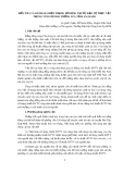
Screening of patients’ sera
Sera from patients with a history of adverse reactions to
tomato were investigated by immunoblotting. Special
attention was drawn to IgE reactions to protein bands in
the high molecular mass range frequently found to be
glycoproteins with ubiquitous carbohydrate epitopes [8,22].
Out of 49 sera from German patients with tomato-related
symptoms such as OAS, nausea, urticaria, abdominal pain
and dyspnea (Table 1), 18 (37%) recognized several bands
above 20 kDa (Fig. 1A).
From the Spanish group, 10 out of 29 (34.5%) sera showed
reactivity in the high molecular mass range (Fig. 1B).
Hence, there was no significant difference in IgE reacti-
vity to glycoproteins between both groups. Besides binding
to protein bands larger than 20 kDa we also observed
reactivity to proteins with a molecular mass of 15 and
9 kDa. IgE binding to carbohydrates was confirmed by blot
inhibition of a patient’s serum with known sensitization
against CCD. Tomato extract as well as the glycopeptide
MUXF obtained from pineapple stem bromelain almost
completely inhibited the IgE reactivity except for one band
at 55 kDa assuming that either this protein does not contain
such glycosylation or the IgE reactivity is based on the
protein backbone alone. No inhibition was observed with
the fibrin glycopeptide MM and extract from low fat milk
(data not shown). These results indicated that the IgE-
binding to most of the tomato proteins in the high molecular
mass range is mediated by the cross-reactive glycan
structure MUXF typically existing in plants but not in
mammals.
The 28 patients showing IgE reactivity in the high
molecular mass range were selected for further studies on
the IgE-binding capacity of Lyc e 2.
Two step purification of Lyc e 2 from tomato extract
The elution profile of the first chromatographic step (HIC)
is shown in Fig. 2A. A 50-kDa band corresponding
to Lyc e 2 was detected in the four water elution fractions
E1–4. After size exclusion chromatography of pooled
fractions E3 and E4 the proteins were nearly homogeneous.
In the elution fractions 30–33 Lyc e 2 with a molecular mass
of 50 kDa was eluted, fractions 34–37 contained a band of
36 kDa and the fractions 38–41 a protein with a molecular
mass of about 20 kDa (Fig. 2B).
Immunoblot analysis with a polyclonal anti-profilin
serum from rabbit confirmed that another important
tomato allergen, profilin, did not contaminate the puri-
fied Lyc e 2-fractions. In contrast to tomato extract that
showed a profilin band at 14 kDa, no bands were visible in
the fractions 30–33 from the second purification step (not
shown).
N-Terminal amino acid sequencing
For N-terminal sequencing fraction E3 from the HIC step
was used. The sequence of the 50 kDa band excised
from the poly(vinylidene difluoride) membrane was
YAXSNAMLXX. A search in the protein database
1330 S. Westphal et al. (Eur. J. Biochem. 270)FEBS 2003
revealedthisproteintobeb-fructofuranosidase (YAW
SNAMLSW). From the N-terminal sequence we were not
able to distinguish between the two isoforms of the protein,
only the molecular mass of 50 kDa would suggest that we
had purified the truncated isoform.
Cloning of the cDNA of two isoforms of tomato
b-fructofuranosidase and recombinant expression
in
E. coli
For protein expression in E. coli, only the cDNA coding for
the mature proteins without signal peptide sequence was
amplified and cloned in the pET100D expression vector.
Because the proteins completely accumulated in insoluble
inclusion bodies, they were purified by electroelution and
refolded. The truncated isoform, designated as Lyc e 2.01
had an apparent molecular mass of 51 kDa. The other
isoform, Lyc e 2.02 migrated as a 60-kDa band. Both
proteins were highly pure (Fig. 3). The CD spectra of
natural Lyc e 2 and recombinant Lyc e 2.02 (rLyc e 2.02)
were highly superimposable and clearly showed the exist-
ence of secondary structures (not shown).
Comparison of IgE-reactivities of the purified natural
and recombinant Lyc e 2
HIC-purified natural and electroeluted recombinant pro-
teins (both isoforms) were separated by SDS/PAGE (0.5 lg
Fig. 1. IgE binding to glycoproteins in tomato extract. IgE-binding of sera from German (A) and Spanish (B) patients to glycosylated tomato extract
proteins separated by SDS/PAGE and transferred to nitrocellulose (20 lg protein per cm). N, negative control, serum from nonallergic subject.
FEBS 2003 Allergenic glycoprotein Lyc e 2 (Eur. J. Biochem. 270) 1331


![Vaccine và ứng dụng: Bài tiểu luận [chuẩn SEO]](https://cdn.tailieu.vn/images/document/thumbnail/2016/20160519/3008140018/135x160/652005293.jpg)