RESEARC H Open Access
Small interfering RNA mediated Poly (ADP-ribose)
Polymerase-1 inhibition upregulates the heat
shock response in a murine fibroblast cell line
Rajesh K Aneja
1*
, Hanna Sjodin
2
, Julia V Gefter
3
, Basilia Zingarelli
4
, Russell L Delude
3
Abstract
Poly (ADP-ribose) polymerase-1 (PARP-1) is a highly conserved multifunctional enzyme, and its catalytic activity is
stimulated by DNA breaks. The activation of PARP-1 and subsequent depletion of nicotinamide adenine
dinucleotide (NAD
+
) and adenosine triphosphate (ATP) contributes to significant cytotoxicity in inflammation of
various etiologies. On the contrary, induction of heat shock response and production of heat shock protein 70
(HSP-70) is a cytoprotective defense mechanism in inflammation. Recent data suggests that PARP-1 modulates the
expression of a number of cellular proteins at the transcriptional level. In this study, small interfering RNA (siRNA)
mediated PARP-1 knockdown in murine wild-type fibroblasts augmented heat shock response as compared to
untreated cells (as evaluated by quantitative analysis of HSP-70 mRNA and HSP-70 protein expression). These
events were associated with increased DNA binding of the heat shock factor-1 (HSF-1), the major transcription
factor of the heat shock response. Co-immunoprecipitation experiments in nuclear extracts of the wild type cells
demonstrated that PARP-1directly interacted with HSF-1. These data demonstrate that, in wild type fibroblasts,
PARP-1 plays a pivotal role in modulating the heat shock response both through direct interaction with HSF-1 and
poly (ADP-ribosylation).
Introduction
Poly (ADP-ribose) polymerase-1 (PARP-1) is a highly
conserved chromatin bound enzyme [1,2] and plays an
important role in DNA repair, gene transcription, cell-
cycle progression, cell death, and maintenance of geno-
mic integrity [3-5]. PARP-1 is activated by DNA breaks
and cleaves nicotinamide adenine dinucleotide (NAD
+
)
into nicotinamide resulting in ADP-ribose moieties;
these moieties covalently attach to various acceptor pro-
teins including PARP itself. The continued activation of
PARP leads to depletion of its substrate NAD
+
with
consequent depletion of ATP, energy failure and cell
death [6].
In addition to its influence on chromatin structure
and stability, recent studies indicate PARP-1 plays a role
in gene-specific transcription [7-9]. PARP-1 regulates
transcription by modifying chromatin-associated
proteins and acts as a cofactor for transcription factors,
most notably NF-B and AP-1 [10,11]. Genetic deletion
of PARP-1 attenuates tissue injury after ischemia and
reperfusion, streptozocin-induced diabetes, endotoxic
and hemorrhagic shock, heat stroke and localized colo-
nic inflammation [12-19]. The benefits conferred by
pharmacological inhibitors of poly (ADP-ribosylation) in
diverse experimental disease models further reiterate the
importance of PARP-1 as an important pharmacological
target [20,21]
Oxidative injury and ATP depletion also leads to
activation of heat shock factor (HSF)-1, a major tran-
scription factor responsible for increased transcription
of genes encoding heat shock proteins, particularly
heat shock protein-70 [22,23]. HSP-70 provides cyto-
protection from a variety of inflammatory insults,
including oxidative stress, viral infections and ische-
mia-reperfusion injury [24,25]. Previously in an in vivo
model of myocardial ischemia/reperfusion injury, we
showed that cardioprotection conferred on PARP-1
-/-
mice is associated with enhanced HSF-1 activity and
* Correspondence: rajaneja@pol.net
1
Departments of Critical Care Medicine and Pediatrics, University of
Pittsburgh School of Medicine and Children’s Hospital of Pittsburgh,
Pittsburgh, PA 15213, USA
Full list of author information is available at the end of the article
Aneja et al.Journal of Inflammation 2011, 8:3
http://www.journal-inflammation.com/content/8/1/3
© 2011 Aneja et al; licensee BioMed Central Ltd. This is an Open Access article distributed under the terms of the Creative Commons
Attribution License (http://creativecommons.org/licenses/by/2.0), which permits unrestricted use, distribution, and reproduction in
any medium, provided the original work is properly cited.

increased expression of HSP-70 as compared to wild-
type mice [26].
Similarly, Fossati et al. documented increased HSP-70
expression in murine PARP-1 deficient fibroblasts as
compared to wild type fibroblasts [27]. In gene knockout
cell lines, unexpected compensatory or redundant
mechanisms develop in response to the missing gene
and can confound experimental observations. To verify
that the upregulation of the heat shock response in
PARP-1deficientmiceisnotacompensatoryresponse
to the missing PARP-1 gene, we employed post-
transcriptional gene silencing technology by RNA inter-
ference. Specifically, we utilized small interfering RNA
(siRNA) to silence PARP-1 gene and hypothesized that
the heat shock response is negatively modulated by
PARP-1 activation in fibroblasts; therefore siRNA
mediated PARP-1 inhibition would lead to augmentation
of the heat shock response.
Material and methods
Cell culture
Mouse fibroblasts from wild-type mice were created by
immortalization by a standard 3T3 protocol [28]. Unless
noted otherwise, all reagents were from Sigma-Aldrich
(St. Louis MO). Cell monolayers were grown at 37°C in
5% CO
2
air in Dulbecco’s modified Eagle medium
(DMEM) (Gibco Technologies, Grand Island, NY) con-
taining 10% fetal bovine serum (FBS), penicillin (100 U/
ml), and streptomycin (100 μg/ml). At 75-80% conflu-
ence, fibroblasts were subjected to heat shock at 43°C
for 45 min followed by recovery at 37°C up to 4 h. If
needed, cells were pretreated with PARP inhibitor 1, 5
dihydroxyisoquinoline (DIQ, 100 μM; Sigma, St. Louis,
MO) for 45 min in all experiments.
Nuclear protein extraction
All nuclear protein extraction procedures were per-
formed on ice with ice-cold reagents. Cells were washed
twice with phosphate-buffered saline (PBS) and har-
vested by scraping. Cells were pelleted in 1 ml of PBS at
14,000 rpm for 1 min. The pellet was washed twice with
PBS and resuspended in lysis buffer [10 mM Tris-HCl
(pH 7.8), 10 mM KCl, 1 mM ethylene glycol tetra acetic
acid (EGTA), 5 mM MgCl
2
, 1 mM dithiothreitol (DTT),
and 0.5 mM phenylmethylsulfonyl fluoride (PMSF)].
The suspension was incubated on ice for 15 min and
Nonidet P-40 was added followed by centrifugation at
4°Cat2,000rpmfor5min.Thesupernatantwasdis-
carded and the cell pellet was dissolved in extraction
buffer(20mMTris-HCl,pH7.8,32mMKCl,0.2mM
EGTA, 5 mM MgCl
2
,1mMDTT,0.5mMPMSFand
25% v/v glycerol) was added to the nuclear pellet and
incubated on ice for 15 min. Nuclear proteins were iso-
lated by centrifugation at 14,000 rpm at 4°C for 10 min.
Protein concentrations of the resultant supernatants
were determined using the Bradford assay. Nuclear pro-
teins were stored at -70°C until used for electromobility
gel shift assays (EMSA).
EMSA
EMSA were performed as previously described [29]. An
oligonucleotide probe corresponding to an HSF-1 con-
sensus sequence (5’-GCC TCG ATT GTT CGC GAA
GTT TCG-3’) was labeled with g-[
32
P] ATP using T4
polynucleotide kinase (Promega) and purified in Bio-Spin
chromatography columns (GE Healthcare, Buckingham-
shire, UK). For each sample 4 μg of nuclear proteins were
incubated with Bandshift buffer (10 mM Tris, 40 mM
KCl, 1 mM (ethylene diamine tetra acetic acid) EDTA,
1 mM DTT, 50 ng/ml poly d(I-C), 10% glycerol) at room
temperature with subsequent addition of the radiolabeled
oligonucleotide probe for 30 min. Protein-nucleic acid
complexes were resolved using a nondenaturing polya-
crylamide gel consisting of 5% acrylamide (29:1 ratio of
acrylamide: bisacrylamide) and run in 0.25 X Tris/
Borate/EDTA (TBE) (45 mM Tris, 45 mM boric acid,
1mMEDTA)for1hat30mAconstantcurrent.Gels
were transferred to Whatman 3 MM paper, dried under
a vacuum at 80°C for 1 h, and used to expose to X-ray
film at -70°C with an intensifying screen.
Real-time reverse transcriptase-PCR analysis
Fibroblasts were subjected to heat shock at 43°C for
45 min followed by recovery at 37°C for 120 min. Cells
were harvested in 1 ml of TRI-Reagent as directed by the
manufacturer (Molecular Research Center, Cincinnati,
OH). Bromochloropropane was used for the extraction.
The final RNA pellet was dissolved in nuclease - free
water and quantified using a GeneQuant Pro UV spectro-
photometer (GE Healthcare). Extracted RNA (1 μg/reac-
tion) was converted to single-stranded cDNA in a 20 μl
reaction using the Reverse Transcriptase System Kit
(Promega) as directed by the manufacturer. The mixture
was heated to 70°C for 10 min, maintained at 42°C for
30 min, and then heated to 95°C for 5 min using a Gene
Amp PCR System 9700 (Applied Biosystems, Foster City,
CA). TaqMan Gene Expression Assays for HSP-70
(GENBANK accession no. NM 010479), 18 S RNA
(endogenous control) and real-time PCR reagents were
purchased from Applied Biosystems (Foster City, CA).
Reaction mixtures for PCR were assembled as follows:
10 μl TaqMan Universal PCR Master Mix, 1 μl of each
Gene Expression Assay mix, 1 μl cDNA template and
7μl of water. PCR reactions were performed in an
Applied Biosystems thermocycler 7300 Real Time PCR
System by incubating at 50°C for 2 min, 95°C for 10 min,
95°C for 15 s, and 60°C for 1 min; the two final condi-
tions were repeated for 40 cycles. Each sample was
Aneja et al.Journal of Inflammation 2011, 8:3
http://www.journal-inflammation.com/content/8/1/3
Page 2 of 9

assayed in duplicate and the values were averaged. A ΔΔ
C
t
relative quantification method was used to calculate
mRNA levels for HSP-70 in the samples. Results were
normalized relative to 18 S rRNA expression.
SiRNA-mediated inhibition of PARP expression
Stealth small interference RNA (siRNA) sequences for
PARP (sequences) were designed using Invitrogen on
line software (Block-iT™RNAi Express) to target PARP-
1 mRNA (accession number NM007415). Small interfer-
ing RNA (siRNA)-mediated silencing of the PARP-1
gene was performed using 21-bp siRNA duplexes pur-
chased from Ambion (Austin, TX). The coding strand
for PARP-1 siRNA was 5’-AUG UCG GCA AAG UAG
AUC CCU UUC C-3’. An unrelated siRNA sequence
(catalog number 12935-113) was used as a control. In
this experiment, cells were incubated for 6 h and trans-
fected at approximately 40% confluency with 20nm
siRNA duplexes using Lipofectamine™2000 (Invitrogen,
Carlsbad, CA) according to the manufacturer’sinstruc-
tions. All the experiments were performed 18 h after
transfection. The efficiency and specificity of siRNA
gene knockdown of PARP-1 was determined by real
time PCR for PARP-1 mRNA and Western blotting for
PARP-1 expression.
Western blot analysis
Western blot analyses were performed as previously
described [29]. Briefly, whole cell lysates containing
30 μg of protein were boiled in equal volumes of loading
buffer (125 mM Tris, pH 6.8, 4% sodium dodecyl sulfate
(SDS), 20% glycerol, and 10% b-mercaptoethanol). Pro-
teins were separated on 8-16% polyacrylamide gels and
subsequently transferred to polyvinylidene difluoride
(PVDF) membranes (GE Healthcare, Buckinghamshire,
UK). For immunoblotting, membranes were blocked
with 5% non-fat dried milk in PBS for 1 h. Primary anti-
bodies against the inducible isoform of HSP-70 (Stress-
gen,Victoria,BC,Canada)wereappliedat1:2500
dilution for 1 h. After washing twice with PBS contain-
ing 0.5% Tween 20 (PBST), secondary antibody (horse
radish peroxidase-conjugated goat anti- rabbit immuno-
globulin G, Stressgen, Victoria, British Columbia) was
applied at 1:4,000 dilution for 1 h. Blots were washed in
PBST thrice for 10 min, incubated in Enhanced Chemi-
luminescence Reagent (GE Healthcare), and used to
expose X-ray film (GE Healthcare).
Immunoprecipitation
Nuclear extracts were incubated with normal mouse IgG-
AC (20 μl Santa Cruz, sc-2343) and incubated for 30 min
at 4°C. Anti-PARP antibody (10 μl Biomol, SA-250) or
HSF-1 antibody (Stressgen, SPA-950) and non-specific
IgG was added to the supernatant for 1 h at 4°C. There-
after, protein A/G PLUS-Agarose beads were added (20
μl Santa Cruz Biotechnology, sc2003) and the samples
were incubated overnight at 4°C. Beads were washed
three times in volume 1xPBS and resuspended in 2 X
SDS- polyacrylamide gel electrophoresis (PAGE) sample
buffersandanalyzedby8%SDS-PAGE.Theproteins
were then transferred onto PVDF membranes (GE
Healthcare, Buckinghamshire,UK).Themembranes
were blocked in 1X PBST containing 5% nonfat dry milk
and incubated with an HSF-1 antibody (Stressgen, SPA-
950) or Anti-PARP antibody (10 μl Biomol, SA-250). The
membranes were washed and incubated with a polyclonal
rabbit anti -rat antibody conjugated to horseradish per-
oxidase (Stressgen, SAB-200). Immunoreaction was
visualized by chemiluminescence.
Data analysis
All values in the figures and text are expressed as mean
±SEM.Theresultswereexaminedbyanalysisof
variance followed by the Bonferroni’s correction post
hoc ttest. A p-value less than 0.05 were considered
significant.
Results
Inhibition of PARP-1 expression by RNA interference
augments HSP-70 protein expression
To investigate the biological consequences of PARP-1
activation and its effect on the heat shock response, we
employed a siRNA based approach to selectively inhibit
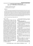
PARP-1 expression. As a first step, we treated fibroblasts
with various siRNA concentrations (10 nm to 100 nm)
and evaluated PARP-1 mRNA and protein expression 18
h after transfection. The lowest concentration of PARP-
1 siRNA resulting in efficient PARP-1 gene knockdown,
as evidenced by a decrease in PARP-1 mRNA and
protein expression, was 20nm (Figure 1A and 1B).
This concentration was employed in all subsequent
experiments.
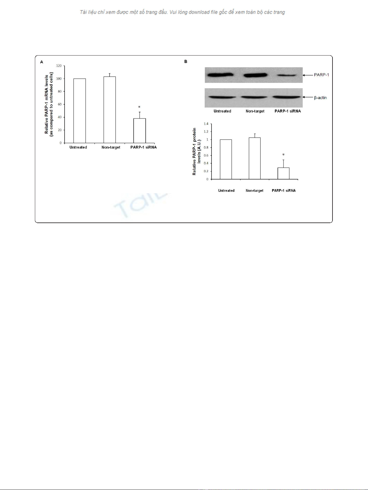
Cells were transfected with siRNA for 12 h, subjected
to heat shock for 45 min and allowed to recover for 4 h.
The expression of HSP-70 was determined by immuno-
blotting. After heat shock, naïve cells demonstrated a
significant increase in HSP-70 protein expression (Figure
2A). HSP-70 protein expression in cells transfected with
non-target siRNA was comparable to naïve cells after
heat shock (Figure 2A and 2B). Using siRNA to silence
PARP-1, we observed that HSP-70 protein expression in
PARP-1 siRNA-transfected cells was markedly upregu-
lated as compared to naïve or non-target siRNA trans-
fected cells (Figure 2A and 2B). These data support the
view that PARP-1 gene silencing leads to augmentation
of HSP-70 protein expression after heat shock.
Aneja et al.Journal of Inflammation 2011, 8:3
http://www.journal-inflammation.com/content/8/1/3
Page 3 of 9
HSP-70 mRNA expression is increased with inhibition of
PARP-1 expression
To further ascertain that PARP-1 inhibition augments
the heat shock response by enhancing HSP-70 gene
transcription, we next determined its effect on HSP-70
mRNA using real time RT-PCR. HSP-70 mRNA was
examined at 60 min after heat shock in transfected and
naïve wild-type cells. After heat shock, both naïve cells
and wild-type cells transfected with non-target siRNA
had comparable HSP-70 mRNA levels (Figure 3). In
contrast, PARP-1 directed siRNA led to a significant
increase in HSP-70 transcripts as compared to cells
transfected with non-target siRNA levels (140 ± 12 vs.
105 ± 7 A.U.). These data reinforce the notion that
PARP-1 knockdown leads to a robust heat shock
response as evidenced by increase in HSP-70 mRNA
and protein expression in PARP-1 knockdown cells
(Figure 3).
HSF-1 DNA-binding activity is increased with inhibition of
PARP-1
HSF-1 is a key transcription factor that regulates HSP-
70 gene expression [22,23]. Hence, we sought to deter-
mine if PARP-1 knockdown increased DNA binding of
HSF-1. We subjected both naïve and siRNA transfected
cells to heat shock and evaluated DNA binding of HSF-
1 by EMSA. After heat shock, both naïve and non-target
siRNA transfected cells demonstrated comparable DNA
binding activity of HSF-1. Using EMSA, we found that
nuclear extracts from cells transfected with PARP-
1siRNA displayed increased binding of HSF-1 to nuclear
DNA as compared with naïve and non-target transfected
cells (Figure 4A and 4B). Collectively, these experiments
suggest that PARP-1 negatively modulates the heat
shock response i.e. knockdown of PARP-1 led to aug-
mentation of the heat shock response by increasing
HSF-1 activation, subsequently leading to increased
HSP-70 gene expression.
PARP-1 interacts with HSF-1
Because previous studies reported that PARP-1 may reg-
ulate transcription factors by a direct physical associa-
tion [7,8,30] we next explored the possibility of a
protein-protein interaction between PARP-1 and HSF-1.
First, we confirmed that the modulation of the heat
shock response by DIQ, a PARP-1 inhibitor is similar to
the increase noted after siRNA mediated PARP-1 inhibi-
tion. Wild-type cells were subjected to heat shock at
43°C for 45 min and allowed to recover for 4 h. The
expression of HSP-70 was determined by immunoblot-
ting. DIQ pretreatment of wild-type cells not subjected
to heat shock did not induce HSP-70 protein expression
(data not shown). After heat shock, DIQ pretreated
wild-type cells demonstrated significant increase in
HSP-70 expression as compared to untreated cells
(Figure 5A). Thus, similar to siRNA mediated PARP-1
inhibition, pharmacologic inhibition of PARP-1 also
increases HSP-70 protein expression.
Figure 1 Naïve, non target siRNA and PARP-1 siRNA transfected cells were tested for PARP-1 gene expression 18 h after transfection.
Figure 1A-Quantitative Real time PCR of PARP-1 mRNA normalized for 18 S mRNA expression. Figure 1B-Representative Western blot analysis for
PARP-1 expression in naïve, non target siRNA and PARP-1 siRNA transfected cells (* Represents p< 0.05 versus naïve cells at the same time
point).
Aneja et al.Journal of Inflammation 2011, 8:3
http://www.journal-inflammation.com/content/8/1/3
Page 4 of 9
To determine if there is protein-protein interaction
between PARP-1 and HSF-1, nuclear lysates were
immunoprecipitated with antibodies against HSF-1. As
shown in Figure 5B, HSF-1 was efficiently immunopreci-
pitated with HSF-1 antibody and no signal was observed
when mouse IgG was used as a control for immunopre-
cipitation. Immunoblotting of the HSF-1immunoprecipi-
tated proteins with PARP-1 antibody demonstrated the
presence of PARP-1 suggesting that HSF-1and PARP-1
physically interact with each other. Wild-type cells trea-
ted with DIQ (PARP-1inhibitor) that had not been
subjected to heat shock demonstrated a slight increase
in nuclear HSF-1 content as compared to control cells.
Cells pretreated with DIQ and subsequently exposed to
heat shock demonstrated increased HSF-1 binding to
PARP-1 as compared to untreated control heat shocked
wild-type cells (Figure 5B).
To confirm the results obtained by HSF-1 immuno-
precipitation, we conducted the reverse experiment by
immunoprecipitating with a PARP-1 antibody and sub-
sequent analysis of the immunoprecipitate for HSF-1.
Similar to our results above, cells pretreated with a
Figure 2 Representative Western blot analysis for HSP-70 expression in naïve, non target siRNA and PARP-1 siRNA transfected cells.
Radiographs of Western blot analyses in whole cell extracts are representative of three similar separate experiments. Cells were subjected to
heat shock at 43°C for 45 min followed by recovery at 37°C for 4 h. In panel 2B the Western blot was quantitated by PhosphorImager analysis
and the mean ± SEM plotted from three independent experiments (* Represents p< 0.05 versus heat shocked naïve cells at the same time
point).
Aneja et al.Journal of Inflammation 2011, 8:3
http://www.journal-inflammation.com/content/8/1/3
Page 5 of 9