
465
ALI = acute lung injury; ARDS = acute respiratory distress syndrome; PaCO2= arterial partial pressure of CO2; PEEP = positive end-expiratory pres-
sure.
Available online http://ccforum.com/content/9/5/465
Abstract
Mechanical ventilation is indispensable for the survival of patients
with acute lung injury and acute respiratory distress syndrome.
However, excessive tidal volumes and inadequate lung recruitment
may contribute to mortality by causing ventilator-induced lung
injury. This bench-to-bedside review presents the scientific
rationale for using adjuncts to mechanical ventilation aimed at
optimizing lung recruitment and preventing the deleterious
consequences of reduced tidal volume. To enhance CO2
elimination when tidal volume is reduced, the following are
possible: first, ventilator respiratory frequency can be increased
without necessarily generating intrinsic positive end-expiratory
pressure; second, instrumental dead space can be reduced by
replacing the heat and moisture exchanger with a conventional
humidifier; and third, expiratory washout can be used for replacing
the CO2-laden gas present at end expiration in the instrumental
dead space by a fresh gas (this method is still experimental). For
optimizing lung recruitment and preventing lung derecruitment
there are the following possibilities: first, recruitment manoeuvres
may be performed in the most hypoxaemic patients before
implementing the preset positive end-expiratory pressure or after
episodes of accidental lung derecruitment; second, the patient can
be turned to the prone position; third, closed-circuit endotracheal
suctioning is to be preferred to open endotracheal suctioning.
Introduction
Mechanical ventilation is indispensable for the survival of
patients with acute lung injury (ALI) and acute respiratory
distress syndrome (ARDS). However, inappropriate ventilator
settings may contribute to mortality by causing ventilator-
induced lung injury. Tidal volumes greater than 10 ml/kg have
been shown to increase mortality [1-5]. High static
intrathoracic pressures may overdistend and/or overinflate
parts of the lung that remain well aerated at zero end-
inspiratory pressure [6-8]. Cyclic tidal recruitment and
derecruitment experimentally produces bronchial damage
and lung inflammation [9]. Although the clinical relevance of
these experimental data has been challenged recently [10,11],
the risk of mechanical ventilation-induced lung biotrauma
supports the concept of optimizing lung recruitment during
mechanical ventilation [12]. It has to be mentioned that the two
principles aimed at reducing ventilator-induced lung injury may
be associated with deleterious effects and require specific
accompanying adjustments. Reducing the tidal volume below
10 ml/kg may increase the arterial partial pressure of CO2
(PaCO2) and impair tidal recruitment [13]. Optimizing lung
recruitment with positive end-expiratory pressure (PEEP) may
require a recruitment manoeuvre [14] and the prevention of
endotracheal suctioning-induced lung derecruitment [15]. This
bench-to-bedside review presents the scientific rationale
supporting the clinical use of adjuncts to mechanical ventilation
aimed at optimizing lung recruitment and preventing the
deleterious consequences of reduced tidal volume.
Adjuncts aimed at increasing CO2elimination
Increase in respiratory rate
In patients with ARDS, increasing the ventilator respiratory
rate is the simplest way to enhance CO2elimination when
tidal volume is reduced [5,16,17]. However, an uncontrolled
increase in respiratory rate may generate intrinsic PEEP
[18,19], which, in turn, may promote excessive intrathoracic
pressure and lung overinflation [20]. If the inspiratory time is
not decreased in proportion to the increase in respiratory
rate, the resulting intrinsic PEEP may even cause right
ventricular function to deteriorate [21]. In addition to
inappropriate ventilator settings – high respiratory rate
together with high inspiratory to expiratory ratio – airflow
limitation caused by bronchial injury promotes air trapping
[22,23]. Acting in the opposite direction, external PEEP
reduces intrinsic PEEP and provides a more homogeneous
Review
Bench-to-bedside review: Adjuncts to mechanical ventilation in
patients with acute lung injury
Jean-Jacques Rouby1and Qin Lu2
1Professor of Anesthesiology and Critical Care Medicine, Director of the Surgical Intensive Care Unit Pierre Viars, La Pitié-Salpêtrière Hospital,
University of Paris, Paris, France
2Praticien Hospitalier, Surgical Intensive Care Unit Pierre Viars, Department of Anesthesiology, Research Coordinator, La Pitié-Salpêtrière Hospital,
Paris, France
Corresponding author: Jean-Jacques Rouby, jjrouby.pitie@invivo.edu
Published online: 28 June 2005 Critical Care 2005, 9:465-471 (DOI 10.1186/cc3763)
This article is online at http://ccforum.com/content/9/5/465
© 2005 BioMed Central Ltd
466
Critical Care October 2005 Vol 9 No 5 Rouby and Lu
alveolar recruitment [24,25], whereas lung stiffness tends to
accelerate lung emptying [16,26]. As a consequence, in a
given patient, it is impossible to predict intrinsic PEEP
induced by a high respiratory rate and no ‘magic number’ can
be recommended. At the bedside, the clinician should
increase the ventilator respiratory rate while looking at the
expiratory flow displayed on the screen of the ventilator: the
highest ‘safe respiratory rate’ is the rate at which the end of
the expiratory flow coincides with the beginning of the
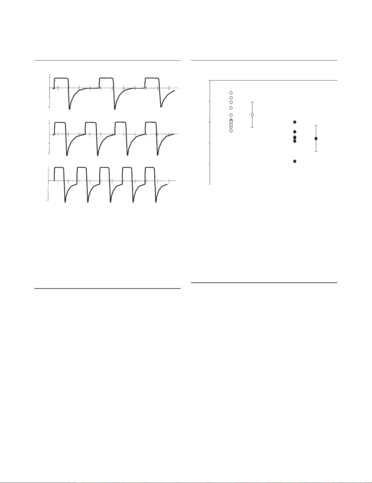
inspiratory phase (Fig. 1).
Decrease in instrumental dead space
When CO2elimination is impaired by tidal volume reduction,
the CO2-laden gas present at end expiration in the
physiological dead space is readministered to the patient at
the beginning of the following inspiration. The physiological
dead space consists of three parts: first, the instrumental dead
space, defined as the volume of the ventilator tubing between
the Y piece and the distal tip of the endotracheal tube;
second, the anatomical dead space, defined as the volume of
the patient’s tracheobronchial tree from the distal tip of the
endotracheal tube; and third, the alveolar dead space, defined
as the volume of ventilated and nonperfused lung units. Only
the former can be substantially reduced by medical
intervention. Prin and colleagues have reported that replacing
the heat and moisture exchanger by a conventional heated
humidifier positioned on the initial part of the inspiratory limb
induces a 15% decrease in PaCO2by reducing CO2
rebreathing [27] (Fig. 2). With a conventional humidifier, the
temperature of the inspired gas should be increased at 40°C
at the Y piece so as to reach 37°C at the distal tip of the
endotracheal tube [27]. In sedated patients, the tubing
connecting the Y piece to the proximal tip of the endotracheal
tube can also be removed to decrease instrumental dead
space [16]. For the same reason, if a capnograph is to be
used, it should be positioned on the expiratory limb, before the
Y piece. Richecoeur and colleagues have shown that
optimizing mechanical ventilation by selecting the appropriate
respiratory rate and minimizing instrumental dead space
allows a 28% decrease in PaCO2[16] (Fig. 2).
Expiratory washout
The basic principle of expiratory washout is to replace, with a
fresh gas, the CO2-laden gas present at end expiration in the
Figure 1
Recommendations for optimizing respiratory rate in patients with acute
respiratory failure/acute respiratory distress syndrome. The clinician
should increase respiratory rate while looking at inspiratory and
expiratory flows displayed on the screen of the ventilator. In (a) too low
a respiratory rate has been set: the expiratory flow ends 0.5 s before
the inspiratory flow. In (b) the respiratory rate has been increased
without generating intrinsic positive end-expiratory pressure: the end of
the expiratory flow coincides with the beginning of the inspiratory flow.
In (c) the respiratory rate has been increased excessively and causes
intrinsic positive end-expiratory pressure: the inspiratory flow starts
before the end of the expiratory flow. The optimum respiratory rate is
represented in (b).
Flow (l/min)
seconds
40
0
40
40
40
seconds
40
0
40
Flow (l/min)
Flow (l/min)
seconds
0
(a)
(b)
(c)
Figure 2
Optimization of CO2elimination in patients with severe acute
respiratory distress syndrome (ARDS). Open circles, reduction of
arterial partial pressure of CO2(PaCO2) obtained by replacing the
heat and moisture exchanger (HME) placed between the Y piece and
the proximal tip of the endotracheal tube by a conventional heated
humidifier (HH) on the initial part of the inspiratory limb in 11 patients
with ARDS (reproduced from [27] with the permission of the
publisher); filled circles, reduction of PaCO2obtained by combining
the increase in respiratory rate (without generating intrinsic end-
expiratory pressure) and the replacement of the HME by a conventional
HH in six patients with ARDS [16]. ConMV, conventional mechanical
ventilation (low respiratory rate with HME); OptiMV, optimized
mechanical ventilation (optimized respiratory rate with HH). Published
with kind permission of Springer Science and Business Media [27].
–50
–40
–30
–20
–10
0
Effect of replacing
HME by HH
Effect of replacing
ConMV by OptiMV
Decrease in PaCO2 (% of control value)

467
instrumental dead space [28]. It is aimed at further reducing
CO2rebreathing and PaCO2without increasing tidal volume
[29]. In contrast to tracheal gas insufflation, in which the
administration of a constant gas flow is continuous over the
entire respiratory cycle, gas flow is limited to the expiratory
phase during expiratory washout. Fresh gas is insufflated by a
gas flow generator synchronized with the expiratory phase of
the ventilator at flow rates of 8 to 15 L/min through an
intratracheal catheter or, more conveniently, an endotracheal
tube positioned 2 cm above the carina and incorporating an
internal side port opening in the internal lumen 1 cm above
the distal tip [16,29]. A flow sensor connected to the
inspiratory limb of the ventilator gives the signal to interrupt
the expiratory washout flow when inspiration starts. At
catheter flow rates of more than 10 L/min, turbulence
generated at the tip of the catheter enhances distal gas
mixing, and a greater portion of the proximal anatomical dead
space is flushed clear of CO2, permitting CO2elimination to
be optimized [30,31]. Expiratory washout can be applied
either to decrease PaCO2while maintaining tidal volume
constant or to decrease tidal volume while keeping PaCO2
constant. In the former strategy, expiratory washout is used to
protect pH, whereas in the latter it is used to minimize the
stretch forces acting on the lung parenchyma, to minimize
ventilator-associated lung injury.
Two potential side effects should be taken into consideration
if expiratory washout is used for optimizing CO2elimination.
Intrinsic PEEP is generated if the expiratory washout flow is
not interrupted a few milliseconds before the beginning of
the inspiratory phase [16,29]. As a consequence, inspiratory
plateau airway pressure may increase inadvertently, exposing
the patient to ventilator-induced lung injury. If expiratory
washout is to be used clinically in the future, the software
synchronizing the expiratory washout flow should give the
possibility of starting and interrupting the flow at different
points of the expiratory phase. A second critical issue
conditioning the clinical use of expiratory washout is the
adequate heating and humidification of the delivered
washout gas.
Currently, expiratory washout is still limited to experimental
use. It is entering a phase in which overcoming obstacles to
clinical implementation may lead to the development of
commercial systems included in intensive-care-unit ventilators
that may contribute to optimizing CO2elimination [30], in
particular in patients with severe acute respiratory syndrome
associated with head trauma [32].
Adjuncts aimed at optimizing lung recruitment
Sighs and recruitment manoeuvres
Periodic increases in inspiratory airway pressure may
contribute to the optimization of alveolar recruitment in
patients with ALI and ARDS. Sighs are characterized by
intermittent increases in peak airway pressure, whereas
recruitment manoeuvres are characterized by sustained
increases in plateau airway pressures. The beneficial impact
of sighs and recruitment manoeuvres on lung recruitment is
based on the well-established principle that inspiratory
pressures allowing reaeration of the injured lung are higher
than the expiratory pressures at which lung aeration vanishes.
At a given PEEP, the higher the pressure that is applied to
the respiratory system during the preceding inspiration, the
greater the lung aeration. In patients with ALI, the different
pressure thresholds for lung aeration at inflation and deflation
depend on the complex mechanisms regulating the removal
of oedema fluid from alveoli and alveolar ducts [33,34], the
reopening of bronchioles externally compressed by cardiac
weight and abdominal pressure [35], and the preservation of
surfactant properties.
Reaeration of the injured lung basically occurs during
inspiration. The increase in airway pressure displaces the
gas–liquid interface from alveolar ducts to alveolar spaces
and increases the hydrostatic pressure gradient between the
alveolar space and the pulmonary interstitium [36]. Under
these conditions, liquid is rapidly removed from the alveolar
space, thereby increasing alveolar compliance [37] and
decreasing the threshold aeration pressure. Surfactant
alteration, a hallmark of ALI, results from two different
mechanisms: direct destruction resulting from alveolar injury,
and indirect inactivation in the distal airways caused by a loss
of aeration resulting from external lung compression [38]. By
preventing expiratory bronchiole collapse, PEEP has been
shown to prevent surfactant loss in the airways and avoid
collapse of the surface film [38]. As a consequence, alveolar
compliance increases and the pressure required for alveolar
expansion decreases. The time scale for alveolar recruitment
and derecruitment is within a few seconds [39,40], whereas
the time required for fluid transfer from the alveolar space to
the pulmonary interstitium is of the order of a few minutes
[36]. It has been demonstrated that the beneficial effect of
recruitment manoeuvres on lung recruitment can be obtained
only when the high airway pressure (inspiratory or incremental
PEEP) is applied over a sufficient period [41,42], probably
preserving surfactant properties and increasing alveolar
clearance [14].
In surfactant-depleted collapse-prone lungs, recruitment
manoeuvres increase arterial oxygenation by boosting the
ventilatory cycle onto the deflation limb of the pressure–
volume curve [42]. However, in different experimental models
of lung injury, recruitment manoeuvres do not provide similar
beneficial effects [43]. In patients with ARDS, recruitment
manoeuvres and sighs are effective in improving arterial
oxygenation only at low PEEP and small tidal volumes
[44,45]. When PEEP is optimized, recruitment manoeuvres
are either poorly effective [46] or deleterious, inducing
overinflation of the most compliant lung regions [47] and
haemodynamic instability and worsening pulmonary shunt by
redistributing pulmonary blood flow towards non-aerated lung
regions [48]. However, after a recruitment manoeuvre, a
Available online http://ccforum.com/content/9/5/465

468
sufficient PEEP level is required for preventing end-expiratory
alveolar derecruitment [49]. Furthermore, recruitment
manoeuvres are less effective when ALI/ARDS is due to
pneumonia or haemorrhagic oedema [43].
Different types of recruitment manoeuvre have been
proposed for enhancing alveolar recruitment and improving
arterial oxygenation in the presence of ALI [50]. A plateau
inspiratory pressure can be maintained at 40 cmH2O for 40 s.
Stepwise increases and decreases in PEEP can be
performed while maintaining a constant plateau inspiratory
pressure of 40 cmH2O [42]. Pressure-controlled ventilation
using high PEEP and a peak airway pressure of 45 cmH2O
can be applied for 2 min [51]. The efficacy and
haemodynamic side effects have been compared between
three different recruitment manoeuvres in patients and
animals with ARDS [49,51]. Pressure-controlled ventilation
with high PEEP seems more effective in terms of oxygenation
improvement, whereas a sustained inflation lasting 40 seconds
seems more deleterious to cardiac output [49,51].
Studies reporting the potential deleterious effects of
recruitment manoeuvres on lung injury of regions remaining
fully aerated are still lacking. As a consequence, the
administration of recruitment manoeuvres should be
restricted to individualized clinical decisions aimed at improv-
ing arterial oxygenation in patients remaining severely
hypoxaemic. As an example, recruitment manoeuvres are
quite efficient for rapidly reversing aeration loss resulting from
endotracheal suctioning [52] or accidental disconnection
from the ventilator. In patients with severe head injury,
recruitment manoeuvres may cause cerebral haemodynamics
to deteriorate [53]. As a consequence, careful monitoring of
intracranial pressure should be provided in case of severe
hypoxaemia requiring recruitment manoeuvres.
Prone position
Turning the patient into the prone position restricts the
expansion of the cephalic and parasternal lung regions and
relieves the cardiac and abdominal compression exerted on
the lower lobes. Prone positioning induces a more uniform
distribution of gas and tissue along the sternovertebral and
cephalocaudal axis by reducing the gas/tissue ratio of the
parasternal and cephalic lung regions [54,55]. It reduces
regional ventilation-to-perfusion mismatch, prevents the free
expansion of anterior parts of the chest wall, promotes PEEP-
induced alveolar recruitment [56], facilitates the drainage of
bronchial secretions and potentiates the beneficial effect of
recruitment manoeuvres [57], all factors that contribute to
improving arterial oxygenation in most patients with early
acute respiratory failure [55] and may reduce ventilator-
induced lung overinflation.
It is recommended that the ventilatory settings be optimized
before the patient is turned into the prone position [35]. If
arterial saturation remains below 90% at an inspiratory
fraction of oxygen of at least 60% and after absolute
contraindications such as burns, open wounds of the face or
ventral body surface, recent thoracoabdominal surgical
incisions, spinal instability, pelvic fractures, life-threatening
circulatory shock and increased intracranial pressure have
been ruled out [56], the patient should be turned to prone in
accordance with a predefined written turning procedure [56].
The optimum duration of prone positioning remains uncertain.
In clinical practice, the duration of pronation can be
maintained for 6 to 12 hours daily and may be safely increased
to 24 hours [58]. The number of pronations can be adapted to
the observed changes in arterial oxygenation after supine
repositioning [55]. Whether the abdomen should be
suspended during the period of prone position is still debated
[56]. Complications are facial oedema, pressure sores and
accidental loss of the endotracheal tube, drains and central
venous catheters. Despite its beneficial effects on arterial
oxygenation, clinical trials have failed to show an increase in
survival rate by prone positioning in patients with acute
respiratory failure [59,60]. Whether it might reduce mortality
and limit ventilator-associated pneumonia in the most severely
hypoxaemic patients [59,60] requires additional study.
Closed-circuit endotracheal suctioning
Endotracheal suctioning is routinely performed in patients
with ALI/ARDS. A negative pressure is generated into the
tracheobronchial tree for the removal of bronchial secretions
from the distal airways. Two factors contribute to lung
derecruitment during endotracheal suctioning: the
disconnection of the endotracheal tube from the ventilator
and the suctioning procedure itself. Many studies have shown
that the sudden discontinuation of PEEP is the predominant
factor causing lung derecruitment in patients with ALI
[52,61]. During a suctioning procedure lasting 10 to
30 seconds, the high negative pressure generated into the
airways further decreases lung volume [15]. A rapid and long-
lasting decrease in arterial oxygenation invariably results from
open endotracheal suctioning [62]. It is caused by a lung
derecruitment-induced increase in pulmonary shunt and a
reflex bronchoconstriction-induced increase in venous
admixture; both factors increase the ventilation/perfusion ratio
mismatch [52]. The decrease in arterial oxygenation is
immediate and continues for more than 15 min despite the re-
establishment of the initial positive end-expiratory level. A
recruitment manoeuvre performed immediately after the
reconnection of the patient to the ventilator allows a rapid
recovery of end-expiratory lung volume and arterial
oxygenation [62]. However, in the most severely hypoxaemic
patients the open suctioning procedure itself may be
associated with dangerous hypoxaemia [62].
Closed-circuit endotracheal suctioning is generally advocated
for preventing arterial oxygenation impairment caused by
ventilator disconnection [63,64]. However, a loss of lung
volume may still be observed, resulting from the suctioning
procedure itself and appearing dependent on the applied
Critical Care October 2005 Vol 9 No 5 Rouby and Lu
469
negative pressure [15,63]. Both experimental studies and
clinical experience suggest that closed-circuit endotracheal
suctioning is less efficient than open endotracheal suctioning
for removing tracheobronchial secretions [64,65]. As a
consequence, the clinician is faced with two opposite goals:
preventing lung derecruitment and ensuring the efficient
removal of secretions [66]. Further clinical studies are
needed to evaluate an optimum method that takes both goals
into account.
In patients with ALI/ARDS, closed-circuit endotracheal
suctioning should be considered the clinical standard. In
severe ARDS, endotracheal suctioning should be optimized
by pre-suction hyperoxygenation and followed by post-
suction recruitment manoeuvres. In addition to the methods
described above, two other types of recruitment manoeuvre
have been proposed to prevent a loss of lung volume and
reverse atelectasis resulting from endotracheal suctioning:
the administration of triggered pressure-supported breaths at
a peak inspiratory pressure of 40 cmH2O during suctioning
[15] and the administration of 20 consecutive hyperinflations
set at twice the baseline tidal volume immediately after
suctioning [52].
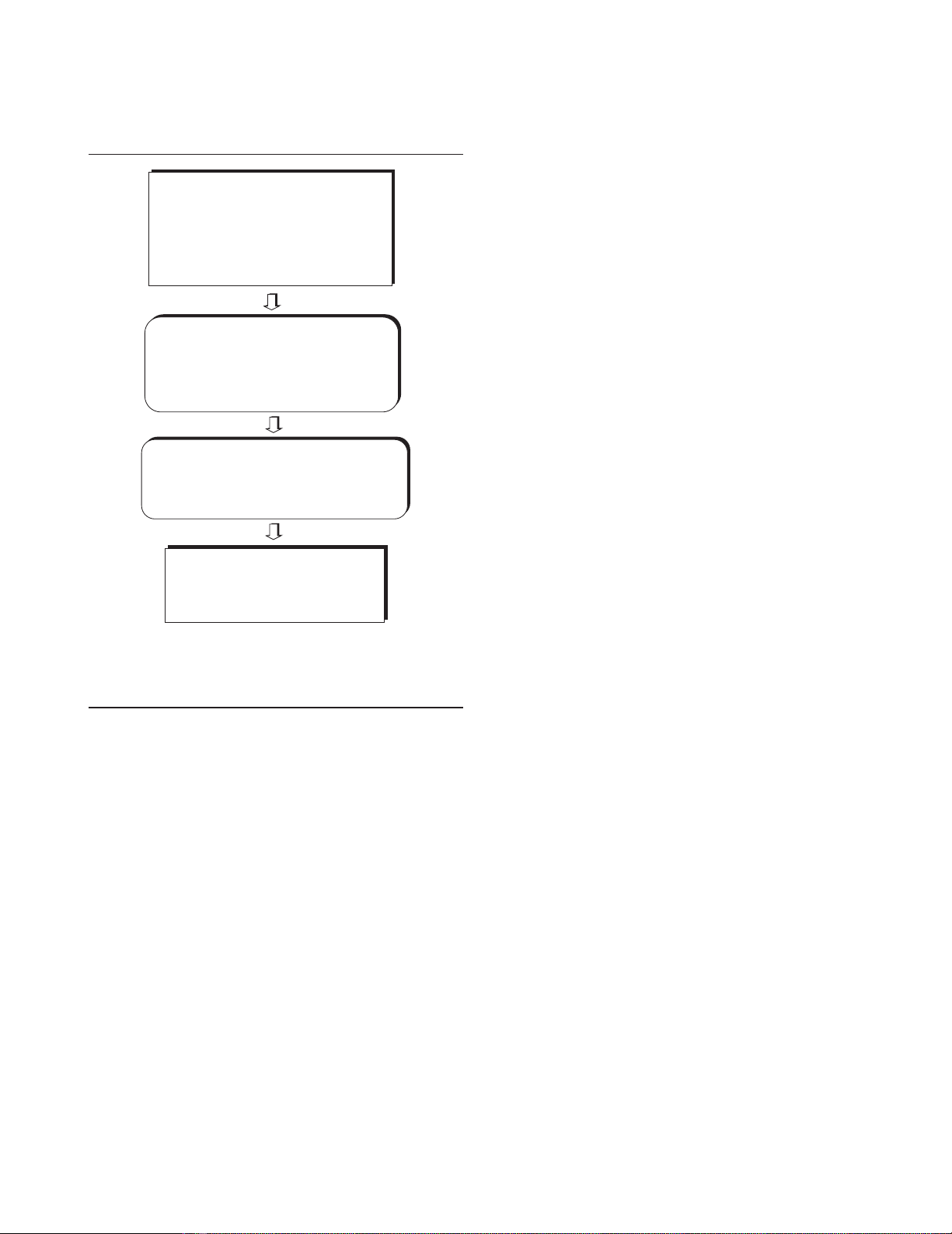
There is as yet no guideline for endotracheal suctioning in
patients with severe ARDS. An algorithm is proposed in
Fig. 3 aimed at preventing lung derecruitment and deteriora-
tion of gas exchange during endotracheal suctioning in hypox-
aemic patients receiving mechanical ventilation with PEEP.
Conclusion
Mechanical ventilation in patients with ALI/ARDS requires
specific adjustments of tidal volume and PEEP. Clinical use
of adjuncts to mechanical ventilation allows optimization of
alveolar recruitment resulting from PEEP and prevention of
deleterious consequences of reduced tidal volume. Appro-
priate increases in respiratory rate, replacement of heat and
moisture exchanger by a conventional humidifier. administra-
tion of recruitment manoeuvre in case of accidental episode
of derecruitment, prone positioning and closed-circuit endo-
tracheal suctioning all contribute to optimization of arterial
oxygenation and O2elimination
Competing interests
The author(s) declare that they have no competing interests.
References
1. Amato MB, Barbas CS, Medeiros DM, Magaldi RB, Schettino GP,
Lorenzi-Filho G, Kairalla RA, Deheinzelin D, Munoz C, Oliveira R,
et al.: Effect of a protective-ventilation strategy on mortality in
the acute respiratory distress syndrome. N Engl J Med 1998,
338:347-354.
2. Brochard L, Roudot-Thoraval F, Roupie E, Delclaux C, Chastre J,
Fernandez-Mondejar E, Clementi E, Mancebo J, Factor P, Matamis
D, et al.: Tidal volume reduction for prevention of ventilator-
induced lung injury in acute respiratory distress syndrome.
The Multicenter Trial Group on Tidal Volume reduction in
ARDS. Am J Respir Crit Care Med 1998, 158:1831-1838.
3. Stewart TE, Meade MO, Cook DJ, Granton JT, Hodder RV, Lapin-
sky SE, Mazer CD, McLean RF, Rogovein TS, Schouten BD, et
al.: Evaluation of a ventilation strategy to prevent barotrauma
in patients at high risk for acute respiratory distress syn-
drome. Pressure- and Volume-Limited Ventilation Strategy
Group. N Engl J Med 1998, 338:355-361.
4. Brower RG, Shanholtz CB, Fessler HE, Shade DM, White P Jr,
Wiener CM, Teeter JG, Dodd-o JM, Almog Y, Piantadosi S:
Prospective, randomized, controlled clinical trial comparing
traditional versus reduced tidal volume ventilation in acute
respiratory distress syndrome patients. Crit Care Med 1999,
27:1492-1498.
5. The Acute Respiratory Distress Syndrome Network: Ventilation
with lower tidal volumes as compared with traditional tidal
volumes for acute lung injury and the acute respiratory dis-
tress syndrome. N Engl J Med 2000, 342:1301-1308.
6. Vieira SR, Puybasset L, Lu Q, Richecoeur J, Cluzel P, Coriat P,
Rouby JJ: A scanographic assessment of pulmonary morphol-
ogy in acute lung injury. Significance of the lower inflection
point detected on the lung pressure-volume curve. Am J
Respir Crit Care Med 1999, 159:1612-1623.
7. Puybasset L, Gusman P, Muller JC, Cluzel P, Coriat P, Rouby JJ:
Regional distribution of gas and tissue in acute respiratory
distress syndrome. III. Consequences for the effects of posi-
tive end-expiratory pressure. CT Scan ARDS Study Group.
Adult Respiratory Distress Syndrome. Intensive Care Med
2000, 26:1215-1227.
8. Nieszkowska A, Lu Q, Vieira S, Elman M, Fetita C, Rouby JJ: Inci-
dence and regional distribution of lung overinflation during
mechanical ventilation with positive end-expiratory pressure.
Crit Care Med 2004, 32:1496-1503.
9. Dos Santos CC, Slutsky AS: Invited review: mechanisms of
ventilator-induced lung injury: a perspective. J Appl Physiol
2000, 89:1645-1655.
Available online http://ccforum.com/content/9/5/465
Figure 3
Recommendations concerning endotracheal suctioning in patients with
severe acute respiratory distress syndrome. FIO2, inspiratory fraction of
oxygen; I/E ratio, inspiratory to expiratory ratio; PEEP, positive end-
expiratory pressure; RR, respiratory rate; TV, tidal volume.
Return to pre-endotracheal suctioning
FiO2, ventilatory mode and peak airway
pressure alarm
VENTILATORY SETTINGS
Closed-circuit endotracheal
suctioning system
Suctioning negative pressure ≤ –400 cmH2O
1–3 suctioning procedures lasting 10 sec each
FiO2=1, 10 min before endotracheal suctioning
Assisted volume controlled ventilatory mode
Peak airway pressure alarm = 70 cmH2O
Same PEEP, TV, RR and I/E ratio
VENTILATORY SETTINGS
Trigger sensitivity set between –1 and –2 cmH2O
Postsuctioning recruitment manoeuvre
20 consecutive TV= 2 × pre-set TV


![PET/CT trong ung thư phổi: Báo cáo [Năm]](https://cdn.tailieu.vn/images/document/thumbnail/2024/20240705/sanhobien01/135x160/8121720150427.jpg)


