
TECH N I C AL INN O V A T ION S Open Access
Early results on the use of biomaterials as
adjuvant to abdominal wall closure following
cytoreduction and hyperthermic intraperitoneal
chemotherapy
Cherif Boutros, Ponandai Somasundar, N Joseph Espat
*
Abstract
Background: Hyperthermic chemotherapy applies thermal energy to both abdominal wall as well as the intra-
abdominal viscera. The combination of the hyperthemia, chemotherapy and cytoreductive surgery (CRS) is
associated with a defined risk of abdominal wall and intestinal morbidity reported to be as high as 15%,
respectively to date, no studies have evaluated the use of biomaterial mesh as adjuvant to abdominal wall closure
in this group of patients. In the present report, we hypothesized that post HIPEC closure with a biomaterial can
reduce abdominal wall morbidity after CRS and hyperthermic intraperitoneal chemotherapy.
Materials and methods: All patients treated with HIPEC in a tertiary care center over 12 months (2008-2009)
period were included. Eight patients received cytoreductive surgery followed by HIPEC for 90 minutes using
Mitomycin C (15 mg q 45 minutes × 2). Abdominal wall closure was performed using Surgisis (Cook Biotech.)
mesh in an underlay position with 3 cm fascial overlap-closure. Operative time, hospital length of stay (LOS) as well
as postoperative outcome with special attention to abdominal wall and bowel morbidity were assessed.
Results: Eight patients, mean age 59.7 ys (36-80) were treated according to the above protocol. The primary
pathology was appendiceal mucinous adenocarcinoma (n = 3) colorectal cancer (n = 3), and ovarian cancer (n =
2). Four patients (50%) presented initially with abdominal wall morbidity including incisional ventral hernia (n = 3)
and excessive abdominal wall metastatic implants (n = 1). The mean peritoneal cancer index (PCI) was 8.75. Twenty
eight CRS were performed (3.5 CRS/patient). The mean operating time was 6 hours. Seven patients had no
abdominal wall or bowel morbidity, the mean LOS for these patients was 8 days. During the follow up period
(mean 6.3 months), one patient required exploratory laparotomy 2 weeks after surgery and subsequently
developed an incisional hernia and enterocutaneous fistula.
Conclusion: The use of biomaterial mesh in concert with HIPEC enables the repair of concomitant abdominal wall
hernia and facilitates abdominal wall closure following the liberal resection of abdominal wall tumors. Biomaterial
mesh prevents evisceration on repeat laparotomy and resists infection in immunocompromised patients even
when associated with bowel resection.
Introduction
Hyperthermic intraperitoneal chemotherapy (HIPEC)
has emerged as an effective method of managing perito-
neal carcinomatosis for different abdominal malignan-
cies particularly of colorectal and ovarian origin [1-3].
Most HIPEC treated patients have had prior abdominal
surgeries and a substantial group of them present with
abdominal wall morbidities including incisional hernia
prior to HIPEC therapy.
Multiple studies have reported that chemotherapy
administration impairs wound healing and that there is
associated increase in wound complications in che-
motherapy exposed patients [4,5]. The effects of HIPEC
* Correspondence: jespat@hepaticsurgery.com
Hepatobiliary and Surgical Oncology, Roger Williams Medical Center,
Providence, RI, USA
Boutros et al.World Journal of Surgical Oncology 2010, 8:72
http://www.wjso.com/content/8/1/72 WORLD JOURNAL OF
SURGICAL ONCOLOGY
© 2010 Boutros et al; licensee BioMed Central Ltd. This is an Open Access article distributed under the terms of the Creative Commons
Attribution License (http://creativecommons.org/licenses/by/2.0), which permits unrestricted use, distribution, and reproduction in
any medium, provided the original work is properly cited.

on wound healing are multiple (Figure 1): first HIPEC
requires the concomitant use of chemotherapy and
hyperthermia which are both known to increase cellular
death and induce apoptosis [6,7]; second, tumor cytore-
duction may require surgical resection of involved
abdominal wall surfaces potentially compromising
abdominal wall closure and strength.
HIPEC impairment of wound healing is not limited to
the abdominal wall, but also anastomosed segments of
the gastrointestinal tract after cytoreductive surgery
bowel resections. In small animal models, HIPEC was
found to significantly decrease both colonic anastomotic
bursting pressure and abdominal wall strength in asso-
ciation with decreased local protein production [8,9]. In
clinical studies, HIPEC was associated with a non negli-
gible percentage (up to 15%) of abdominal wall morbid-
ity including; wound infection, dehiscence, evisceration
and bowel morbidity including; anastomotic leak and
intra-abdominal abscess (Table 1) [10,11].
Moreover, postoperative bowel morbidity often
required reoperation [10], in this case abdominal wound
healing already inhibited by HIPEC will be impaired by
the infected milieu.
Biomaterial mesh has emerged as an attractive option
for complex abdominal wall reconstruction providing an
additional reinforcement to the abdominal wall with an
absorbable material resistant to infection and with
potential remodeling to host own tissue. Therefore, we
used a biomaterial mesh to reinforce the abdominal wall
closure at the end of CRS-HIPEC procedure. We
hypothesized that this approach could minimize the rate
of postoperative abdominal wall complications reported
after CRS-HIPEC.
Methods
Under institutional IRB approval, all patients treated
with HIPEC at a tertiary care center over a12 months
interval (2008-2009) period were identified using a pro-
spectively maintained departmental database. Records
were reviewed for preoperative, operative and postopera-
tive data. Pertinent information for analysis included
gender, age, primary malignancy, number of previous
abdominal operations, peritoneal cancer index [12],
operative time and cytoreductive procedures performed.
Follow-up data were obtained by review of clinic notes.
Abdominal wall and bowel/intra-abdominal morbidities
were ascertained during the immediate and postopera-
tive period and subsequent clinic visits.
Data was recorded in a Microsoft Excel® (Microsoft,
Redmond, WA) database. Descriptive statistics, includ-
ing means, standard deviations or counts and percen-
tages were calculated.
Operative technique
Abdominal exploration was performed through midline
laparotomy incision. Lysis of adhesions, when needed
was performed, followed by assessment of the intra-
abdominal extent of the disease and the peritoneal can-
cer index (PCI). Appropriate cytoreductive surgery was
then performed with an attempt for complete removal
of all macroscopic tumor deposits on parietal and visc-
eral peritoneal surfaces and resection of involved viscera.
After completion of the CRS, HIPEC was immediately
performed during the same surgical procedure for all
patients using a closed-abdomen technique (Figure 2).
Two inflow catheters were placed under direct vision to
the right and left upper quadrants above the liver and
the spleen respectively and two outflow catheters were
placed under direct vision on the right and left para-
colic gutters. The inflow and outflow tubes were secured
to the skin and connected to a cardiopulmonary bypass
pump (Figure 3). A temperature needle probe was
placed into one of the outflow catheters and the skin
was closed using a running locked heavy nylon stitch,
while the fascia was left open.
After priming the circuit, a flow of 2.0-2.4 L/minute of
DIANEAL PD-2, dextrose 1.5%® (Baxter, Deerfield, IL)
solution containing Mitomycin C was obtained and
maintained during the whole HIPEC time. Mitomycin C
was used at a standard dose of 15 mg q 45 minutes × 2
Figure 1 The association of hyperthermia, cytoreductive
surgery and chemotherapy carry a considerable risk of
abdmonial wall and bowel morbidity.
Table 1 Selected studies reporting abdominal wall
morbidity (AWM) and bowel/intra-abdominal morbidity
(Bowel/IA M)
Study (year) N AWM Bowel/IA M
Franko (2008) [10] 65 10.7% 15.4%
Kianmanesh (2007) [2] 43 11.6% 13.9%
Stewart (2006) [11] 110 15.4% 6.3%
Sugarbaker (2006) [29] 356 3% 5.47%
Witkamp (2001) [30] 29 3% 3%
Boutros et al.World Journal of Surgical Oncology 2010, 8:72
http://www.wjso.com/content/8/1/72
Page 2 of 7
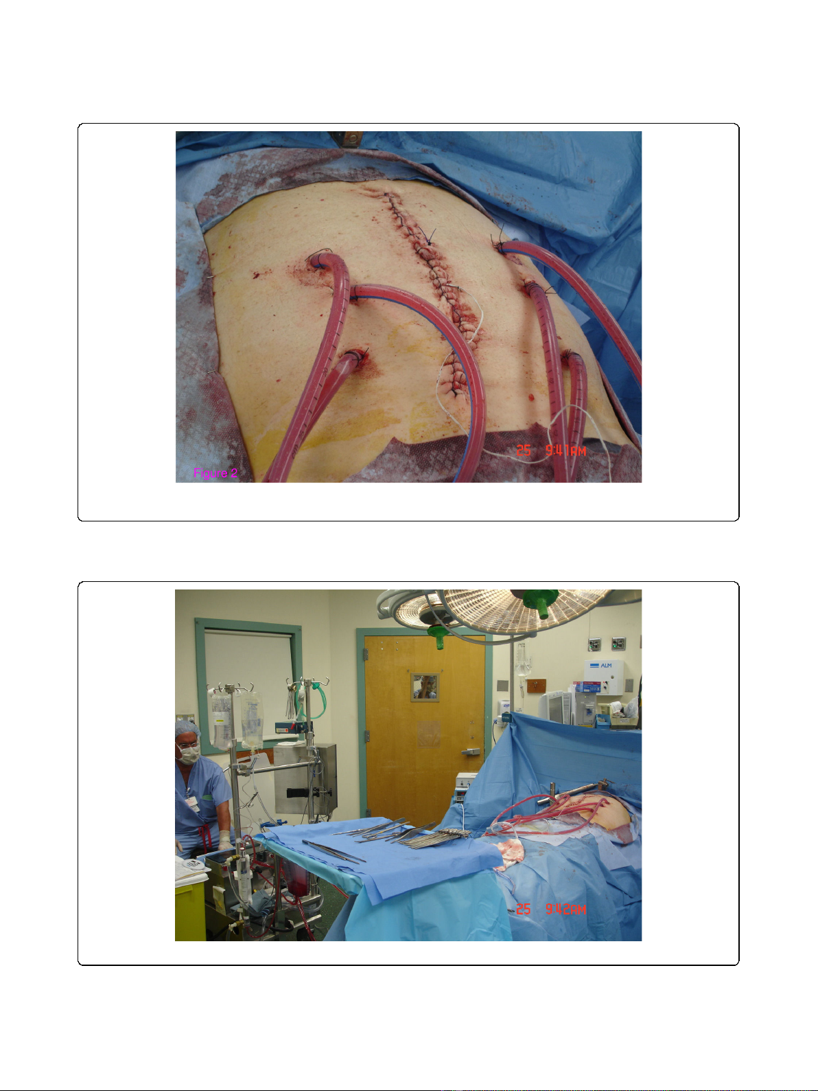
Figure 2 Using a closed technique: Two inflow and four outflow catheters were used for HIPEC administration. Only the skin of the
surgical wound is temporarily closed.
Figure 3 Using a cardiopulmonary bypass machine, The HIPEC circuit is completed.
Boutros et al.World Journal of Surgical Oncology 2010, 8:72
http://www.wjso.com/content/8/1/72
Page 3 of 7

for all patients. Hyperthermia was obtained by a heating
unit in the cardiopulmonary bypass pump maintaining
inflow temperature at 42°C, while the abdominal tem-
perature was continuously checked by the needle probe
to confirm a minimu of 41°C in the outflow. A total
HIPEC time of 90 minutes was applied to all patients.
At the end of the 90 minutes dwell time, the HIPEC
solution was retrieved through the cardiopulmonary
pump outflow catheters and disposed; the abdominal
cavity was flushed with the effluent removed via the
outflow catheters and the abdominal incision was
reopened.
The abdominal wall was closed using a 20 × 20 cm
piece of Surgisis® mesh (Cook Biotech, West Lafayette,
IN). Abdominal wall closure was performed by the same
surgeon performing the CRS-HIPEC procedure. Surgisis
mesh was placed in underlay position and secured to
the anterior abdominal wall using circumferential trans-
fascial absorbable sutures (#1 PDS) 2 cm apart. When
possible, the native fascia was closed over the Surgisis
mesh using absorbable sutures and the skin was closed
using skin stapler.
Results
The RWMC HIPEC program started on June 2008; over
one year period eleven patients received HIPEC, includ-
ing three patients who received totally laparoscopic
HIPEC for persistent PET scan evident activity from col-
orectal cancer in the mesenteric or retroperitoneal
lymph nodes after standard adjuvant chemotherapy.
Eight patients received exploratory laparotomy, CRS and
HIPEC and were included in this study (Table 2).
Patients’mean age was 59.7 years (36-80), M: F ratio
was 1:1 and the origin of the primary malignant disease
was colorectal (n = 4), appendiceal (n = 2) and ovarian
(n = 2). All patients had prior abdominal surgeries and
three patients had > two prior abdominal explorations.
Peritoneal cancer index varied from 0-18 with a mean
of 8.75. Prior to HIPEC therapy, four patients (4/8, 50%)
presented with abdominal wall morbidities including
incisional hernias (n = 3) and abdominal wall metastatic
implants (n = 1).
A total 28 CRS procedures were performed (average
3.5/patient). One third of the CRS included bowel resec-
tion-anastomosis (9/28, 32%) all performed by linear
staplers.
Mean total surgical procedure time was 5 h 49 min-
utes ± 1 h 10 minutes including 90 minutes devoted for
HIPEC (Table 3). There was no peri-operative mortality
and no postoperative neutropenia. Seven patients had
neither abdominal wall nor bowel/intra-abdominal mor-
bidities with a mean length of stay of 8 days (range 4-
16). All patients were followed in subsequent clinic vis-
its, the mean follow up was 6.3 months.
One patient required re-exploration two weeks after
HIPEC procedure and subsequently developed incisional
hernia and enterocutaneous fistula. This patient is a 59
year old patient with ovarian cancer and three prior
abdominal surgeries including two debulking proce-
dures. Prior to HIPEC procedure, the patient physical
examination was significant for extensive abdominal
wall metastatic implants.
The patient PCI was 12 and CRS included bowel
resection/anastomosis x2, splenectomy and abdominal
wall resection. The patient post-operative period was
marked by respiratory failure requiring reintubation. On
post-operative day 15, a brown discharge was noted
from the surgical incision and a decision for surgical re-
exploration was made.
Upon re-exploration, the previously placed Surgisis
mesh was intact and easily dissected from the underly-
ing bowel, prior anastomoses were intact and there was
no evidence of gastrointestinal leak. Blood culture was
obtained when the postoperative wound discharge was
initially noted and intravenous antibiotic therapy was
Table 2 Patient characteristics
Age Sex ASA Pathology Prior surgery PCI
53 M 2 MAC
Appendix
Appendectomy 18
70 M 2 Colorectal
cancer
Open colectomy and ventral
hernia repair
3
59 F 3 Ovarian
Cancer
Three midline Ex Lap. 12
57 F 2 MAC
Appendix
Appendectomy 0
72 M 3 Colorectal
Cancer
Open colectomy 9
80 F 3 Ovarian
Cancer
Hysterectomy 10
36 F 2 MAC
Appendix
TAHBSO 15
51 M 3 Colorectal
cancer
Open colectomy + 4 Ex. Lap. 3
PCI: Peritoneal cancer index; MAC: Mucinous adenocarcinoma; TAHBSO: total
abdominal hysterectomy and bilateral salpingo-oopherectomy; Ex. Lap.:
Exploratory laparotomy.
Table 3 Outcome of patients after HIPEC surgery
Surgical time (mn) AWM Bowel/IA M LOS (D)
410 ø ø 9
310 ø ø 4
380 Dehiscence ECF 120
280 ø ø 6
370 ø ø 5
430 ø ø 16
230 ø ø 10
380 ø ø 6
AWM: Abdominal wall morbidity; Bowel/IA M: Bowel and intra-abdominal
morbidity; LOS: Length of stay; ECF: Enterocutaneous fistula
Boutros et al.World Journal of Surgical Oncology 2010, 8:72
http://www.wjso.com/content/8/1/72
Page 4 of 7

started imperatively with the hypothesis of sepsis. Dur-
ing re-exploration cultures from the peritoneal fluid
were also obtained; both blood and peritoneal cultures
did not support an infectious etiology. A dramatic lysis
of residual tumor implants was noted producing a
melted brown discharge. A new Surgisis mesh was
placed and the fascia was closed as previously described.
Subsequently, the patient developed wound dehiscence
with no evisceration as well as enterocutaneous fistula.
A negative pressure dressing was applied to the abdom-
inal wound and the patient was discharged home three
months after the HIPEC procedure.
Discussion
The introduction of biomaterial mesh had revolutio-
nized the field of abdominal wall closure [13,14].
Broadly grouped, biomaterial mesh is either human allo-
graft or xenograft and dermal or non dermal in origin.
Specific guidelines for specific biomaterial mesh selec-
tion for a given case remain to be defined; however in
general it is accepted that for complex and contami-
nated cases, biomaterial mesh offersaviablesubstitute
to the patient’s own tissue. In particular the use of bio-
material mesh has been described to be clinically mean-
ingful when the host native abdominal fascia is
insufficient for closure without tension, ie (loss of
abdominal domain), when there is a lack of viable tissue
and a components separation is not technically feasible,
or the field is contaminated or potentially contaminated
and permanent synthetic mesh is relatively contraindi-
cated [15]. Biomaterial meshes are known to be resistant
to infection[16] and overcome the limitations of syn-
thetic mesh for use in contaminated or potentially con-
taminated wounds, provide a tissue remodeling matrix,
for host tissues and fibroblasts [17]. In this series, the
potential role of biomaterial mesh as adjuvant to
abdominal wall closure in the setting of significantly
potential impaired abdominal wall wound healing fol-
lowing HIPEC, with or without prior incisional hernia
or after cytoreductive surgery of abdominal wall meta-
static implants was investigated. In cases, there was a
clinical indication for mesh reinforcement due to wea-
kened, lacking or non viable abdominal wall fascia; the
choice of biomaterial mesh was supported by the pre-
sence of potential contamination or frank contamination
subsequent to a procedure entering the gastrointestinal
tract.
Surgisis mesh was utilized in all open HIPEC proce-
dures. This biomaterial mesh is composed of lyophilized
porcine small intestinal submucosa, is known to attract
cells to the wound area and signaling surrounding tis-
sues to grow across the scaffold [18]. The choice of this
particular biomaterial mesh was based on the senior
authors previous published experience with Surgisis
[14,19]as well as the reports of others observing that
Sugisis remodels into vascularized host tissue [17], thus
allowing resistance to infection. Additionally, Surgisis is
predominantley composed of collagen rather than elas-
tin compared to dermal-based biomaterials; thus it is
expected to result in less abdominal diathesis or hernia
recurrence overtime [20].
All Surgisis meshes were placed in underlay position
with a minimum of a bilateral 3 cm fascial overlap-clo-
sure using absorbable number one PDS transfascial
sutures placed circumferentially no more than two cm
apart. Underlay repairs, such as Rivers-Stoppa retro-rec-
tus repair, have been reported to result in improved
recurrence rate and allow for re-approximation of the
midline, thus potentially improving the mechanical func-
tion of the abdominal wall [21,22].
The HIPEC protocol employed was the well-described
regimen of single agent (Mitomycin C) at a dose (15 mg
q 45 minutes x2) with a cumulative dwell time of 90
minutes, for all patients regardless of the origin of the
primarymalignancyandthebodysurfaceareaofthe
patient [23]. The present protocol does not include a
measurement of serum Mitomycin C levels thus we are
unable to discuss its pharmakodynamics in this setting,
these data have been previously described by others
[24,25]. Postoperatively, none of the patients in this ser-
ies developed neutropenia which has reported to occur
in up to 39% of HIPEC patients using Mitomycin C at a
higher dose[26]. It should be emphasized that though
there are multiple series reporting the use of different
chemotherapeutic agent (s) and different doses during
HIPEC, there is no consensus statement or general
agreement on a single universal protocol. Currently,
efforts are undergoing to create a registry database for
all active HIPEC programs in USA allowing outcome
analysis to elucidate this still evolving topic.
Seven of eight patients included in this study did not
develop abdominal wall or bowel/intra-abdominal mor-
bidities postoperatively and were discharged home after
a mean length of stay of eight days. A single patient did
sustain the complication of suspicious for enterocuta-
neous fistula wound discharge with associated with
respiratory failure 7 days after HIPEC necessitating re-
exploration. Upon re-exploration, the integrity of the
abdominal wall and gastrointestinal were verified. The
Surgisis mesh was found intact and the bowel was easily
dissected from the mesh as has been previously
described in experimental models [27,28]. At operation,
the patient was found to have extensive tumor necrosis
from unresectable pelvic mass; because of potential
compromised rectal wall, a loop diverting ostomy was
created. The abdominal wall fascia was closed again
with a new Surgisis mesh to prevent evisceration. Not
unexpectedly, the native fascia later dehisced, and the
Boutros et al.World Journal of Surgical Oncology 2010, 8:72
http://www.wjso.com/content/8/1/72
Page 5 of 7