
JOURNAL OF
Veterinary
Science
J. Vet. Sci. (2008), 9(2), 183
191
*Corresponding author
Tel: +91-33-24733469; Fax: +91-33-24730957
E-mail: biswa_kundu@rediffmail.com
Efficacy of nano-hydroxyapatite prepared by an aqueous solution
combustion technique in healing bone defects of goat
Samit Kumar Nandi1, Biswanath Kundu2,*, Samir Kumar Ghosh2, Dipak Kumar De1, Debabrata Basu2
1Department of Veterinary Surgery and Radiology, West Bengal University of Animal and Fishery Sciences, Kolkata, India
2Bioceramics and Coating Division, Central Glass and Ceramic Research Institute, Kolkata, India
The present study was undertaken to evaluate porous
hydroxyapatite (HAp), the powder of which was prepared
by a novel aqueous solution combustion technique, as a
bone substitute in healing bone defects in vivo, as assessed
by radiologic and histopathologic methods, oxytetracycline
labeling, and angiogenic features in Bengal goat. Bone
defects were created in the diaphysis of the radius and
either not filled (group I) or filled with a HAp strut (group
II). The radiologic study in group II showed the presence
of unabsorbed implants which acted as a scaffold for new
bone growth across the defect, and the quality of healing
of the bone defect was almost indistinguishable from the
control group, in which the defect was more or less
similar, although the newly formed bony tissue was more
organized when HAp was used. Histologic methods
showed complete normal ossification with development of
Haversian canals and well-defined osteoblasts at the
periphery in group II, whereas the control group had
moderate fibro-collagenization and an adequate amount
of marrow material, fat cells, and blood vessels. An
oxytetracycline labeling study showed moderate activity of
new bone formation with crossing-over of new bone
trabeculae along with the presence of resorption cavities
in group II, whereas in the control group, the process of
new bone formation was active from both ends and the
defect site appeared as a homogenous non-fluoroscent
area. Angiograms of the animals in the control group
showed uniform angiogenesis in the defect site with
establishment of trans-transplant angiogenesis, whereas in
group II there was complete trans-transplant shunting of
blood vessel communication. Porous HAp ceramic
prepared by an aqueous combustion technique promoted
bone formation over the defect, confirming their biologic
osteoconductive property.
Keywords: angiogenesis, bone healing, goat, hydroxyapatite
Introduction
Although bone tissues are capable of regenerative growth,
the repair process is inadequate in many clinical and
pathologic situations, including massive bone loss caused by
trauma and tumor resection, as well as the reconstructive
surgery required to correct developmental deformities. The
lost bone can be replaced by endogenous or exogenous bone
tissues, which is associated with several disadvantages. The
properties required for ideal bone substitutes include
biocompatibility, biodegradability, ability to provide struc-
tural support, capacity to serve as drug carriers, ease of use in
clinical practice, and an affordable cost/benefit ratio
[8,22,31]. Due to the limited availability and donor site
morbidity of bone autografts, and the risk of possible
immune responses, disease transmission, and the cost of
allografts, the use of synthetic bioactive materials opens new
possibilities for clinical application, mainly in orthopaedics
and dentistry [5,31,38,42].
A number of materials, such as metals, metal alloys,
collagen, carbon-based materials, polymers, ceramics, and
composites of the above materials have been recommended
to fill and reconstruct bone defects, but none have been
shown to be ideal. However, metals are being widely used for
major load-bearing orthopedic applications [28]. The
materials have many limitations, though, due to unfavour-
able corrosion properties, wear, encapsulation by dense
fibrous tissues to develop improper stress distribution,
and/or adverse tissue reactions [17]. Several non-metallic
materials have been proposed for reconstruction of bone, but
none have been found to be suitable for wide application in
clinical conditions. Biocompatibility, along with biodegra-
dability and suitable mechanical properties of materials, are
essential prerequisites for mimicking natural bone, which
unfortunately exists in a small group of materials. Although
autogenous bone grafts are still considered the gold standard
for bone replacement, and allogenic bone grafts are widely
used, several ceramic biomaterials have been developed as
synthetic bone substitutes, thus challenging their supremacy.
184 Samit Kumar Nandi et al.
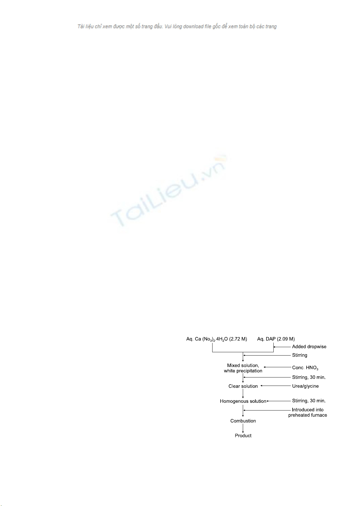
Fig. 1. Flow chart for the aqueous solution combustion technique
for
p
re
p
aration of the nano-HA
p
.
For the current study, calcium-phosphate ceramics, such as
hydroxyapatite (HAp), have been used because their
chemical composition is closely related to that of the mineral
phase of bone [19]. These ceramics are adequately
biocompatible [10] and do not induce adverse local tissue
reactions, immunogenicity, or systemic toxicity. Furthermore,
because this material is osteoconductive, it acts as a support
for new bone formation within the pore sites [24], which are
deliberately generated in the structure. However, depending
on the preparation technique, the material exhibits gross
different powder characteristics, microstructure, and
associated mechanical and biologic properties. When
nano-sized particles below 100 nm of HAp are concerned, it
is still a challenge to synthesize the same via a simple
method. Moreover, for repair and reconstruction of diseased
or damaged bones or tissues, a biphasic calcium phosphate
(BCP) composed of a suitable percentage of HAp and β-
tri-calcium phosphate (β-TCP) are thought to be near the
ideal solution for this remodeling of bone. The first studies of
LeGeross et al. [23] on BCP with varying HAp/β-TCP
demonstrated that the bioactivity of these ceramics may be
controlled by manipulating the HAp/β-TCP ratios. Although
various routes have been developed to synthesize HAp
powders [16], only a few reports are available concerning the
production of β-TCP [20]. For the synthesis of both
materials, the most commonly adapted technique is wet
chemical precipitation [2], followed by calcinations. We
have successfully synthesized a series of BCP composition
with varied HAp and β-TCP content by using a novel
aqueous combustion technique. This processing technique is
often adapted for the rapid preparation of a variety of oxide
ceramic powders [20]. The process involves an exothermic,
usually very rapid and self-sustaining chemical reaction
between the desired metal salts (oxidizer), preferably
nitrates, and a suitable organic fuel, such as urea, glycine,
carbohydrazide, and citric acidin an aqueous solution. The
reaction is initiated at a fairly low temperature followed by
rapid cooling, and this in turn leads to nucleation of
crystallites without much growth. The reaction between the
oxidizer and fuel releases large amounts of reaction heat that
is utilized to synthesize the desired materials in situ and the
large volume of gas evolved disintegrates the high purity
products to friable agglomerates of very fine particulates.
The purpose of the present study was to evaluate porous
HAp, the powders of which were prepared by a novel
aqueous solution combustion technique, as a bone substitute
in healing bone defects.
Materials and Methods
Synthesis of nano-crystalline HAp by an aqueous
solution combustion method
Calcium nitrate tetrahydrate (S.D. Fine-Chem, India) and
di-ammonium hydrogen ortho-phosphate (DAP; S.D.
Fine-Chem, India) were used as raw materials for the
preparation of calcium phosphate powders. Urea (Glaxo,
India) and glycine (Glaxo, India), both A.R. grade, were
used as the fuel. For synthesizing HAp, aqueous stock
solutions of calcium nitrate tetrahydrate (2.72 M) and DAP
(2.09 M) were first mixed slowly with continuous stirring;
subsequently concentrated nitric acid was added dropwise to
dissolve the resulting white precipitate. A predetermined
amount of solid fuel was added to the clear solution and
homogenized by stirring with a magnetic stirrer for 30 min.
at room temperature. One glass ceramic-coated mild steel
(dia. ∼80 mm, volume 130 ml) container containing the
solution was introduced into a muffle furnace preheated to
the desired temperature (300-700oC). A stainless steel wire
mesh was put on the reaction container to reduce particle loss
through aerosol formation. Immediately after placement in
the furnace, the mixed solution started to boil, followed by
the evolution of a large volume of gases. The mass then
frothed and swelled to yield foam, from where a flame
appeared and burned with incandescence. At the initiation of
ignition, the furnace was switched off. The heat evolved
during the reaction sustained itself and proceed to com-
pletion without requiring any further heat from an external
source. The general flowchart for the process is shown in
Fig. 1. Details of the in vitro characterization of the prepared
powder are beyond the scope of this article, but can be found
elsewhere [12]. This powder has been used for the following
studies.
Fabrication of porous HAp
In the present study, porous (35-40% by volume) HAp was
fabricated by using β-naphthalene and polyvinyl alcohol
(S.D. Fine-Chem, India) as a combustible organic material.
HAp powder was milled separately with oleic acid surfactant
and a pre-calculated amount of β-naphthalene.
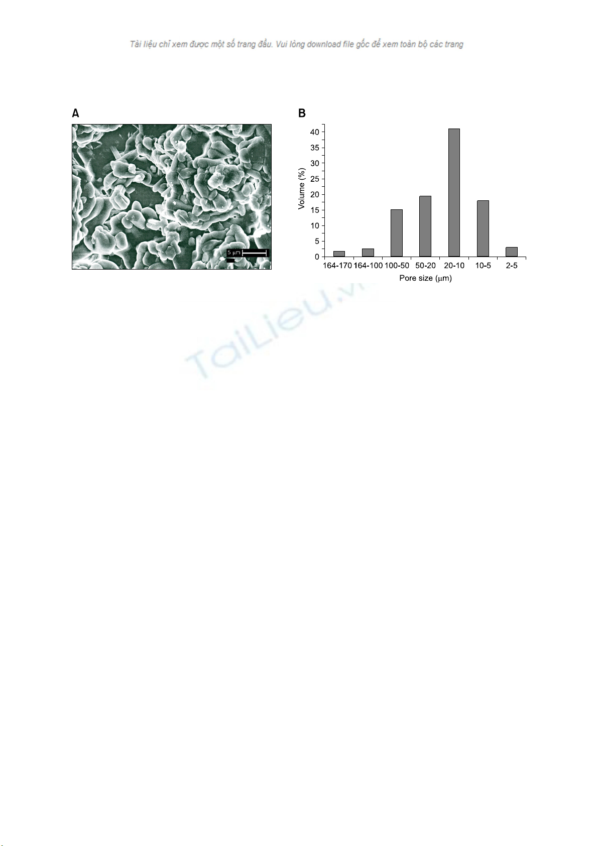
Hydroxyapatite for healing bone defects 185
Fig. 2. (A) Scanning electron micrograph of the porous specimen of HAp before implantation in goats. (B) Histogram of pore size dis-
tribution patterns of the HAp specimen.
Rectangular- shaped (12 × 5 × 3 mm3) blocks were uniaxially
cold-compacted with low pressure, and subsequently cold
iso-statically pressed at 100 MPa for homogeneous
densification. All specimens were slowly dried at 80oC for 3
days. Finally, HAp specimens were sintered at 1,250oC for 2
h. Archimedes’ principle using water as the immersing
medium was used to calculate the density and apparent
porosity of the sintered specimens. Scanning electron
microscopy (SEM) and mercury intrusion porosimetry
(MIP) were used to obtain the pore shape, size, morphology,
and distribution of the specimens. Fig. 2A shows the SEM
photomicrograph of the porous strut with a tag of 5 μm,
while Fig. 2B shows the histogram based on the MIP data for
distribution of the pores in the struts. The porous struts were
initially pasteurized with distilled water and subsequently
autoclaved at 121oC for 30 min. before implantation.
Archimedes' principle using water as the immersing medium
was used to calculate the density and apparent porosity of the
sintered specimens and found to be 2.04 g/ml and 35.2% on
an average, respectively.
Animal experimentation
Animal experimentation was carried out following the
procedures conforming to the standards of the Institutional
Animal Ethical Committee of the West Bengal University of
Animal and Fishery Sciences. Twelve black Bengal goats of
both genders, weighing 10-12 kg, were randomly distributed
into 2 groups of 6 animals each, as follows: control (group I),
in which the bone defect was not treated and the test
specimen (group II), in which porous HAp blocks were
inserted within the bone defect. Under standard aseptic
conditions and sedation with xylazine hydrochloride (0.05
mg/kg body weight; Indian Immunologicals, India) in
animals which had received atropine and local 2% ligno-
caine hydrochloride (Neon Laboratories, India), a 3 cm
longitudinal skin incision was made on the lateral side of the
radius bone. The implant sites (1 × 0.5 cm) were prepared
using a micro-motor dental drill after exposing the cortical
bone followed by irrigation with sterile normal saline. In the
controls (group I), the defect was left as such without any
implant, while in group II, HAp blocks were placed in the
defect sites. The implants were secured in position by
suturing the periosteum, muscle, subcutaneous tissue, and
skin in layers. Postoperatively, all the animals received
cefotaxime sodium (250 mg I/M twice daily; Mapra India,
India) and injectable meloxicam (0.5 ml once daily for 5
days; Intas Pharmaceuticals, India) with daily dressing
changes of the surgical wounds.
Local inflammatory reaction and healing of the wound
Local inflammatory reactions and healing of the wounds
were assessed by visual and manual examinations from the
day of surgery up until the 90th day postoperatively.
Radiological examination
Radiographs were obtained of the operated forelimb
immediately after implantation and subsequently on days
21, 30, 60, and 90 postoperatively to assess the status of the
implant, the host-bone reaction to the implant, and new bone
formation. X-rays were also obtained after light sedation
using xylazine hydrochloride (0.05 mg/kg body weight).
Histological study
The implanted ceramic blocks, along with the surrounding
bones, were collected from the animals on day 90 post-
operatively. The bone sections with both normal and
implanted areas were prepared by decalcification following
a standard technique; 4 μm sections were cut and stained
with hematoxylin and eosin to observe the status of the bone
implants and the cellular response of host bone to the
186 Samit Kumar Nandi et al.
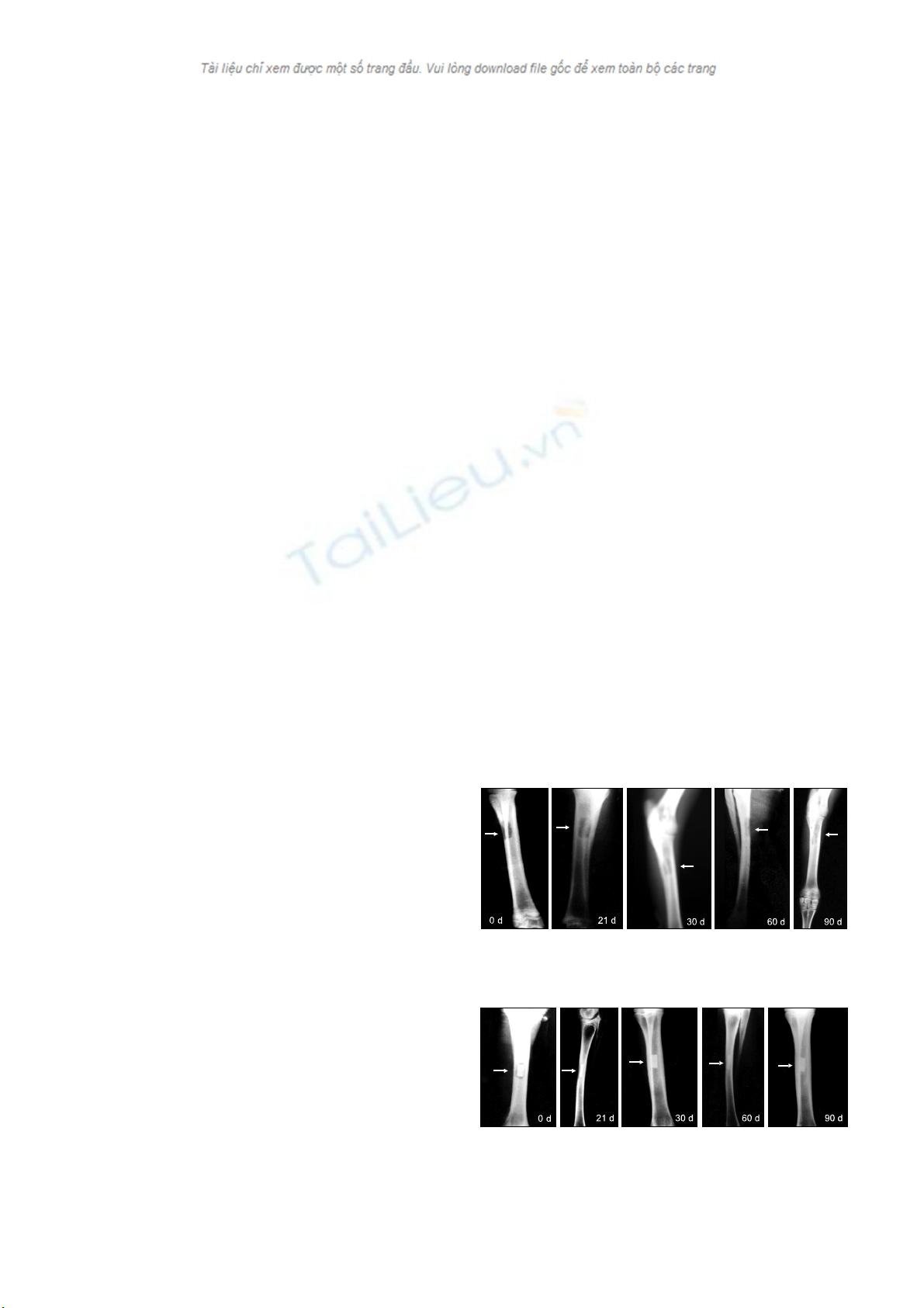
Fig. 3. Radiographs of the control site obtained on day 0, 21, 30,
60
,
and 90
p
ost-o
p
erativel
y
.
Fig. 4. Radiographs of the HAp-implanted site obtained on day 0,
21, 30, 60, and 90 post-operatively.
implants.
Oxytetracycline labeling study
Fluorochrome (oxytetracycline dehydrate; Pfizer India,
India), at a dose of 50 mg/kg body weight, was given on days
77, 78, 85, and 86 (2-6-2 i.e. two injections on day 77 and 78
and after 6 days another two injections on days 85 and 86)
post-operatively for double-toning of new bone. Undecalcified
ground sections were prepared [27] from the implanted
segments of bone and the sections were ground to 20 μm
thickness using different grades of sand paper. The ground-
undecalcified sections were observed under ultraviolet
incidental light with an Orthoplan microscope (Excitation
filter, BP- 400 range; Leitz, USA) for tetracycline labeling to
determine the amount and source of newly formed bone.
Angiographic study
Radial angiography was performed by making a 4-5 cm
skin incision aseptically on the medial aspect of the thigh
under xylazine hydrochloride sedation and local infiltration
analgesia with 2% lignocaine hydrochloride on day 90
postoperatively. The radial arteries were located, exteriorized,
and catheterized using polyethylene catheters connected to a
syringe containing 15 ml sodium iothalmate (Mallinckradt,
USA). The contrast material was infused with regular gentle
digital pressure and radiographs were taken at 14 mAs, 50
kVP, and 90 cm FFD. The catheter was removed and the
puncture of the artery was sutured with 4-0 chromic catgut,
and finally the skin wound was closed. For better visuali-
zation of the arteries, one test limb from each group was
collected after euthanizing the animal at the end of the
experiment; the limb was perfused with lead oxide suspen-
sion (20% W/V) in a manner similar to that used to examine
the vascular response of host bone and surrounding tissues in
the implanted area and visualization of the implant.
Results
Local inflammatory reactions and healing of the
wound
No marked inflammatory reactions were observed in the
control and experimental groups following placement of
bioceramic implants up to the 90th day postoperatively.
Weight-bearing capacity in each animal gradually improved,
as signs of inflammation subsided (within 10 days). There
was no adverse local effect, such as marked hematoma or
edema, during the early postoperative period. Wound
healing was uneventful in all cases and the sutures were
removed on the 10th postoperative day. The implants were
clinically stable in the bone.
Radiological observations
On day 0 in group I (control), the radiographs showed the
cortical defect devoid of any implant, resulting in a
radiolucent gap. On day 21, the radiographs revealed a
minimal periosteal reaction and smoothing edges with
oval-shaped corners of the cortical bone defects. On day 30,
radiographs indicated a substantial reduction in the gap size,
which was in the process of obliteration by hard tissue
materials of similar density to that of host bone. On day 60,
the defect was not totally obliterated by newly grown bony
tissue. On day 90, radiographs showed that the defect was
similar to what was observed after day 60, except that the
newly formed bony tissue was more organized and the
fractured end became smooth and round. Representative
radiographs are shown in Fig. 3.
Radiographs obtained on day 0 of group II (HAp) of the
defect site showed a rectangular-shape mid-shaft diaphyseal
defect with a well-placed HAp block and a radio-density of
the implant, similar to that of the host bone. On day 21, the
diagram showed a well-established periosteal reaction with
narrowing of the gap between the bone and implant without
any signs of implant resorption. On day 30, the radiographs
showed the presence of the implant and radiologically-
detectable newly grown host tissue. On day 60, the implant
was noted to have a reduced density in comparison to the
radiographs of previous days. On day 90, there was complete
bridging of the cortical defect along the axis of the radius
with a similar radio-dense bony material to that of normal
bone. The presence of the implant could be identified by a
radio-dense shadow in the implanted site and the implant
was not absorbed, rather it had undergone structural changes
by a host graft interaction. Representative radiographs are
shown in Fig. 4.
Hydroxyapatite for healing bone defects 187
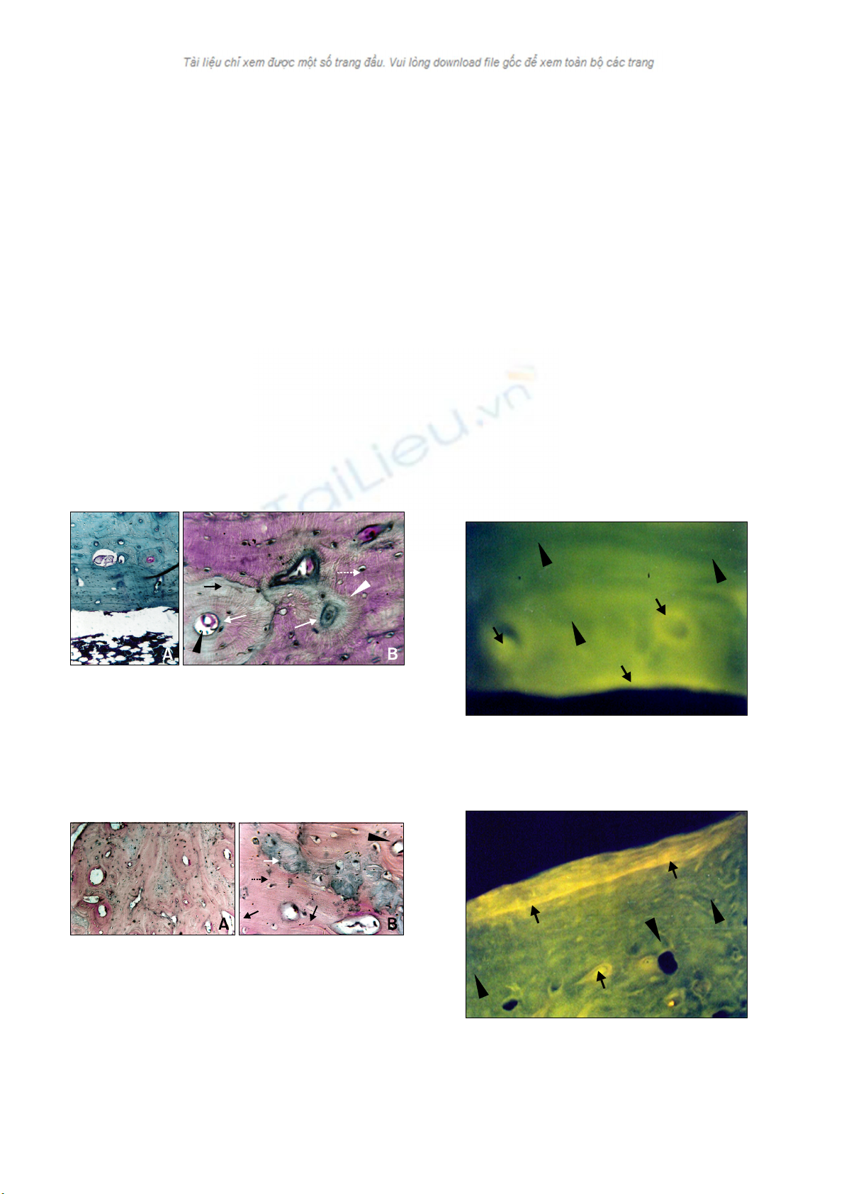
Fig. 5. Histologic sections of the control site. (A) The section
showed an adequate amount of marrow material, fat cells, and
blood vessels, along with a lamellar appearance of bone in the
cortical area of the control bone. H&E stain, ×10. (B) The sectio
n
showed the presence of woven bone at the cortex of the control
bone. Woven bone (white arrows), Haversian canal (black arrow-
head), Haversian system (white arrowhead), new bone (white ar-
row with dotted line) and host bone (black arrow). H&E stain, ×45.
Fig. 6. Histologic sections of the HAp-implanted bone. (A) The
section showed well-developed Haversian canals with defined
osteoblasts at the periphery along with the presence of non-ab-
sorbed materials. H&E stain, × 10. (B) Histologic section
showed well-developed lamellar bone (black arrowhead).
Cortical area along with unabsorbed biodegradable material as a
refractile crystalloid body (black arrow). New bone (white ar-
row) and host bone (black arrow with dotted line). H&E stain,
×45.
Fig. 7. Photomicrograph showing the presence of homogenous
non-fluoroscent area of cancellous bone at the defect site. New
bone (arrows) and host bone (arrowheads). ×63.
Fig. 8. Photomicrograph on day 90 showing presence of fluo-
rescent osteoid tissue in the interspace of the HAp implant. Ne
w
bone (arrows) and host bone (arrowhead). ×63.
Histological study
Tissue sections from group I (control) showed mild
inflammatory reactions with moderate fibro-collagenization.
The cortex showed a lamellar appearance of the bone along
with the presence of woven bone in some places. The
marrow space showed an adequate amount of marrow
material, fat cells, and blood vessels (Figs. 5A and B).
Tissue sections of group II (HAp) showed complete normal
ossification with development of Haversian canals and
well-defined osteoblasts at the periphery. The blood vessels
in the Haversian spaces were well-developed. The marrow
space showed development of blood vessels with very little
amount of marrow material. Non-absorbed biodegradable
material was also noted in the lamellar cortical bone and in
the marrow space as a refractile, crystalloid structure (Figs.
6A and B).
Oxytetracycline labeling study
In group I (control), the process of new bone formation was
active from both ends. Newly formed osseous tissues
originating from the periosteal, as well as the endosteal,
surface of the bone were seen, however, the intensity was
dominant on the periosteal side. The defect was completely
filled with newly formed cancellous bone and appeared as a
homogenous non-fluorescent area. However, a narrow linear
zone near the periosteum revealed a golden-yellow fluorescence,
suggestive of new bone formation in the area (Fig. 7). Union
in the defect site of the bone was complete in most of the
animals.
In group II (HAp) under fluorescent microscopy, the defect
line was visualized as a line of golden-yellow fluorescence,
whereas the host bone evinced a dark, sea green homogenous
colour. In this group, the activity of new bone formation was
moderate. Within this new osteoid tissue, which completely
filled the bone defect; crossing-over of the new bone
trabeculae was evident. Resorption cavities were present,
indicating that the resorption and replacement of bone were
well under progress (Fig. 8).