JOURNAL OF
Veterinary
Science
J. Vet. Sci. (2008), 9(4), 425
427
Short Communication
*Corresponding author
Tel: +82-53-950-5975; Fax: +82-53-950-5955
E-mail: jeongks@knu.ac.kr
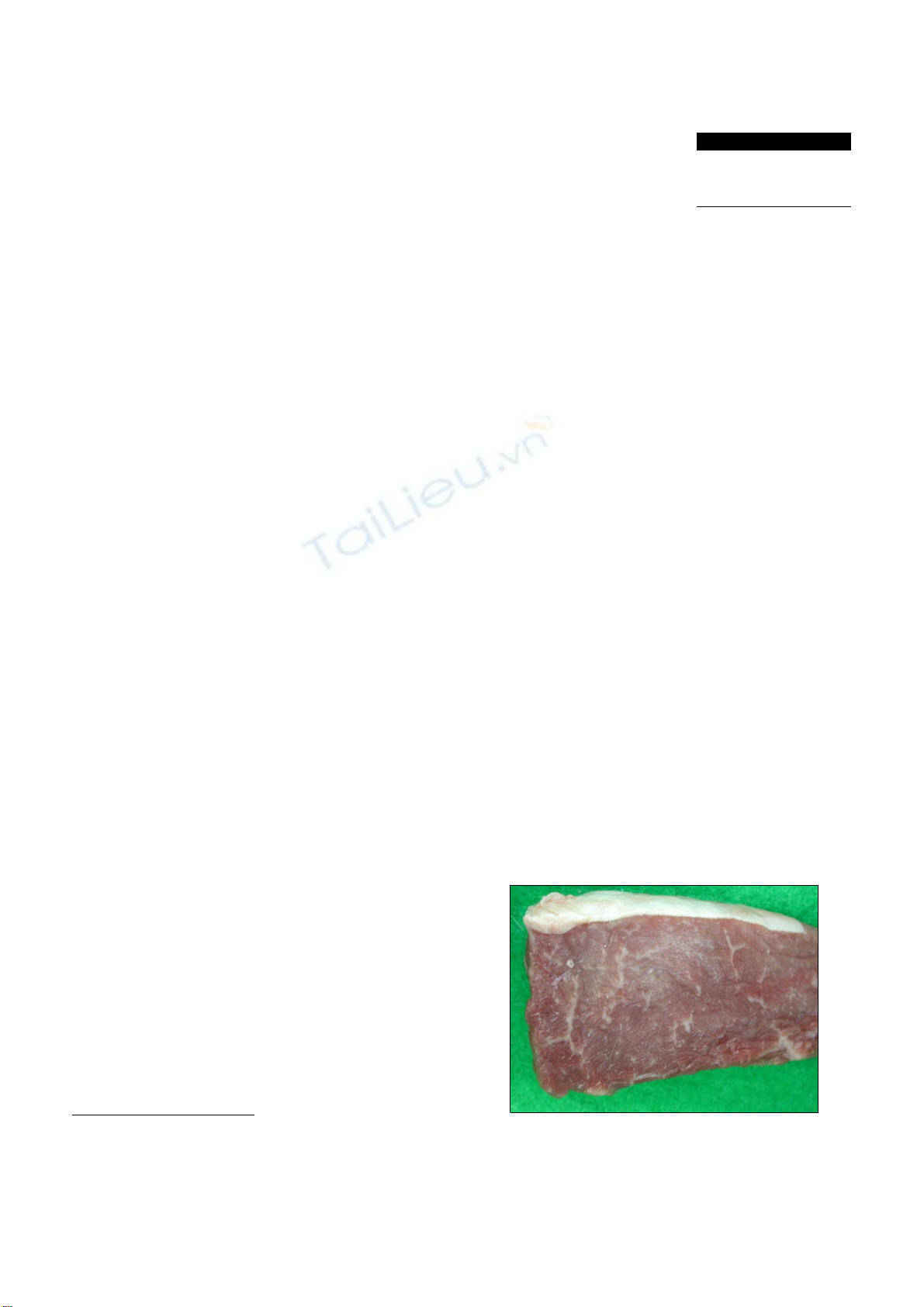
Fig. 1. Appearance of the skeletal muscle of the Korean native
cow. The muscle was generally whitish and pale with a white
streak.
Eosinophilic myositis in a slaughtered Korean native cattle
Sun Hee Do1,2, Da-Hee Jeong1, Jae-Yong Chung1, Jin-Kyu Park1, Hai-Jie Yang1, Dong-Wei Yuan1, Kyu-Shik
Jeong1,*
1Department of Veterinary Pathology, College of Veterinary Medicine, Kyungpook National University, Daegu 702-701,
Korea
2College of Veterinary Medicine, Konkuk University, Seoul 143-701, Korea
Histopathological findings of eosinophilic myositis in the
carcass of a slaughtered Korean native cow are presented.
Lesions contained massive fibrous septae with vacuolar
changes in some lesions, and the hypercontraction and
rupturing of muscle bundles, with replacement by eosinophils.
Necrosis and severe eosinophil infiltration were observed.
Sarcoplasmic fragmentation and atrophy developed. Typical
of granuloma, calcified myofibers were focally surrounded
by macrophages and numerous inflammatory cells, and
multinucleated giant cell formation was evident.
Keywords:
eosinophilic myositis, granuloma, Korean native
cow, myofiber
Eosinophilic myositis (EM) is a collective term used in
meat inspection to designate diseases of clinically healthy
animals that have focal, greencolored muscular lesions of
unknown origin [7,10]. EM is a relatively rare condition in
cattle and sheep of all ages [7,8]. Previous reports have
provided evidence that Sarcocystis are directly associated
with the lesions and contribute to rejection and down-
grading of carcasses at meat-processing plants [1,2,5].
Bovine EM has been only rarely described. The cause of
EM remains unknown since most cases were detected
during a routine postmortem inspection and because there
is little tissue reaction in the intermediate host, cattle, in
which asexual development of Sarcocystis occurs.
Presently, we describe a case of EM in a 3-year-old cow
with a normal history and no pre-existing clinical
conditions at the time of slaughter. A routine postmortem
inspection of the carcass revealed generalized whitish and
pale skeletal muscles in the cervical region (Fig. 1).
Carcasses with locally whitish and pale lesions that had
been rejected by inspectors were acquired for comparative
purposes. Representative sections of cervical skeletal
muscles were fixed immediately in 10% neutral buffered
formalin, processed routinely, and embedded in paraffin.
Tissue sections 4 μm in thickness were cut and stained with
hematoxylin and eosin and periodic acid-Schiff (PAS) for
detection of pathogen.
Histopathological examination of the lesions revealed
interstitial fibrosis, more developed fibrous septa, and
augmented numbers of fibroblasts between the myofibers
as compared with normal skeletal muscles (Fig. 2). The
carcass was whitish and pale (Fig. 1). The skeletal muscles
showed histopathological features of eosinophilic myositis
that contained focal granulomatous lesions with multifocal
calcified necrotic fibers, mixed inflammatory cells mainly
consistent with eosinophils, and the infiltration of a
multinucleated giant cell. Massive intramuscular
infiltration of the eosinophils was identified in the
specimens; as well, most myofibers were hypercontracted,
ruptured, and were replaced by eosinophils (Fig. 3).
Eosinophils were present within and adjacent to the
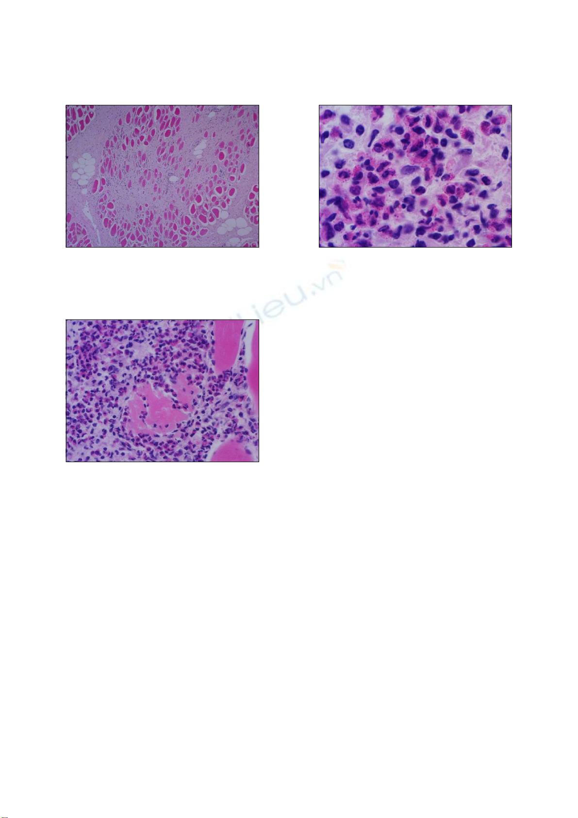
426 Sun Hee Do et al.
Fig. 4. High magnification of Fig. 3. ×66.Fig. 2. Histochemical examination of the skeletal muscle of the
Korean native cow. The whitish and pale muscle exhibited
augmented interstitial fibrous septa and the strong activation o
f
fibroblasts between the myofibers. H&E stain. ×12.
Fig. 3. Eosinophil examination of the skeletal muscle of the
Korean native cow. Massive eosinophils are present within and
adjacent to the affected myofibers. H&E stain. ×33.
affected myofibers (Fig. 4). There was no evidence of
infection from parasites, or sarcocystosis. Especially
considering the appearance of tissues following PAS
staining, a diagnosis of bovine idiopathic EM was made.
EM is a group of three inflammatory disorders -
eosinophilic polymyositis, eosinophilic perimyositis and
focal eosinophilic myositis - characterized pathologically
by an eosinophilic cell infiltrate in the skin and skeletal
muscles [4,6]. The pathogenesis of EM varies; a
correlation between serum interleukin-5 levels and disease
severity [9] suggests a role for this interleukin species in
eosinophil activation.
Sarcocystis spp. has long been suspected as an etiological
agent of EM [7]. Recent reports have provided evidence that
sarcocysts are directly associated with lesions and
contribute to the rejection and down grading of carcasses at
meat- processing plants [1,2,5,10]. In one study, up to 5%
of carcasses rejected in the United States were positive for
sarcocysts [5]. Presently, generalized inflammatory
reactions were common in sections of all the acquired
rejected carcasses, and sections of all lesions revealed
granulomatous reactions. Most of the granulomas observed
in the EM carcasses may have resulted from sarcocysts that
served as chronic inflammatory stimuli. It is not surprising
that sarcocysts were found in only a small proportion of
granulomas.
Currently, there are no tests that can predict the presence
of EM prior to slaughter. Immunoglobulins that are
specific to species of Sarcocystis have been found in both
affected and unaffected cattle [2]. Speciation of the
responsible pathogen in cattle is unclear. Regarding the
respective occurrence of EM and sarcocytosis, EM has
little economic importance while sarcocystosis has great
economic importance to the meat-producing and meat
packing industries, and to the overall public health.
There have been a few reports of carcasses with numerous
disseminated lesions that were rejected as human food
sources and presented a public health threat. Although
carcasses with a few localized or excisable lesions can be
approved, nearly all grossly affected carcasses are assumed
to contain disseminated lesions, even though such lesions
may be difficult to find.
In the present study, a cow with pale and whitish skeletal
muscles grossly evident in the loggisimus capitus was
observed at the slaughter house. Histosections of carcass
tissue revealed lesions that were characterized by a
massive infiltration of eosinophils between the myofibers,
hypercontraction and rupture of the muscle bundles, and
augmented fibrous septae. As well, focally granulomatous
inflammatory lesions were observed. Our case may well
represent Sarcocystis spp. EM, although the direct cause
was not ascertained.
When confronted by EM, we suggest that not only
rejected carcass should be examined, but the entire animal
stock of the farm as well. While it is likely that the cause of
EM a Sarcocystis spp. infection, livestock officials can

Bovine eosinophilic myositis 427
prevent or reduce sarcocystosis by controlling the
movement of working dogs and cats, and eliminating stray
and wild animals from cattle and sheep pastures, feedlots,
and feedmills to avoid feed and water-being contaminated
with sporocysts. Eliminating bovine and ovine raw
muscles and viscera from the diets of dogs and cats is
prudent to prevent infecting the definitive hosts. Moreover,
human defecation in or near feed or water that could be
consumed by cattle should be restricted so as to avoid the
transmission of Sarcocystis spp. oocysts to cattle.
Acknowledgements
The authors wish to thank the veterinary meat inspectors
who collected the material. This work was supported by the
faculty research fund of Konkuk University in 2007.
References
1. Bundza A, Feltmate TE. Eosinophilic myositis/lymphade-
nitis in slaughter cattle. Can Vet J 1989, 30, 514-516.
2. Gajadhar AA, Yates WD, Allen JR. Association of eosino-
philic myositis with an unusual species of Sarcocystis in a
beef cow. Can J Vet Res 1987, 51, 373-378.
3. Granstrom DE, Ridley RK, Baoan Y, Gershwin LJ.
Immunodominant proteins of Sarcocystis cruzi bradyzoites
isolated from cattle affected or nonaffected with eosinophilic
myositis. Am J Vet Res 1990, 51, 1151-1155.
4. Hall FC, Krausz T, Walport MJ. Idiopathic eosinophilic
myositis. QJM 1995, 88, 581-586.
5. Jensen R, Alexander AF, Dahlgren RR, Jolley WR,
Marquardt WC, Flack DE, Bennett BW, Cox MF, Harris
CW, Hoffmann GA, Troutman RS, Hoff RL, Jones RL,
Collins JK, Hamar DW, Cravans RL. Eosinophilic
myositis and muscular sarcocystosis in the carcasses of
slaughtered cattle and lambs. Am J Vet Res 1986, 47,
587-593.
6. Pickering MC, Walport MJ. Eosinophilic myopathic
syndromes. Curr Opin Rheumatol 1998, 10, 504-510.
7. Reiten AC, Jensen R, Griner LA. Eosinophilic myositis
(sarcosporidiosis; sarco) in beef cattle. Am J Vet Res 1966,
27, 903-906.
8. Thomas JH. Muscle and tendon. In: Jubb KVF, Kennedy
PC, Palmer N (eds.). Pathology of Domestic Animals. Vol.
1. 3rd ed. pp. 245-252, Academic Press, Orlando, 1985.
9. Trüeb RM, Lubbe J, Torricelli R, Panizzon RG, Wüthrich
B, Burg G. Eosinophilic myositis with eosinophilic
cellulitislike skin lesions. Association with increased serum
levels of eosinophil cationic protein and interleukin-5. Arch
Dermatol 1997, 133, 203-206.
10. Wouda W, Snoep JJ, Dueby JP. Eosinophilic myositis due
to Sarcocystis hominis in a beef cow. J Comp Pathol 2006,
135, 249-253.