
BioMed Central
Page 1 of 4
(page number not for citation purposes)
World Journal of Surgical Oncology
Open Access
Case report
Primary hepatic embryonal sarcoma masquerading as metastatic
ovarian cancer
Peter Kullar1, Christopher Stonard2, Neville Jamieson1, Emmanuel Huguet1,
Raaj Praseedom1 and Asif Jah*1
Address: 1Department of Hepatobiliary and Transplant Surgery, Addenbrooke's Hospital, Cambridge University Hospitals NHS Foundation Trust,
Cambridge CB2 2QQ, UK and 2Department of Histopathology, Chesterfield Royal Hospital NHS Foundation Trust, Chesterfield, Derbyshire DE4
3GJ, UK
Email: Peter Kullar - peterkullar@hotmail.com; Christopher Stonard - Christopher.Stonard@chesterfieldroyal.nhs.uk;
Neville Jamieson - neville.jamieson@addenbrookes.nhs.uk; Emmanuel Huguet - Emmanuel.Huguet@addenbrookes.nhs.uk;
Raaj Praseedom - raaj.praseedom@addenbrookes.nhs.uk; Asif Jah* - asif.jah@addenbrookes.nhs.uk
* Corresponding author
Abstract
Background: Hepatic embryonal sarcoma (HES) is a rare but aggressive primary tumor of the
liver occurring most frequently in childhood.
Case presentation: We report a case of a 52 year old woman having previously undergone
treatment for ovarian serous papillary carcinoma who subsequently presented with a large solitary
mass in the liver. Initially this was presumed to be metastasis from the ovarian primary however,
on further examination it was shown to be a primary hepatic embryonal sarcoma.
Conclusion: Primary liver tumors should be considered in differential diagnoses in patients with
ovarian cancer who subsequently present with liver tumors. This is particularly important when
there is no direct evidence of recurrence of ovarian cancer.
Background
Hepatic embryonal sarcoma (HES) is rare primary tumor
of the liver usually occurring in childhood. It is generally
considered an aggressive tumor with a poor prognosis.
Approximately 60 cases have been reported in adults. On
the contrary, ovarian carcinoma frequently tends to
metastasize to the liver [1]. We report a case of a woman
who had previously undergone treatment for bilateral
ovarian serous papillary carcinoma and subsequently pre-
sented with a large solitary mass in the liver which turned
out to be a primary hepatic embryonal sarcoma. We also
review the current literature, diagnosis and treatment of
HES.
Case presentation
A 52 year old woman presented with pain and a palpable
mass in the right upper quadrant of the abdomen 18
months after completing treatment for ovarian carci-
noma. She had previously undergone bilateral salpingo-
oophorectomy and en bloc subtotal colectomy for bilateral
high-grade ovarian serous papillary carcinoma infiltrating
the sigmoid colon. She also had peritoneal and diaphrag-
matic seedlings which were debulked concurrently. After
the resection, the residual disease was estimated to be less
than 0.5 cm2. Subsequently, she received 6 cycles of adju-
vant chemotherapy with carboplatin and taxol. Her
CA125 level which was elevated at 149 U/L prior to resec-
Published: 23 June 2009
World Journal of Surgical Oncology 2009, 7:55 doi:10.1186/1477-7819-7-55
Received: 9 April 2009
Accepted: 23 June 2009
This article is available from: http://www.wjso.com/content/7/1/55
© 2009 Kullar et al; licensee BioMed Central Ltd.
This is an Open Access article distributed under the terms of the Creative Commons Attribution License (http://creativecommons.org/licenses/by/2.0),
which permits unrestricted use, distribution, and reproduction in any medium, provided the original work is properly cited.
World Journal of Surgical Oncology 2009, 7:55 http://www.wjso.com/content/7/1/55
Page 2 of 4
(page number not for citation purposes)
tion later returned to 8 U/L (normal range = 0 – 25 U/L).
Apart from chemotherapy, there was no past history of
exposure to radiotherapy or any carcinogens.
During this presentation, the only abnormality on routine
hematology and biochemistry was a slightly elevated alka-
line phosphatase at 178 U/L (normal range = 35–130 U/
L). The CA125 level was within the reference range (7 U/
L) and no other tumor markers were assayed. A Compu-
terized Tomography (CT) scan revealed a large heteroge-
neous mass almost completely replacing the right lobe of
the liver (Figure 1). No other abdominal, pelvic or lung
lesions were identified. Due to the recent history of ovar-
ian cancer, this lesion was suspected to be a solitary met-
astatic deposit from the ovarian cancer. The liver lesion
was not biopsied in order to avoid any needle tract seed-
ing with malignant cells. In the absence of any extrahe-
patic disease she proceeded to undergo an extended right
hepatectomy. During hepatectomy the tumor was found
to be adherent to the diaphragm, a part of which was
removed en bloc. Post-operatively, she made a good recov-
ery without any complications.
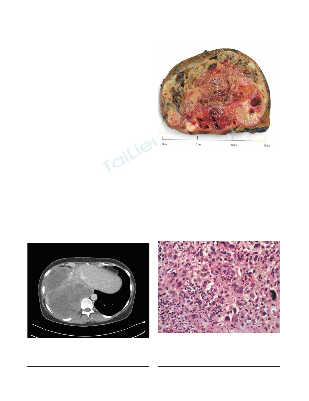
Macroscopically, the tumor was a large, solid and cystic
mass that included areas of necrosis and cystic degenera-
tion measuring 18 × 12 × 8 cm (Figure 2). Microscopi-
cally, it consisted of pleomorphic malignant spindle cells
including rhabdoid and bizarre giant cell forms on a
chrondromyxoid stromal background (Figure 3) with
focal Periodic Acid Schiff-positive cytoplasmic granules.
There was no evidence of epithelial differentiation which
excluded the possibility of sarcomatoid hepatocellular
carcinoma. Immunohistochemical staining was positive
for alpha1-antitrypsin but showed no other indicators of
differentiation. This contrasted with the serous papillary
morphology of the previously resected ovarian tumor that
showed no sarcomatoid areas and no rhabdoid differenti-
ation. The liver tumor was therefore judged to be a second
primary rather than a metastatic deposit of the resected
ovarian carcinoma. The lesion was classified as primary
HES on the basis of its morphology and immunohisto-
Computerized Tomography scan of the abdomen, the tumor is shown to be occupying almost the whole the right lobe of the liverFigure 1
Computerized Tomography scan of the abdomen,
the tumor is shown to be occupying almost the
whole the right lobe of the liver.
Cut section of the resected hepatic tumorFigure 2
Cut section of the resected hepatic tumor.
Representative section of a more cellular area showing plump spindle cells, bizarre giant cell forms and scattered apoptotic bodiesFigure 3
Representative section of a more cellular area show-
ing plump spindle cells, bizarre giant cell forms and
scattered apoptotic bodies. (Haematoxylin and eosin
stain, photographed at ×200 magnification).

World Journal of Surgical Oncology 2009, 7:55 http://www.wjso.com/content/7/1/55
Page 3 of 4
(page number not for citation purposes)
chemical staining pattern with complete excision noted at
the hepatic resection margins.
After remaining well for approximately 6 months she
developed progressive shortness of breath and a follow-up
CT scan demonstrated a large recurrent tumor contiguous
across the right hemithorax and abdomen. A percutane-
ous biopsy of this mass confirmed the recurrence of the
primary hepatic sarcoma. Palliative chemotherapy with
ifosfamide and doxorubicin was commenced but had to
be stopped after 3 cycles due to severe side effects. She
received palliative care for gradual deterioration and died
6 months later.
Discussion
Ovarian cancer usually presents with widespread intra-
peritoneal metastasis [2]. A minority of patients present
with aggressive disease manifested by liver, lung or brain
metastases [3]. An autopsy study of 428 patients with
ovarian cancer reported that over 40% of the patients with
ovarian cancer had evidence of liver metastases at the time
of death [1]. Amongst the ovarian cancers, the stromal
tumors were the most likely histological subtype to metas-
tasize to the liver [1].
Approximately 20% of the patients with epithelial type of
ovarian cancer have been reported to have normal levels
of CA125 [4,5]. During second presentation of this
patient, the liver function tests were slightly deranged but
the CA125 level was within the reference range.
Primary liver sarcomas are rare tumors and represent only
0.2% of all primary liver tumors [6]. This group includes
a number of different histological types such as angiosar-
coma, leiomysarcoma, fibrosarcoma, HES, and malignant
fibrous histiocytoma [7]. HES are most commonly
reported in a pediatric age group, with the peak incidence
between the ages of 6 and 10 years. Only 68 cases have
been reported in the adult population [8]. Although the
etiology of primary liver sarcomas is unclear, there is an
increased risk associated with radiation therapy and high
doses of alkylators or anthracyclines [8]. The use of Thoro-
trast as a radiological contrast medium in the 1950s was
associated with a very high incidence of hepatic angiocsar-
comas [9]. Our patient had received taxol and carboplatin
as adjuvant chemotherapy after resection of ovarian can-
cer but there is no documented association of these agents
with development of primary liver sarcomas.
The presentation of liver sarcomas is usually non-specific
with symptoms such as abdominal discomfort, anorexia,
fever or weight loss [10]. Hepatomegaly may be present
with large tumors and liver function tests may be
deranged although frank jaundice is rare [7,10]. The non-
specific nature of the presenting symptoms makes clinical
diagnosis extremely difficult without imaging or biopsy.
Although the lesion can be identified on ultrasonography
and CT scan, contrast-enhanced Magnetic Resonance
Imaging (MRI) scan is the best imaging modality for char-
acterization of primary liver tumors [11].
There are a number of radiographic features which distin-
guish HES from mesenchymal and other primary hepatic
tumors. The myxoid stroma typical of HES appear as large
central areas of hypointense signal on T1-weighted images
that have high intensity signal on T2-weighted images.
Similarly, in the solid peripheral areas of the mass hetero-
geneous contrast enhancement is seen on both CT and
MRI scans [12].
HES have previously been associated with poor outcome.
In 1978 Stocker reported a series of 31 patients with HES
with mean survival of less that one year after diagnosis
[10]. Although radical surgery remains the mainstay of
treatment, recent studies have shown improved survival
with radical surgery and use of ifosfamide-based multi-
agent chemotherapy. Almogy et al reported on a series of
8 primary liver sarcomas including two HES managed
with surgical resection and adjuvant chemotherapy [13].
Following a liver resection, one patient with a satellite
lesion and a second patient with recurrent HES were
treated with chemotherapy which led to tumor shrinkage
and enabled a second hepatic resection. There is mount-
ing evidence to suggest that neo-adjuvant chemotherapy
would allow down-sizing of HES to be followed by resec-
tional surgery [14,15]. Bisogno et al identified 17 children
with HES treated with initial conservative surgery and
multi-agent chemotherapy followed by second-look sur-
gery for any residual disease [14]. They report 70% (12/
17) survival at follow up (ranging from 2.4 to 20 years).
Thus, in most cases, the accepted standard treatment
would consist of aggressive surgical resection with combi-
nation chemotherapy either in neoadjuvant or adjuvant
setting.
Conclusion
In this patient HES masqueraded as a metastatic ovarian
carcinoma. This case illustrates that the possibility of a pri-
mary liver tumor should be considered in patients with
ovarian cancer who subsequently present with liver
tumors, particularly when there is no direct evidence of
recurrence of ovarian cancer.
Consent
Written informed consent was obtained from the patient's
next of kin for publication of this case report and accom-
panying images. A copy of the written consent is available
for review by the Editor-in-Chief of this journal.

Publish with BioMed Central and every
scientist can read your work free of charge
"BioMed Central will be the most significant development for
disseminating the results of biomedical research in our lifetime."
Sir Paul Nurse, Cancer Research UK
Your research papers will be:
available free of charge to the entire biomedical community
peer reviewed and published immediately upon acceptance
cited in PubMed and archived on PubMed Central
yours — you keep the copyright
Submit your manuscript here:
http://www.biomedcentral.com/info/publishing_adv.asp
BioMedcentral
World Journal of Surgical Oncology 2009, 7:55 http://www.wjso.com/content/7/1/55
Page 4 of 4
(page number not for citation purposes)
Competing interests
The authors declare that they have no competing interests.
Authors' contributions
PK and AJ wrote the manuscript, CS reviewed the histol-
ogy and contributed to the manuscript, NJ, EH critically
reviewed the manuscript and RP performed the hepatec-
tomy. All authors read and approved the manuscript.
References
1. Rose PG, Piver MS, Tsukada Y, Lau TS: Metastatic patterns in his-
tologic variant of ovarian cancer: An autopsy study. Cancer
1989, 64:1508-13.
2. Chobanian N, Dietrich CS: "Ovarian cancer". Surg Clin North Am
2008, 88(2):285-99.
3. Geisler JP, Geisler HE: Brain metastases in epithelial ovarian
carcinoma. Gynecol Oncol 1995, 57:246-249.
4. Rosen DG, Wang L, Atkinson JN, Yu Y, Lu KH, Diamandis EP, Hell-
strom I, Mok SC, Liu J, Bast RC Jr: Potential markers that com-
plement expression of CA125 in epithelial ovarian cancer.
Gynecol Oncol 2005, 99(2):267-77.
5. Niloff JM, Bast RC Jr, Schaetzl EM, Knapp RC: Predictive value of
CA 125 antigen levels in second-look procedures for ovarian
cancer. Am J Obstet Gynecol 1985, 151(7):981-6.
6. Primary liver cancer in Japan. Clinicopathologic features and
results of surgical treatment. Liver Cancer Study Group of
Japan. Ann Surg 1990, 211(3):277-87.
7. Weitz J, Klimstra DS, Cymes K, Jarnagin WR, D'Angelica M, La
Quaglia MP, Fong Y, Brennan MF, Blumgart LH, Dematteo RP: Man-
agement of primary liver sarcomas. Cancer 2007,
109(7):1391-6.
8. Lenze F, Birkfellner T, Lenz P, Hussein K, Länger F, Kreipe H, Dom-
schke W: Undifferentiated embryonal sarcoma of the liver in
adults. Cancer 2008, 112(10):2274-82.
9. Silpananta P, Illescas FF, Sheldon H: Multiple malignant neo-
plasms 40 years after angiography with Thorotrast. Can Med
Assoc J 1983, 128(3):289-92.
10. Stocker JT, Ishak KG: Undifferentiated (embryonal) sarcoma of
the liver: report of 31 cases. Cancer 1978, 42:336-348.
11. Marti-Bonmati L, Ferrer D, Menor F, Galant J: Hepatic mesenchy-
mal sarcoma: MRI findings. Abdom Imaging 1993, 18(2):176-179.
12. Mortelé KJ, Ros PR: Cystic focal liver lesions in the adult: differ-
ential CT and MR imaging features. Radiographics 2001,
21(4):895-910.
13. Almogy G, Lieberman S, Gips M, Pappo O, Edden Y, Jurim O, Simon
Slasky B, Uzieli B, Eid A: Clinical outcomes of surgical resections
for primary liver sarcoma in adults: results from a single cen-
tre. Eur J Surg Oncol 2004, 30(4):421-7.
14. Bisogno G, Pilz T, Perilongo G, Ferrari A, Harms D, Ninfo V, Treuner
J, Carli M: Undifferentiated sarcoma of the liver in childhood:
a curable disease. Cancer 2002, 94(1):252-7.
15. Kim DY, Kim KH, Jung SE, Lee SC, Park KW, Kim WK: Undifferen-
tiated (embryonal) sarcoma of the liver: Combination treat-
ment by surgery and chemotherapy. J Pediatr Surg 2002,
37:1419-1423.