BioMed Central
Page 1 of 7
(page number not for citation purposes)
Acta Veterinaria Scandinavica
Open Access
Research
In vitro analysis of expression vectors for DNA vaccination of
horses: the effect of a Kozak sequence
Guðbjörg Ólafsdóttir1, Vilhjálmur Svansson*1, Sigurður Ingvarsson1,
Eliane Marti2 and Sigurbjörg Torsteinsdóttir1
Address: 1Institute for Experimental Pathology, University of Iceland, Keldur, Reykjavík, Iceland and 2Department of Clinical Veterinary Medicine,
Vetsuisse Faculty, University of Berne, Switzerland
Email: Guðbjörg Ólafsdóttir - gudbjol@hi.is; Vilhjálmur Svansson* - vsvanss@hi.is; Sigurður Ingvarsson - siguring@hi.is;
Eliane Marti - eliane.marti@itz.unibe.ch; Sigurbjörg Torsteinsdóttir - sibbath@hi.is
* Corresponding author
Abstract
One of the prerequisite for developing DNA vaccines for horses are vectors that are efficiently
expressed in horse cells.
We have analysed the ectopic expression of the human serum albumin gene in primary horse cells
from different tissues. The vectors used are of pcDNA and pUC origin and include the
cytomegalovirus (CMV) promoter. The pUC vectors contain CMV intron A whereas the pcDNA
vectors do not.
Insertion of intron A diminished the expression from the pcDNA vectors whereas insertion of a
Kozak sequence upstream of the gene in two types of pUC vectors increased significantly the in
vitro expression in primary horse cells derived from skin, lung, duodenum and kidney.
We report for the first time the significance of full consensus Kozak sequences for protein
expression in horse cells in vitro.
Background
DNA vaccines have attracted great interest since they
induce strong and lasting humoral and cellular immune
response in experimental animals. Their ability to modu-
late the immune response and to shift it from Th2 to Th1
holds a promise for treatment of allergies and cancer [1,2].
In large animals and humans DNA vaccines have, how-
ever, not lived up to this expectation. Their major draw-
back is low and short lived immune response [3,4]. One
of the reasons for this is thought to be due to limited
expression of the gene product involved and few activated
antigen presenting cells. It is therefore important to
improve the efficacy of expression in the cells of the rele-
vant animal [5,6].
Virus-based vector vaccines have been quite effective in
attaining protection against several viral diseases in horses
such as influenza [7,8], West Nile fever [9-12] and equine
viral arteritis [12,13]. Some of those vaccines have been
licensed [7,9]. With plasmid based DNA vaccination of
horses, protection has been achieved against West Nile
virus with a single immunisation [14]. However, the
potency of this type of genetic vaccines still needs to be
improved for obtaining an adequate immune response
Published: 4 November 2008
Acta Veterinaria Scandinavica 2008, 50:44 doi:10.1186/1751-0147-50-44
Received: 10 April 2008
Accepted: 4 November 2008
This article is available from: http://www.actavetscand.com/content/50/1/44
© 2008 Ólafsdóttir et al; licensee BioMed Central Ltd.
This is an Open Access article distributed under the terms of the Creative Commons Attribution License (http://creativecommons.org/licenses/by/2.0),
which permits unrestricted use, distribution, and reproduction in any medium, provided the original work is properly cited.
Acta Veterinaria Scandinavica 2008, 50:44 http://www.actavetscand.com/content/50/1/44
Page 2 of 7
(page number not for citation purposes)
without using extreme means of injection such as sensi-
tive sites and too many boosts [9,15].
In vectors used for DNA vaccines strong promoters are
used to give the maximum expression of antigens. The
most commonly used is the cytomegalovirus immediate
early gene promoter (CMV-IE) [16,17]. The strongest
expression is generally obtained when the full length,
enhanced CMV-IE promoter is used, including the first
intron from the IE1 gene (intron A) [18-20].
A Kozak sequence adjacent to the ATG start codon greatly
increases the efficiency of translation and hence overall
expression of the gene product. It functions by slowing
down the rate of scanning by the ribosome and improving
the chance of it recognising the start of translation at the
AUG start codon. For optimal expression it is recom-
mended to use the full consensus (GCC)GCC A/G CC
ATG G [21,22].
Our efforts to Th1 focus the immune response of horses
by vaccinating them with vectors of pcDNA origin
resulted in low immune response [23]. We therefore tried
to improve the expression from the vectors with a Kozak
sequence and an intron A. Insertion of the Kozak
sequence increased the expression in all the cells whereas
addition of the intron A decreased the expression.
Methods
2.1. Construction and purification of vectors
Origin and modification of vectors is shown in table 1 and
figure 1. The HSA gene (1822 nucleotides, database no
NM000477) was amplified by polymerase chain reaction
(PCR) from pcDNA3.1/GS-HSA (G1) (Invitrogen),
digested with EcoRI and XhoI and ligated with T4 DNA
ligase into pcDNA3.1/V5-His (Invitrogen) (H1). The gene
was amplified using primers 5'-GGTGTGAATTCCAT-
GAAGTGGGTAACCTTTAT-3' and 5'-GGTGTCTCGAGCG-
TAAGCCTAAGGCAGCTTGA-3' and cloned in frame with
V5 epitope and polyhistidine tag. The CMV intron A was
amplified by PCR from VR1012 (Vical) (V), using 5'-
CAGTTAAGCTTCGCAGAGCTCGTTTAGTGA-3' and 5'-
CAGTTGGATCCAGTGTCGACGACGGTGAC-3', primers
that included splice sites. The PCR product was digested
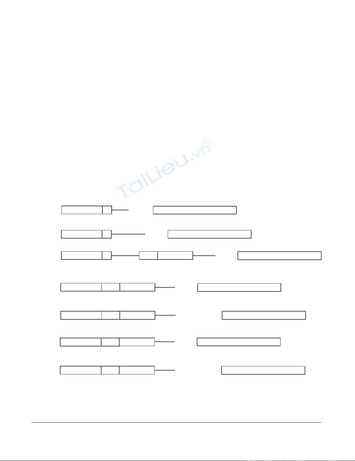
Linearized format of the vectors used in the studyFigure 1
Linearized format of the vectors used in the study. G1: pcDNA3.1/GS-HSA, H1: pcDNA3.1/V5-His+HSA, H2:
pcDNA3.1/V5-His+HSA with Intron A insert from VR1012, W1: gWIZ+HSA, W2: gWIZ+HSA with Kozak, V1: VR1012+HSA
and V2: VR1012+HSA with Kozak. CMV-promoter: Human cytomegalovirus immediate early I promoter/enhancer, T7: T7
promoter priming site, 25–59 bp: Variable number of base pairs in vector backbone, Exon 1: CMV Exon 1, Intron A: CMV
Intron A, HSA gene: Human serum albumin gene. The whole and semi Kozak sequences are shown with capital letters.
-
gcctt
CACC ATG-
T7CMV
-promotor 25 bp
HSA
-gene
-
tggaattcc
CC ATG-
59 bp
HSA
-gene
G1
H1
H2
E
xon
1
-
tggaattcc
CC ATG-
HSA
-gene
I
ntron
A
49 bp 39 bp
T7CMV
-promotor
T7CMV
-promotor
W1
E
xon
1
-
atcgcggccgctt
ATG-
HSA
-gene
I
ntron
ACMV
-promotor
W2
E
xon
1I
ntron
ACMV
-promotor
V1
E
xon
1I
ntron
ACMV
-promotor
V2
E
xon
1
-
atcg
AGCCGCCACC ATG-
HSA
-gene
I
ntron
ACMV
-promotor
29 bp
31 bp
-
atcg
AGCCGCCACC ATG-
HSA
-gene
31 bp
-
atcgcggccgctt
ATG-
HSA
-gene
29 bp

Acta Veterinaria Scandinavica 2008, 50:44 http://www.actavetscand.com/content/50/1/44
Page 3 of 7
(page number not for citation purposes)
with BamHI and HindIII (Fermentas) and ligated into H1
between the promoter and the HSA gene to make vector
H2. Different from the parental vector V there are addi-
tional 111 nucleotides between the CMV promoter and
intron A in vector H2 (Figure 1). The HSA gene, V5
epitope and 6His tag were amplified by PCR from H1
(pcDNA3.1/V5-His+HSA), digested with BamHI and NotI
and ligated into V (VR1012) and gWIZ (W) (Gene Ther-
apy Systems, Inc.) plasmids with or without a typical
Kozak sequence. The translation initiation site of HSA was
modified towards consensus Kozak sequence GCCAC-
CATG when the gene was amplified from H1. The HSA
gene, V5 and His6 tags were amplified using 5'-GGTAT-
GCGGCCGCTTATGAAGTGGGTAACCTTTAT-3' without
Kozak or using 5'-GTATGCGGCCGCCACCATGAAGT-
GGGTAACCTTTAT-3' with Kozak sequence and 5'-
CGCTAGGATCCAATCAATGGTGATGGTGATGATG-3'.
Taq DNA Polymerase (New England BioLabs) was used
for PCR amplifications. The PCR products and DNA
digested with restricted endonucleases were extracted and
purified from agarose gel with QIAEX II kit according to
suppliers protocol (QIAGEN).
Selected clones were grown in LB broth (DIFCO) contain-
ing the appropriate antibiotics. The plasmids were propa-
gated in the DH5α strain of E. coli, harvested and purified
by QIAGEN Plasmid Midi Kits according to the suppliers
protocol (QIAGEN). Verifying the presence of the HSA
gene and the intron A in the plasmids was done with
restriction enzymes; amplified by PCR; and sequenced
with universal and specific primers. The Kozak sequence
was verified by DNA sequencing using BigDye Terminator
v3.1 Cycle Sequencing Kit (Applied Biosystems). The
pBudCE4.1/lacZ/CAT vector was purchased from Invitro-
gen.
2.2. Cell cultures
Primary horse cells were derived from lung and kidney tis-
sue of a horse fetus and skin and duodenum of foals. The
lung, kidney and skin cells were fibroblast like but very
different in morphology and growth rate. The duodenum
cells had endothelium morphology. The lung, kidney,
skin and the African green monkey kidney cells (COS-7)
(ATCC) were propagated in Dulbecco's MEM (DMEM)
(Invitrogen, GIBCO) supplemented with 2 mM
glutamine, 100 IU/ml penicillin, 100 IU/ml streptomycin
and 10% fetal bovine serum (Invitrogen, GIBCO) referred
to as DMEM growth medium. The duodenum was cul-
tured in CS-S medium for endothelial cells (Sigma) sup-
plemented with 2 mM glutamine, 100 IU/ml penicillin,
100 IU/ml streptomycin, 1% endothelial growth factor
(Sigma) and 20% FCS. The primary cells were not used in
higher than 10th passage.
2.3. Transfection
The expression of HSA was tested by transfection of COS-
7 cells using Lipofectamine 2000 (Invitrogen) following
the protocol recommended by the manufacturer. Briefly
the cells were cultured in monolayer to 90–95% conflu-
ency in DMEM growth medium in 12-well plate (NUNC).
Lipofectamine 2000 was diluted 1: 25 in Opti-MEM (Inv-
itrogen, GIBCO) (85 μl) and incubated 5 min at room
temperature (RT). DNA was diluted to 1.35 μg/ml in Opti-
MEM (85 μl) mixed with the Lipofectamine 2000 solu-
tion, incubated 20 min at RT and then added to the cells.
Transfection was performed in culture medium without
antibiotics for 48 hrs (Figure 2). Transfection for 24 hrs
gave similar results (data not shown). Cells treated the
same way with Lipofectamine 2000 but without DNA
served as negative controls. The primary horse cells were
transfected in the same way except two types of plasmids
instead of one were used for transfection. The pBudCE4.1/
lacZ/CAT vector (Invitrogen) was used to control the
transfection. The vectors with the HSA gene, 1,35 μg/ml
and pBudCE4.1/lacZ/CAT 0,6 μg/ml were mixed in 100 μl
Opti-MEM. The vectors were tested at least three times in
the cell lines and for obtaining the results shown in figure
3 the vectors were transfected into all the cells at the same
time point.
2.4. Western blot
The expression of HSA and CAT was monitored in West-
ern blot. SDS-PAGE was done in the Mini-protean II sys-
tem from Bio-Rad according to manufactures instructions.
In short, transfected cells and control cells were boiled
(1:1 vol) in 2× reducing sample buffer and applied to a
denaturing 12% separation gel followed by a transfer to
Immobilon-P membrane (Millipore) using semi-dry Mil-
liBlot Graphite Electroblotter (Millipore). Membranes
were incubated overnight at 4°C with 1:5000 mouse
monoclonal antibody against V5 (Invitrogen) then 1 hr at
RT with goat anti-mouse IgG conjugated to alkaline phos-
phatase (Dako) 1:1000 and nitro blue tetrazolium chlo-
ride and 5-bromo-4-chloro-3-indolyl phosphate (NBT/
BCIP) (Roche) was used to detect bound antibody.
Results
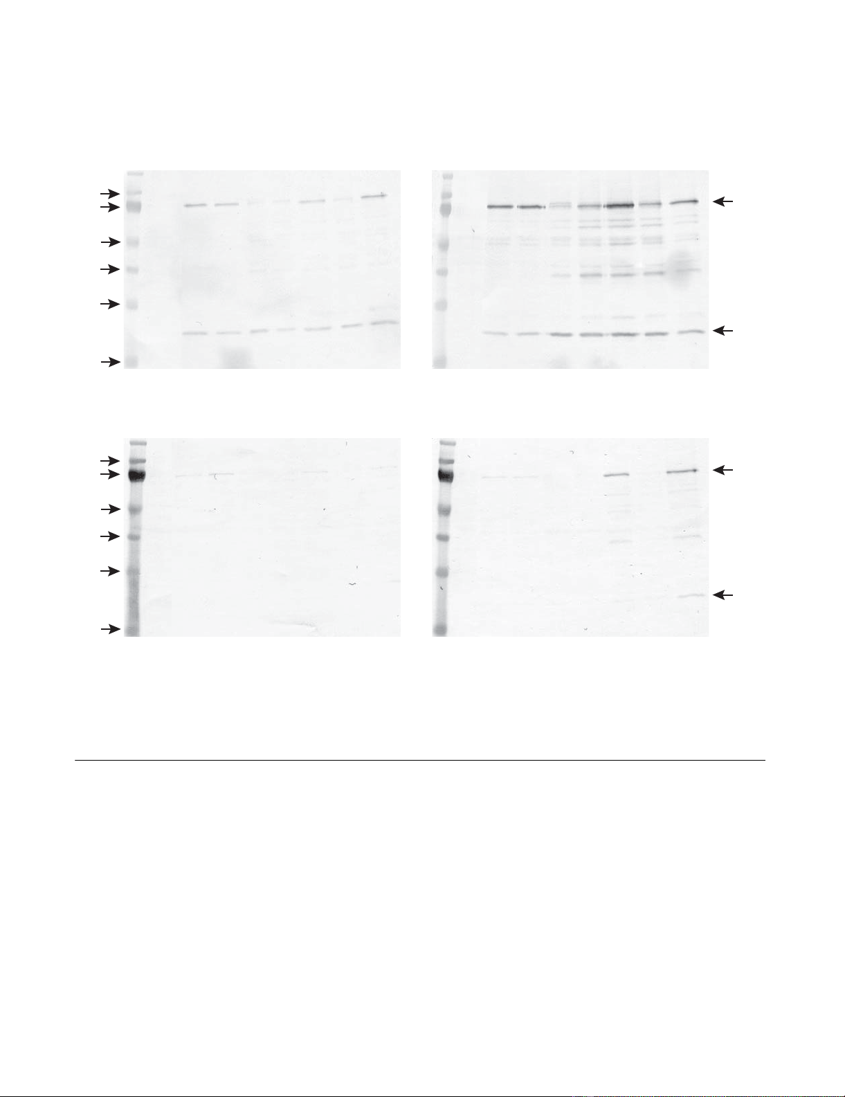
3.1 Effect of Kozak sequence
The translation initiation sites of HSA in the vectors G1
and H1 have semi Kozak sequences, CACCATG and
CCATG, respectively, and are efficiently expressed in COS-
7 (Figure 2) cells and horse lung cells but to a low extent
in horse skin cells and poorly in duodenum and kidney
cells (Figure 3). The wild type translation initiation site of
HSA was replaced by the Kozak consensus sequence,
GCCACCATG, in the two vectors W1 and V1 containing
the intron A to obtain the vectors W2 and V2. In COS-7
cells V2 shows slightly more expression than V1 but the
expression of W2 was diminished as compared to the W1
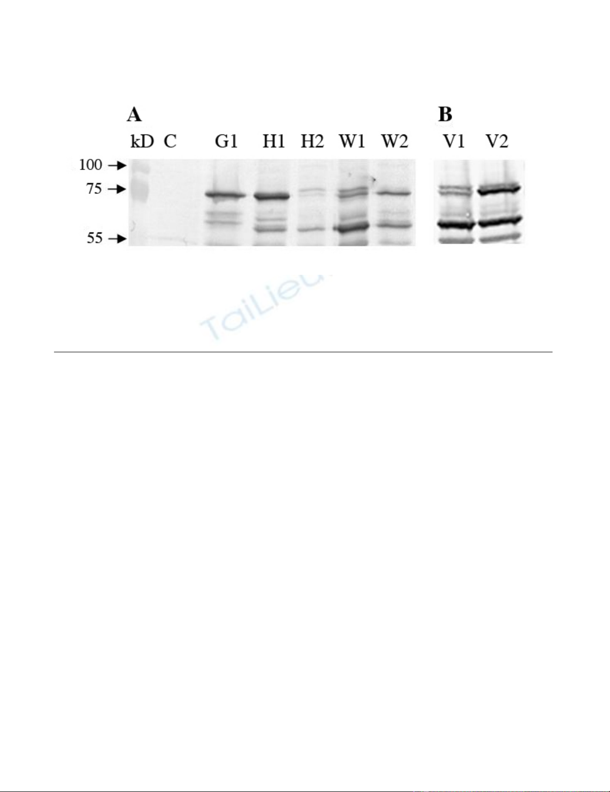
Acta Veterinaria Scandinavica 2008, 50:44 http://www.actavetscand.com/content/50/1/44
Page 4 of 7
(page number not for citation purposes)
parent (Figure 2). W1 and V1 were expressed to a low level
in cells from lung and to a very low level in skin, kidney
and duodenum cells (Figure 3). This was significantly
changed in W2 and V2 as the insertion of the Kozak
sequence increased expression in all four horse cell lines
as compared to the parent vectors W1 and V1. In the skin
and kidney cells the expression from W2 and V2 reached
similar levels to that of G1 and H1 that have a semi Kozak
sequence. In the duodenum cells the expression from
both W2 and V2 exceeded the G1 and H1 expression. In
the lung cells the V2 showed similar level of expression as
the G1 and H1 but W2 slightly higher expression (Figure
3).
3.2 Effect of intron A
The vectors G and H1 that do not contain intron A are
similarly expressed in all the cells. Insertion of intron A
into H1 to make H2 resulted in poorer expression of H2
both in COS-7 cells (Figure 2) and in all four horse cells
as compared to the parental vector H1 (Figure 3). Despite
of containing Intron A the W1 and V1 vectors show less
expression than G and H1 in all the cells. This is presum-
ably because of the lack of a Kozak sequence, as W2 and
V2 vectors that have both intron A and a Kozak sequence
show similar or higher expression than G and H1 in the
horse cells (Figure 3).
The pBudCE4.1/lacZ/CAT plasmid was used as a control
for the transfection. In the skin and lung cells the CAT
expression was similar showing that similar amount of
DNA was transfected and similar amount of cells were
harvested from each well. However, the CAT expression
was hardly or not detected in the kidney and duodenum
cells (Figure 3).
Discussion
Seven different mammalian expression vectors were com-
pared for their ability to drive high levels of HSA protein
expression in four different primary horse cells and COS-
7. Two of the vectors, G1 and H1 with the HSA gene have
been tested for DNA vaccination in horses, and both
induced low immune response [23]. In order to develop
vectors that have a significant expression in horse cells we
investigated the effects of Kozak consensus and intron A
sequences on the levels of expression of the HSA gene.
Sequences flanking the AUG initiation codon within
mRNA have been shown to be important in recognition of
the initial AUG. The consensus sequence surrounding the
start codon is known as the Kozak consensus sequence,
GCCA/GCCAUGG. The G at position +4 and A/G at posi-
tion -3 of the start codon are especially important because
lack of these bases causes reduction in efficiency [22,24].
This translation initiation signal directs the ribosomes to
initiate protein synthesis from mRNAs. It is postulated by
the scanning mechanism of initiation that the 40S ribos-
omal subunits enters at the 5' end of the mRNA and scans
downstream until it comes across the first AUG codon.
Initiation by ribosomes will start at the first AUG codon,
but if there is a weak or no Kozak consensus sequence
some ribosomes bypass and continue to scan downstream
until another AUG start codon has been encountered. This
Expression of HSA gene on different vectors in COS-7 cellsFigure 2
Expression of HSA gene on different vectors in COS-7 cells. COS-7 cells were transfected with HSA vectors using
Lipofectamine 2000, cultured for 48 h, harvested and applied to Western blot. Control (C) cells treated the same way without
DNA. (A) HSA vectors: pcDNA3.1/GS (G1), pcDNA3.1/V5-His (H1), pcDNA3.1/V5-His with intron A (H2), gWIZ (W1) and
gWIZ with Kozak (W2). (B) HSA vectors:VR1012 (V1) and VR1012 with Kozak (V2). The vectors were tested at least three
times.
Acta Veterinaria Scandinavica 2008, 50:44 http://www.actavetscand.com/content/50/1/44
Page 5 of 7
(page number not for citation purposes)
is called leaky scanning [25]. In horses the Kozak
sequence is commonly found as an initiation signal for
gene translation as in other vertebrates [21]. For equine
arteritis virus suboptimal intraleader AUG and not an
optimal Kozak sequence has been shown to be critical for
virus replication [26].
Although the HSA in the vectors G and H1 have only semi
Kozak sequences (bold), TTCACCATGA and AATTC-
CATGA respectively, they are efficiently expressed in COS-
7 (Figure 2) cells and horse lung cells but to a low extent
in skin cells and poorly in duodenum and kidney cells
(Figure 3). The vectors W1 and V1 do not have a Kozak
consensus sequence and were expressed to a low level in
cells from lung and to a very low level in skin, kidney and
duodenum cells (Figure 3).
The Kozak consensus sequence, GCCACCATG, was
inserted into the W1 and V1 vectors that already con-
tained intron A. This significantly changed the expression
of the progeny vectors W2 and V2 in all horse cell lines
(Figure 3). No convincing effect was seen in the COS-7
cells (Figure 2).
Leaky scanning is a likely reason for the bands of lower
molecular weight than 73 kDa HSA band seen in the blots
(Figure 2 and 3) as their sizes match with the positions of
AUG codons downstream in the HSA gene. However,
Expression of HSA gene on different vectors in primary equine skin (a), lung (b), kidney (c) and dudenum (d) cellsFigure 3
Expression of HSA gene on different vectors in primary equine skin (a), lung (b), kidney (c) and dudenum (d)
cells. The cells were transfected simultaneously with HSA vectors and pBudCE4.1/lacZ/CAT control vector using Lipo-
fectamine 2000, cultured for 48 h, harvested and applied to Western blot. Control (C) cells treated the same way without
DNA. HSA vectors: pcDNA3.1/GS (G1), pcDNA3.1/V5-His (H1), pcDNA3.1/V5-His with intron A (H2), gWIZ (W1), gWIZ
with Kozak (W2), VR1012 (V1) and VR1012 with Kozak (V2). The 75 kDa HSA band and the 30 kD CAT band from the
pBudCE4.1/lacZ/CAT plasmid is indicated. The vectors were tested at least 3 times in each cell line.
A: B:
C: D:
kd C G1 H1 H2 W1 W2 V1 V2 kd C G1 H1 H2 W1 W2 V1 V2
kd C G1 H1 H2 W1 W2 V1 V2 kd C G1 H1 H2 W1 W2 V1 V2
skin cells lung cells
kidney cells duodenal cells
100
75
55
40
33
24
100
75
55
40
33
24
HSA
b-gal
HSA
b-gal