
Interaction with calmodulin is important for the secretion
of thimet oligopeptidase following stimulation
Lilian C. Russo
1,2
, Camila N. Gon
˜i
1
, Leandro M. Castro
1
, Amanda F. Asega
3
,
Antonio C. M. Camargo
3
, Cleber A. Trujillo
4
, Henning Ulrich
4
, Marc J. Glucksman
5
,
Cristoforo Scavone
2
and Emer S. Ferro
1
1 Department of Cell Biology and Development, Institute of Biomedical Sciences, University of Sa˜o Paulo, Brazil
2 Department of Pharmacology, Institute of Biomedical Sciences, University of Sa˜o Paulo, Brazil
3 Center for Applied Toxinology, CAT ⁄CEPID, Butantan Institute, Sa˜o Paulo, Brazil
4 Departamento de Bioquı
´mica, Instituto de Quı
´mica, University of Sa˜ o Paulo, Brazil
5 Midwest Proteome Center and Department of Biochemistry and Molecular Biology, Rosalind Franklin University of Medicine and Science/
Chicago Medical School, North Chicago, IL, USA
Keywords
14-3-3e; calmodulin; protein kinase A;
protein–protein interaction; unconventional
secretion
Correspondence
E. S. Ferro, Laborato
´rio de Comunicac¸a˜o
Celular, Departamento de Biologia Celular e
do Desenvolvimento, Instituto de Cie
ˆncias
Biome
´dicas, Universidade de Sa˜o Paulo, Av.
Prof. Lineu Prestes 1524, Sala 431, Sa˜o
Paulo, SP, 05508-900, Brazil
Fax: +55 11 3091 7402
Tel: +55 11 3091 7310
E-mail: eferro@usp.br
(Received 11 May 2009, revised 3 June
2009, accepted 10 June 2009)
doi:10.1111/j.1742-4658.2009.07144.x
Thimet oligopeptidase (EC 3.4.24.15; EP24.15) was originally described as
a neuropeptide-metabolizing enzyme, highly expressed in the brain, kidneys
and neuroendocrine tissue. EP24.15 lacks a typical signal peptide sequence
for entry into the secretory pathway and is secreted by cells via an uncon-
ventional and unknown mechanism. In this study, we identified a novel cal-
cium-dependent interaction between EP24.15 and calmodulin, which is
important for the stimulated, but not constitutive, secretion of EP24.15.
We demonstrated that, in vitro, EP24.15 and calmodulin physically interact
only in the presence of Ca
2+
, with an estimated K
d
value of 0.52 lm. Con-
focal microscopy confirmed that EP24.15 colocalizes with calmodulin in
the cytosol of resting HEK293 cells. This colocalization markedly increases
when cells are treated with either the calcium ionophore A23187 or the
protein kinase A activator forskolin. Overexpression of calmodulin in
HEK293 cells is sufficient to greatly increase the A23187-stimulated secre-
tion of EP24.15, which can be inhibited by the calmodulin inhibitor calmi-
dazolium. The specific inhibition of protein kinase A with KT5720 reduces
the A23187-stimulated secretion of EP24.15 and inhibits the synergistic
effects of forskolin with A23187. Treatment with calmidazolium and
KT5720 nearly abolishes the stimulatory effects of A23187 on EP24.15
secretion. Together, these data suggest that the interaction between
EP24.15 and calmodulin is regulated within cells and is important for the
stimulated secretion of EP24.15 from HEK293 cells.
Structured digital abstract
lMINT-7148420:EP24.15 (uniprotkb:P52888) and Calmodulin (uniprotkb:P62161)bind
(MI:0407)bysurface plasmon resonance (MI:0107)
lMINT-7148437:EP24.15 (uniprotkb:P52888) and Calmodulin (uniprotkb:P62158)colocalize
(MI:0403)bysurface plasmon resonance (MI:0107)
lMINT-7148406:Calmodulin (uniprotkb:P62161)binds (MI:0407)toEP24.15 (uni-
protkb:P52888)bypull down (MI:0096)
Abbreviations
AFU, arbitrary fluorescence units; CaM, calmodulin; CMZ, calmidazolium; EP24.15, thimet oligopeptidase (EC 3.4.24.15); ER-Golgi, rough
endoplasmic reticulum-Golgi apparatus; GST, glutathione S-transferase; HEK293, human embryonic kidney 293 cells; MTT, 3-(4,5-dimethyl-
thiazol-2-yl)-2,5-diphenyl-tetrazolium bromide; PB, phosphate buffer; PKA, protein kinase A; QFS, quenched-fluorescence substrate; TBS,
Tris-buffered saline.
4358 FEBS Journal 276 (2009) 4358–4371 ª2009 The Authors Journal compilation ª2009 FEBS

Introduction
Protein and neuropeptide secretion are crucial biologi-
cal events that provide extracellular access to mole-
cules involved in cell signalling. In the conventional
rough endoplasmic reticulum-Golgi apparatus (ER-
Golgi) secretory pathway, there is dynamic interplay
between cell organelles and their constituents. The ini-
tial step of this pathway is the cotranslational translo-
cation of the protein into the lumen of the ER, and
the pathway culminates with the exocytosis of secre-
tory vesicles containing the molecules. The vast major-
ity of secreted proteins contain a hydrophobic signal
peptide sequence that targets the protein to the secre-
tory pathway [1,2]. However, several proteins that lack
a signal peptide sequence are also secreted, and this
secretion is mediated though the so-called ‘alternative’
or ‘unconventional’ secretory mechanism [3]. The vari-
ous stages involved in protein secretion by this latter
mechanism remain to be identified, and multiple path-
ways for unconventional protein secretion may exist
[3–5].
Several proteins that do not contain signal peptides
are secreted without undergoing early entry into the
ER-Golgi secretory pathway, including neurolysin
[6,7], interleukin-1b[8,9], HIV-tat, Leishmania hydro-
philic acylated surface protein B [10–12], the chroma-
tin-binding proteins HMGB1 and En2, galectin-1,
galectin-3 [13,14], thioredoxin, basic fibroblast growth
factor 1 and 2 [3] and the GRASP proteins [15]. Thi-
met oligopeptidase (EC 3.4.24.15; EP24.15) also lacks
a signal peptide sequence for entry into the secretory
pathway, but has been shown to be secreted from neu-
roendocrine tissues [16–18], as well as from distinct
cell lineages. However, little is known about the
mechanism and major molecular components of the
unconventional secretory pathway.
The secretion of EP24.15 from ATt20 cells, a mouse
pituitary tumour cell line, can be stimulated by
A23187 and corticotrophin-releasing hormone and
blocked by brefeldin A and nocodazole. However,
EP24.15 is not present in the secretory vesicles of
AtT20 cells [5]. Subcellularly, EP24.15 has been shown
to extensively colocalize with syntaxin-6, an integral
trans-Golgi network protein, in the perinuclear region
of AtT20 cells [19]. EP24.15 has also been found to
associate with small vesicular organelles distributed
throughout the cell body, and some, but not all, of
these organelles are also positive for adrenocorticotro-
pic hormone [19,20]. Moreover, ultrastructural experi-
ments using electron microscopy have demonstrated
that EP24.15 is strongly associated with the cytoplas-
mic face of the membranes of neurosecretory elements
in the rat brain, including the ER, Golgi cisternae,
tubulovesicular organelles, synaptic vesicles and endo-
somes [21]. Taken together, these data strongly suggest
that EP24.15 secretion occurs by an unconventional
pathway, but requires components of the classic secre-
tory pathway [4,22].
Many specialized processes (for example, cell signal-
ling, synapse formation and maintenance, neurotrans-
mitter and hormone release, axonal transport and
nerve cell targeting) are tightly regulated by protein–
protein interactions. The interaction of EP24.15 with
the scaffold protein 14-3-3ehas been described previ-
ously to facilitate the secretion of EP24.15 from
human embryonic kidney 293 (HEK293) cells stimu-
lated with forskolin [4]. The interaction of EP24.15
and 14-3-3edramatically increases when EP24.15 is
phosphorylated on Ser644 by protein kinase A (PKA).
Furthermore, EP24.15 secretion induced by the cal-
cium ionophore A23187 can be stimulated in vivo by
overexpressing 14-3-3eor by treating HEK293 cells
with the PKA activator forskolin [4,23]. However, the
molecular mechanisms involved in the alternative
secretion of EP24.15 are not well characterized, and
additional proteins that participate in the unconven-
tional secretory pathway used by EP24.15 remain to
be discovered.
In this study, we demonstrate that EP24.15 and cal-
modulin (CaM) interact both in vitro and in vivo, and
that this interaction is important for the unconven-
tional secretion of EP24.15 following stimulation. Our
results suggest that, in vitro, EP24.15 interacts with
CaM only in the presence of calcium, with an esti-
mated K
d
value of 0.52 lm. The overexpression of
CaM significantly increases the stimulated secretion
of EP24.15 in HEK293 cells, and the colocalization of
CaM and EP24.15 increases when these cells are trea-
ted with either A23187 (1 or 10 lm) or forskolin
(10 lm). These data suggest a novel interaction
between CaM and EP24.15 which has physiological
implications for the unconventional secretion of
EP24.15 following stimulation.
Results
In a yeast two-hybrid screen to identify proteins that
interacted with EP24.15, we found that CaM could
interact with EP24.15 (data not shown). To confirm
these results and further investigate the possible func-
tional relevance of this interaction, we cloned the
L. C. Russo et al. Interaction of calmodulin and EP24.15
FEBS Journal 276 (2009) 4358–4371 ª2009 The Authors Journal compilation ª2009 FEBS 4359
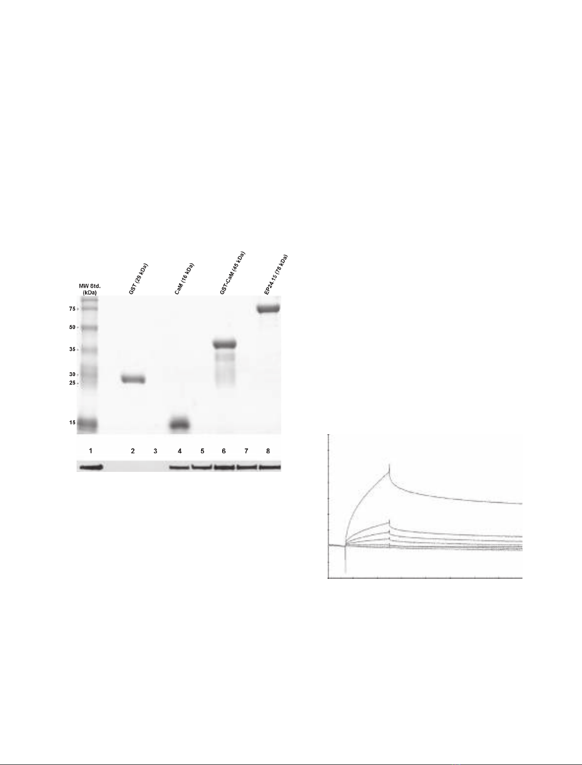
full-length cDNA encoding rat CaM and expressed the
CaM protein in Escherichia coli using the pGEX-4T2
plasmid system (Fig. 1A). CaM was expressed as a
fusion protein with glutathione S-transferase (GST)
(GST-CaM; Fig. 1A), purified, covalently immobilized
onto a glutathione–Sepharose column, as described
previously, and incubated with EP24.15 (10 lg) in
either the absence (Fig. 1B, lane 3) or presence
(Fig. 1B, lanes 4–8) of increasing Ca
2+
concentrations.
After extensive washing to remove nonspecifically
bound proteins, the CaM-associated protein complexes
were eluted by boiling the resin in SDS sample buffer.
A western blot of these complexes demonstrated that
EP24.15 physically interacts with CaM (Fig. 1B). In vitro,
even the lowest Ca
2+
concentration tested (0.12 lm)
was sufficient for the interaction between EP24.15 and
CaM, although these proteins do not associate in the
absence of calcium. Covalently immobilized GST alone
was used as a negative control and, even in the pres-
ence of 1200 lmCa
2+
, EP24.15 did not interact with
control GST (Fig. 1B, lane 2).
The Ca
2+
-dependent interaction between EP24.15
and CaM was also analysed using a surface plasmon
resonance-based biosensor (Biacore T100; GE Health-
care, Uppsala, Sweden), which allows for the real-time
detection and monitoring of molecular binding events
[24,25]. A sensogram from a representative experiment
is shown in Fig. 2. CaM was covalently immobilized
onto a CM5 chip, and association and dissociation
slopes were obtained with the addition of an increasing
concentration of EP24.15. The association slopes
increase proportionally to the EP24.15 concentration
from 0.1 to 5.0 lm. The association between EP24.15
and CaM occurs rapidly (within seconds), whereas the
dissociation occurs more slowly (Fig. 2). Under these
experimental conditions, the interaction between
EP24.15 and CaM was estimated to have a K
d
value
of 0.52 lm.
To investigate the possible in vivo interaction
between EP24.15 and CaM, double-labelling immuno-
cytochemical experiments using confocal microscopy
were conducted in HEK293 cells (Fig. 3). HEK293
cells were transiently transfected with a modified
A
B
Fig. 1. Recombinant protein expression and in vitro protein–protein
interaction assays. (A) SDS–polyacrylamide gel (12%) stained with
Coomassie brilliant blue demonstrating the purity of the proteins
expressed in E. coli. The proteins were purified on a glutathione–
Sepharose column, and free proteins and GST fusion proteins were
eluted from the column using thrombin or glycine, respectively.
The proteins were further purified using size-exclusion centrifuga-
tion. (B) In vitro binding experiments were performed using GST
(5 lg; lane 2) or GST–CaM (5 lg; lanes 3–8) immobilized on a gluta-
thione–Sepharose matrix. The CaM samples were pre-incubated
with 5 mMEGTA. The samples were washed and incubated in
increasing concentrations of calcium (lM): 0 (lane 3), 0.12 (lane 4),
1.2 (lane 5), 12 (lane 6), 120 (lane 7) or 1200 (lane 8). CaM was
incubated with EP24.15 (10 lg) and washed. The associated pro-
teins were eluted by boiling the glutathione–Sepharose matrix in
SDS–PAGE sample buffer. The eluted proteins were separated by
SDS–PAGE, and bound EP24.15 was detected using western blot-
ting with an EP24.15-specific antibody. As a positive control, 0.1 lg
of recombinant EP24.15 was added to the gel (lane 1). The results
are representative of three independent experiments.
Time (s)
5 µM
1 µM
0.5 µM
0.25 µM
0.1 µM
–20
–10
0
10
20
30
40
50
60
70
1000 200 300 400 500 600 700 800
Arbitrary resonance units
Fig. 2. Interaction between CaM and EP24.15 as measured by sur-
face plasmon resonance. CaM was immobilized on a CM5 sensor
chip by amine linkage and increasing concentrations of EP24.15
(0.1, 0.25, 0.5, 1 and 5 lM) were applied in a constant flow. It
should be noted that EP24.15 rapidly interacted with CaM, but dis-
sociation occurred slowly. The K
d
value between these two pro-
teins was 0.52 lM. The results presented are representative of
three independent experiments.
Interaction of calmodulin and EP24.15 L. C. Russo et al.
4360 FEBS Journal 276 (2009) 4358–4371 ª2009 The Authors Journal compilation ª2009 FEBS
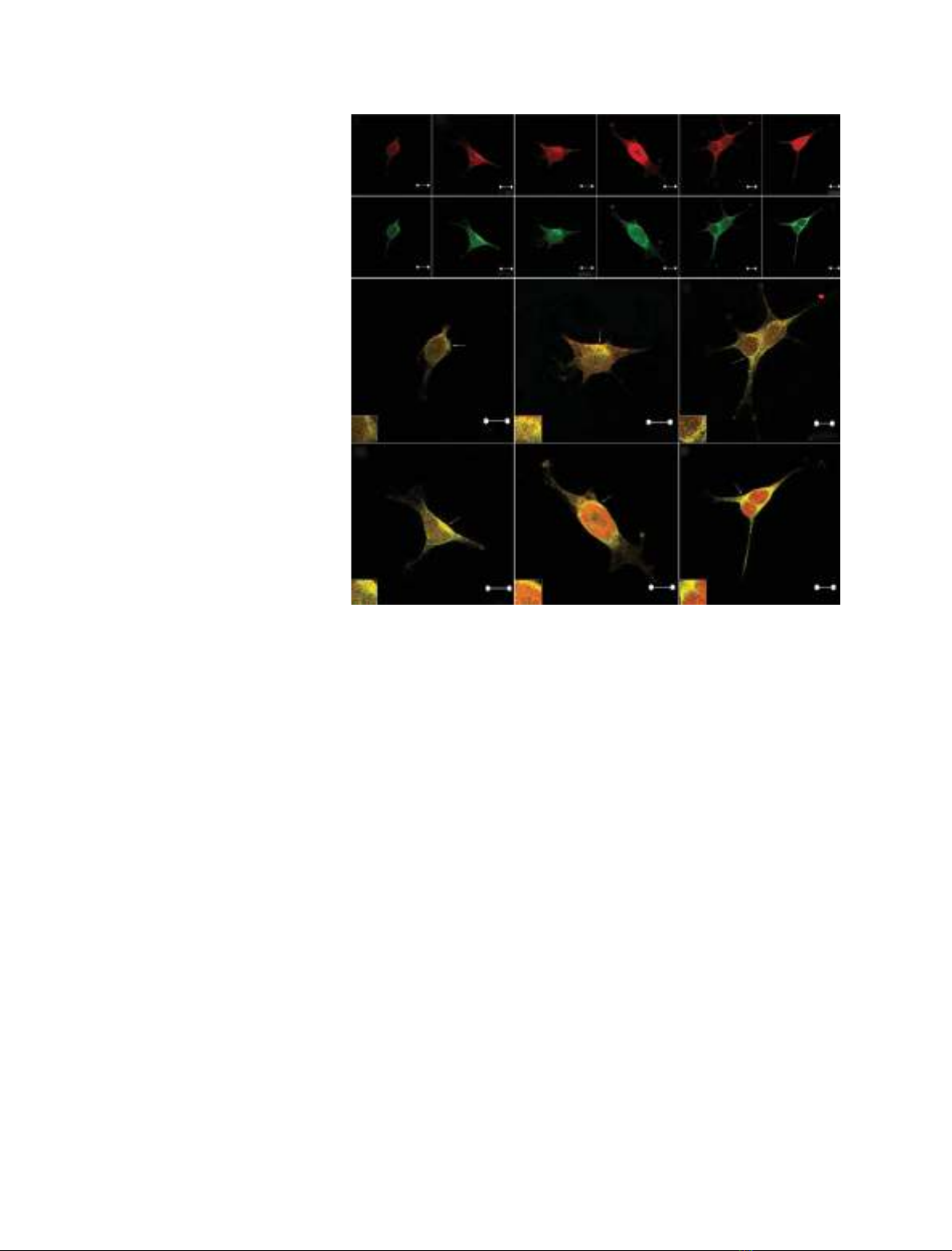
pCMV plasmid that was empty (negative control) or
encoded EP24.15, CaM and ⁄or 14-3-3e. The over-
expression of EP24.15, CaM or 14-3-3ein these cells
was confirmed by western blot (data not shown).
EP24.15 and CaM were distributed throughout the
cells, as analysed by confocal microscopy (Fig. 3).
However, the discrete colocalization of these two pro-
teins could be observed after superimposing the indi-
vidual localization patterns (Fig. 3M,O). Because we
have shown previously that 14-3-3eaffects the uncon-
ventional secretion of EP24.15 [4], we investigated the
effect of the overexpression of 14-3-3ewith both
EP24.15 and CaM on the localization patterns. Fol-
lowing the overexpression of 14-3-3e, the colocalization
of EP24.15 and CaM was more pronounced (Fig. 3Q).
Treatment with the PKA activator forskolin caused an
incremental change in the cellular colocalization of
EP24.15 and CaM (Fig. 3N,P), which was more evi-
dent in cells overexpressing EP24.15, CaM and 14-3-3e
(Fig. 3R). The control experiments, in which the pri-
mary antisera were omitted or pre-absorbed, showed
no specific staining (data not shown). Therefore, these
findings suggest that the EP24.15 and CaM interaction
can be regulated in vivo by PKA activation, particu-
larly in cells that also express 14-3-3e.
CaM is involved in many cellular functions, includ-
ing cell signalling and exocytosis [26]. Therefore, we
were interested in the importance of CaM for the
secretion of EP24.15 in HEK293 cells. We found that
EP24.15 activity and protein expression were not
altered in cells overexpressing CaM or 14-3-3e(data
not shown). Moreover, the constitutive secretion of
EP24.15 was not affected in HEK293 cells overexpress-
ing: (a) CaM, (b) EP24.15, (c) CaM and EP24.15, (d)
CaM and 14-3-3eor (e) CaM, EP24.15 and 14-3-3e
(data not shown). In contrast, CaM overexpression
alone enhanced the A23187-stimulated secretion of
EP24.15 from HEK293 cells (Fig. 4A). Cotreatment of
these cells with both A23187 and the PKA activator
forskolin produced a synergistic effect on EP24.15
secretion (Fig. 4A). As expected, EP24.15 overexpres-
sion in HEK293 cells was sufficient to cause a propor-
tional increase in EP24.15 secretion on A23187
stimulation, which was also potentiated by forskolin
(Fig. 4A). However, the overexpression of both CaM
and EP24.15 in these cells produced no additional
increase in the secretion of EP24.15 following stimula-
tion (Fig. 4), suggesting that the stimulated secretion
of EP24.15 could be regulated by additional proteins.
As mentioned above, 14-3-3ehas previously been
ABCDEF
KJ
I
H
G
MO Q
R
L
P
N
10 µm 10 µm 10 µm 10 µm 10 µm 10 µm
10 µm 10 µm 10 µm 10 µm 10 µm 10 µm
10 µm 10 µm 10 µm
10 µm
10 µm
10 µm
Fig. 3. Colocalization of EP24.15 and CaM
in HEK293 cells. HEK293 cells were
transiently transfected with modified pCMV
vectors. Cells in (A), (B), (G) and (H) were
mock transfected with the empty vector.
Cells in (C), (D), (I) and (J) were cotransfect-
ed with the plasmids expressing EP24.15
and CaM. Cells in (E), (F), (K) and (L) were
cotransfected with the plasmids expressing
EP24.15, CaM and 14-3-3e. The cells in (B),
(D), (F), (H), (J) and (L) were treated with
10 lMforskolin. The transfected cells were
analysed by confocal microscopy. EP24.15
localization is shown in red (A–F), and CaM
localization is shown in green (G–L). (M–R)
Merged images from (A) and (G) (M), (B)
and (H) (N), (C) and (I) (Q), (D) and (J) (P), (E)
and (K) (Q) and (F) and (L) (R). It should be
noted that forskolin treatment causes an
increased colocalization between EP24.15
and CaM (N, P and R), which is more
evident in cells overexpressing EP24.15,
CaM and 14-3-3e(R). These data are repre-
sentative of three independent experiments.
L. C. Russo et al. Interaction of calmodulin and EP24.15
FEBS Journal 276 (2009) 4358–4371 ª2009 The Authors Journal compilation ª2009 FEBS 4361
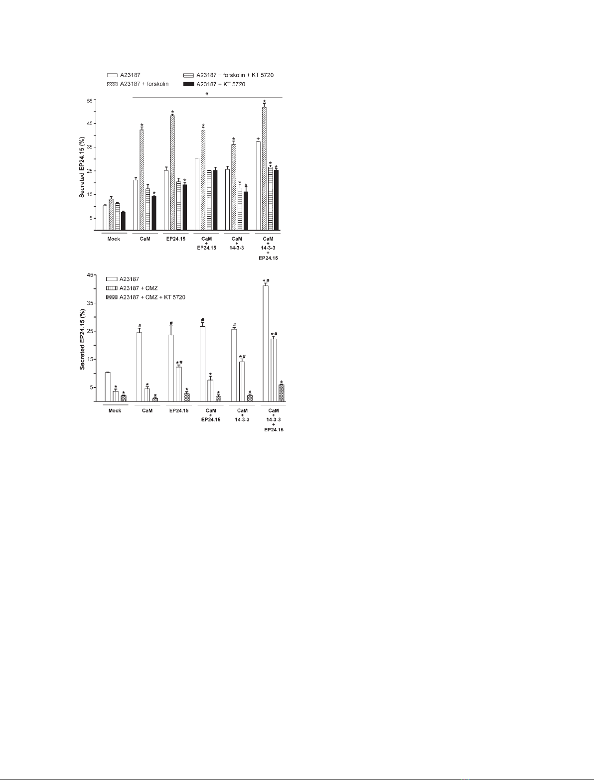
shown to facilitate the secretion of EP24.15 [4]. Indeed,
A231817-stimulated EP24.15 secretion was higher in
HEK293 cells overexpressing CaM, EP24.15 and 14-3-3e
(Fig. 4). However, forskolin showed no further syner-
gistic effects on the A23187-stimulated secretion of
EP24.15, suggesting additional limiting steps in this
unconventional secretory pathway. The synergistic
effects of forskolin and A23187 on EP24.15 secretion
were completely blocked by the specific PKA inhibitor
KT5720, whereas the stimulatory effect of A23187 was
only partially inhibited by this compound (Fig. 4A).
The specific CaM inhibitor calmidazolium (CMZ)
partially blocked the A23187-stimulated secretion of
EP24.15 (Fig. 4B). Interestingly, treatment with both
KT5720 and CMZ essentially abolished the secretion
of EP24.15 following stimulation (Fig. 4B). Impor-
tantly, the cell viability was greater than 99% by both
Trypan blue exclusion and 3-(4,5-dimethylthiazol-2-yl)-
2,5-diphenyl-tetrazolium bromide (MTT) in all experi-
ments, indicating that the EP24.15 secretion measured
in the above experiments was not a result of nonspe-
cific cell leakage (data not shown). Taken together,
these data further suggest that the stimulated secretion
of EP24.15 is a cellular event regulated in part by the
CaM pathway.
Next, we used confocal microscopy to measure the
increase in the intracellular cytosolic calcium concentra-
tion ([Ca
2+
]
i
) in HEK293 cells treated with A23187
(Fig. 5A,B). Although this technique has limitations
(the maximum detectable calcium concentration is
below the maximum level that can be reached within
the cell), we observed an increase in [Ca
2+
]
i
propor-
tional to the A23187 concentration (Fig. 5A,B). In par-
allel, the secretion of EP24.15 following stimulation was
measured at distinct A23187 concentrations (Fig. 5C).
These data indicate that HEK293 cells secrete EP24.15
only when [Ca
2+
]
i
is above 4 lm(Fig. 5C). In addition,
we performed double-labelling immunocytochemical
experiments for confocal microscopy to analyse the
effect of an increase in [Ca
2+
]
i
on the colocalization of
EP24.15 and CaM (Fig. 6). HEK293 cells were tran-
siently transfected with the modified pCMV plasmid
vector expressing EP24.15, CaM and ⁄or 14-3-3e(or an
empty control), and were treated with various concen-
trations of A23187. Interestingly, the intracellular colo-
calization of EP24.15 and CaM increased when the cells
were treated with 1 lmA23187 (Fig. 6O,Q), conditions
that were not sufficient to induce EP24.15 secretion
(Fig. 5C). A large increase in EP24.15 and CaM colo-
calization occurred in cells overexpressing these two
proteins that were treated with 10 lmof A23187
(Fig. 6P), with or without the additional overexpression
of 14-3-3e(Fig. 6R). Therefore, the colocalization of
A
B
Fig. 4. CaM and the secretion of EP24.15 following stimulation. (A,
B) The stimulated secretion of EP24.15 was evaluated in HEK293
cells transfected with plasmid expressing mock (control cells),
CaM, EP24.15, CaM and EP24.15, CaM and 14-3-3e, or CaM,
EP24.15 and 14-3-3e. Following transfection, the cells were equili-
brated for 1 h in serum-free DMEM medium containing 0.1% dialy-
sed BSA. This medium was then replaced with identical fresh
medium that had been previously equilibrated with CO
2
, and the
cells were stimulated with 10 lMA23187. As indicated, the cells
were pre-incubated with 10 lMforskolin, 0.5 lMKT5720 and ⁄or
3lMCMZ for 20 min. These compounds were also present during
A23187 stimulation. The medium was collected, and the EP24.15
activity in the medium was quantified using the fluorogenic sub-
strate QFS. The secreted EP24.15 activity is expressed here as
the percentage of the enzyme secreted based on the total activity
measured in the corresponding cell homogenates. A significant
increase in A23187-stimulated secretion can be observed in all
experimental conditions compared with the mock-transfected cells
(#, P< 0.05). The cells overexpressing CaM, EP24.15 and 14-3-3e,
and stimulated with A23187, secreted more EP24.15 compared
with the other groups (+, P< 0.05). It should be noted that the
PKA activator forskolin significantly increased EP24.15 secretion
(*, P< 0.001). The data are expressed as the mean ± standard
deviation of three independent experiments. The data were analy-
sed by ANOVA with a post hoc Tukey test.
Interaction of calmodulin and EP24.15 L. C. Russo et al.
4362 FEBS Journal 276 (2009) 4358–4371 ª2009 The Authors Journal compilation ª2009 FEBS