
BioMed Central
Page 1 of 13
(page number not for citation purposes)
Virology Journal
Open Access
Research
Occult hepatitis B infection: an evolutionary scenario
Formijn J van Hemert*1, Hans L Zaaijer2, Ben Berkhout1 and
Vladimir V Lukashov1
Address: 1Laboratory of Experimental Virology, Department of Medical Microbiology, Center for Infection and Immunity Amsterdam (CINIMA),
Academic Medical Center, University of Amsterdam, Amsterdam, the Netherlands and 2Laboratory of Clinical Virology, Department of Medical
Microbiology, Center for Infection and Immunity Amsterdam (CINIMA), Academic Medical Center, University of Amsterdam, Amsterdam, the
Netherlands
Email: Formijn J van Hemert* - f.j.vanhemert@amc.uva.nl; Hans L Zaaijer - h.l.zaaijer@amc.uva.nl; Ben Berkhout - b.berkhout@amc.uva.nl;
Vladimir V Lukashov - v.lukashov@amc.uva.nl
* Corresponding author
Abstract
Background: Occult or latent hepatitis B virus (HBV) infection is defined as infection with
detectable HBV DNA and undetectable surface antigen (HBsAg) in patients' blood. The cause of an
overt HBV infection becoming an occult one is unknown. To gain insight into the mechanism of the
development of occult infection, we compared the full-length HBV genome from a blood donor
carrying an occult infection (d4) with global genotype D genomes.
Results: The phylogenetic analysis of polymerase, core and X protein sequences did not
distinguish d4 from other genotype D strains. Yet, d4 surface protein formed the evolutionary
outgroup relative to all other genotype D strains. Its evolutionary branch was the only one where
accumulation of substitutions suggests positive selection (dN/dS = 1.3787). Many of these
substitutiions accumulated specifically in regions encoding the core/surface protein interface, as
revealed in a 3D-modeled protein complex. We identified a novel RNA splicing event (deleting
nucleotides 2986-202) that abolishes surface protein gene expression without affecting polymerase,
core and X-protein related functions. Genotype D strains differ in their ability to perform this
2986-202 splicing. Strains prone to 2986-202 splicing constitute a separate clade in a phylogenetic
tree of genotype D HBVs. A single substitution (G173T) that is associated with clade membership
alters the local RNA secondary structure and is proposed to affect splicing efficiency at the 202
acceptor site.
Conclusion: We propose an evolutionary scenario for occult HBV infection, in which 2986-202
splicing generates intracellular virus particles devoid of surface protein, which subsequently
accumulates mutations due to relaxation of coding constraints. Such viruses are deficient of
autonomous propagation and cannot leave the host cell until it is lysed.
Background
Occult HBV infections are defined as the presence of HBV
DNA and the absence of HBV surface antigen (HBsAg
encoded by the S gene) in plasma or serum of HBV-
infected patients [1]. This infection may persist in individ-
uals for years without emerging symptoms of overt HBV
Published: 11 December 2008
Virology Journal 2008, 5:146 doi:10.1186/1743-422X-5-146
Received: 24 November 2008
Accepted: 11 December 2008
This article is available from: http://www.virologyj.com/content/5/1/146
© 2008 van Hemert et al; licensee BioMed Central Ltd.
This is an Open Access article distributed under the terms of the Creative Commons Attribution License (http://creativecommons.org/licenses/by/2.0),
which permits unrestricted use, distribution, and reproduction in any medium, provided the original work is properly cited.

Virology Journal 2008, 5:146 http://www.virologyj.com/content/5/1/146
Page 2 of 13
(page number not for citation purposes)
infection. Co-infection [2], drug abuse [3] or immuno-
suppression [4] can trigger an enhancement of HBV DNA
levels without an increase of HBsAg. Transmission of HBV
from individuals with occult HBV infection may occur via
organ transplantation or blood transfusion [5]. It is pres-
ently unclear to what extent occult HBV infection repre-
sents a risk factor for the community other than for the
infected individual [6].
In HBV sequences obtained from serum samples of HBsAg
seronegative carriers, a plethora of mutations has been
observed [7-10]. Point mutations, deletions and splicing
alternatives have been associated with occult HBV, but it
is unclear whether these mutations are a cause or a conse-
quence of an occult HBV infection. Many of these occult
infection associated mutations reside in the S gene and/or
regions governing the regulation of S gene expression, but
they have also been documented for the core (C) and
polymerase (P) genes.
Replication-defective mutants of HBV have been detected
in the circulation of symptom-free individuals as early as
1987, and a notable example showed a deletion in to the
pre-S region [11], which mediates cellular receptor bind-
ing [12]. Subsequently, splicing of viral RNA has been
identified as a major cause of HBV genome and particle
heterogeneity [13-16]. Spliced viral mRNA may become
translated into aberrant HBV proteins with unknown
function [17]. The existence of a potential splice site does
not necessarily mean that it is constitutively used. A region
called PRE (Posttranscriptional Regulatory Element) has
been identified in the HBV genome. The PRE facilitates
the export of PRE-containing transcripts from the nucleus
to the cytoplasm [18-20]. Consequently, viral transcripts
reach the cellular translational machinery along two com-
peting pathways: either being promoted by PRE before
splicing occurs or via the regular export route of spliced
cellular mRNAs. More recently, Hass and coworkers
referred to this competitive feature to demonstrate that
integrity of the 458/459 exon/intron transition is required
for the accumulation of pre-S2/S mRNA ([21] see also edi-
torial). Posttranscriptional reduction of surface protein
and mRNA expression to a background level was due to a
single G458A substitution [21] and could also be caused
by deletion of 30 nucleotides immediately downstream of
this site [22].
Recently, we obtained sequence information for HBV
strains present in occult infections [7]. Based on its analy-
sis, we here propose a novel splicing event of HBV RNA
(deleting the nucleotides from 2986 to 202) that abol-
ishes surface protein expression without affecting other
functions encoded in the virus genome (P, C and X). HBV
strains prone to this splicing opportunity constitute a sep-
arate clade in a phylogenetic tree of the genotype D
polymerase sequences. In this clade, a T-to-G mutation at
position 173 truncates a splice-promoting polypyrimi-
dine tract [23] and also affects the local secondary struc-
ture of the viral RNA [24]. As a result, the splicing activity
at the neighboring 202 splice acceptor site may be down-
regulated. The splicing possibility (2986-202) based on
NetGene2 predictions presently awaits further experimen-
tal support by analysis of liver samples, which are much
more complicated to obtain from healthy occult HBV car-
riers than blood samples.
Results
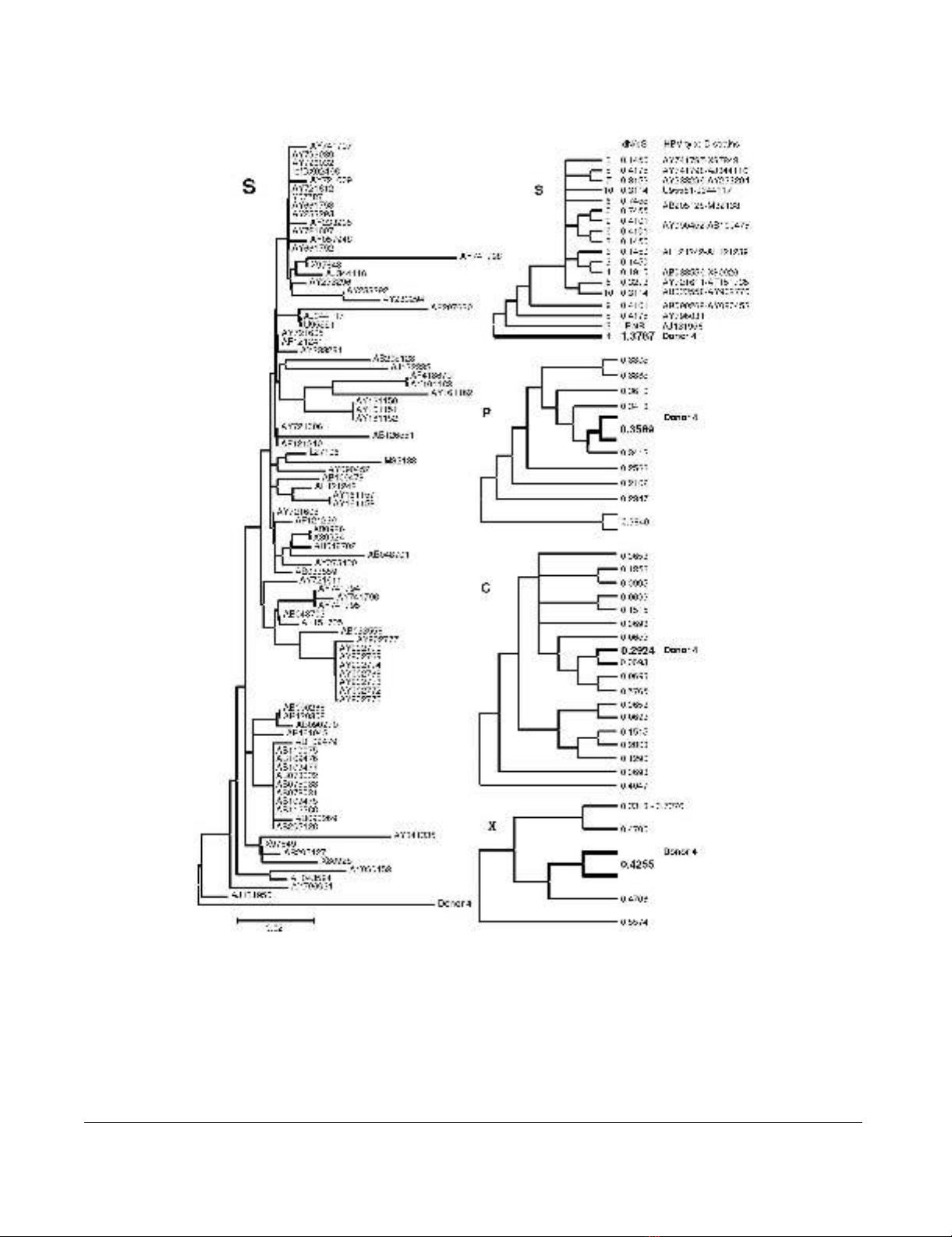
Mutations in occult EU155893 HBV DNA
HBV surface protein of donor 4 with an occult HBV infec-
tion (EU155893, d4) takes the outgroup position in a
bootstrapped phylogenetic tree based on JTT-estimates of
amino acid replacements in genotype D surface proteins
(Fig 1, left panel). The lengths of the branches of the avail-
able surface protein sequences from the other donors with
occult HBV infection (1a, 1b, 2, 3, 5a and 5b) were similar
or even larger than the d4 branch length leading to severe
tree compression and were therefore excluded from the
tree. PAML analysis allowing dN/dS values of clades and
branches to exceed the value of 1 generated a dN/dS value
of 1.3787 for the branch of d4 surface protein gene,
almost a fourfold of the average value of 0.3579 ± 0.1831
(range 0.1450–0.7455) of the other clades and branches
(Fig 1, right panel, S). A likelihood ratio comparison with
a similar analysis limiting dN/dS values to maximally 1
provided statistical support (p < 0.001). In the other HBV
genes, the dN/dS values of d4 DNA were close to the aver-
age values (Fig 1, right panel, P, C and X) – P: 0.3162 ±
0.0656 (range 0.2102–0.3840), C: 0.2180 ± 0.1733
(range 0.0653–0.5765) and X: 0.5136 ± 0.1490 (range
0.3318–0.7376). These data indicate the presence of pos-
itive selection or relaxed selective constraints as a charac-
teristic property of the surface protein gene in this case of
occult infection. During evolution from an overt to the
present occult infection, the surface protein gene of d4
HBV accumulated non-synonymous and synonymous
nucleotide substitutions to approximately equal propor-
tions.
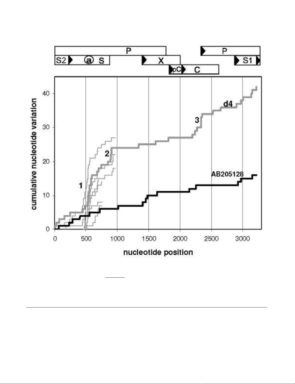
The HBV genome of d4 contains 42 unique nucleotide
substitutions that are not observed in a collection of 89
genotype D HBV species (DQ series [8] were not included,
see below). In control strain AB205128 from a patient
with overt HBV infection, only 16 characteristic mutations
had accumulated in the genome. In order to pinpoint
clusters of d4-specific substitution, we awarded each of
these mutations a value of 1 and plotted the mutational
hits cumulatively along the genome (Fig 2). Steep
increases of the plot indicate regions of enhanced diver-
gence, which is prominent in d4 HBV DNA at the a-deter-
minant region (10/42 substitutions), the oligonucleotide
Virology Journal 2008, 5:146 http://www.virologyj.com/content/5/1/146
Page 3 of 13
(page number not for citation purposes)
HBV strain phylogenyFigure 1
HBV strain phylogeny. A bootstrap consensus tree based on JTT-estimates of amino acid replacements in surface proteins
of HBV genotype D displays the surface protein of donor 4 carrying an occult infection in the outgroup position (left panel).
The scale bar indicates 2% of evolutionary divergence. For phylogenetic analysis by maximum likelihood, the HBV type D
strains were grouped according to their topological position, approximately and provided with labels as indicated next to the
branches of the compressed topology tree (right panel, S). The corresponding values obtained for dN/dS are in between of the
labels and strains columns; PatB means ''parameter at boundary''. Data on donor 4 are in bold-face. The three panels marked
by P(olymerase), C(ore) and X were constructed in a similar fashion, but without mentioning GenBank IDs and clade/branch
labels. In case of P and X, the donor 4 species was combined with its nearest neighbor in order to avoid deviation due to insuf-
ficient branch length.
Virology Journal 2008, 5:146 http://www.virologyj.com/content/5/1/146
Page 4 of 13
(page number not for citation purposes)
895–909 (4/42) and the central part of the core protein
(5/42). As far as sequences are available, accumulation of
nucleotide substitutions specifically at the a-determinant
region is also prominent in strains from other donors with
occult HBV infection (Fig 2, thin lines: 1a, 1b, 2, 3, 5a and
5b). Conservation prevails in X protein, the N-terminal
part of S and in the remaining parts of core and polymer-
ase. S1, S2, and C-terminal parts of S display an interme-
diate degree of variation. In the control strain AB205128,
local accumulation of mutations can hardly be observed
and slopes are similar to those of HBV d4 DNA in the con-
served regions. Enhanced mutational rates at sites are usu-
ally associated with a relaxation of functional constraints
of the regions involved and may indicate a contribution of
Mutational scan along the HBV genomeFigure 2
Mutational scan along the HBV genome. Nucleotide substitutions uniquely present in EU155893 HBV DNA (d4, thick
grey line, occult infection) and in control AB205128 HBV DNA (thick black line, overt infection) are compared with 89 HBV
DNAs of genotype D and plotted cumulatively along the HBV genome. Steep slopes at the a-determinant (1), the oligonucle-
otide 895–909 (2) and the central part of C (3) indicate the relatively high divergence of these regions in d4 HBV. Thin grey
lines represent characteristic mutations in the available HBV sequences from blood samples of the other donors with occult
HBV infection. Numbering starts from the conventional EcoR1 site between S1 and S2. A map of HBV genome organization is
provided on top of the figure.
Virology Journal 2008, 5:146 http://www.virologyj.com/content/5/1/146
Page 5 of 13
(page number not for citation purposes)
these regions to the evolutionary transition from an overt
into an occult HBV infection. A diminished interaction
between core and surface proteins due to the mutations
introduced at the regions 1 and 3 of HBV d4 DNA (Fig 2)
may provide a substantiation of this process, rendering
the transition irreversible.
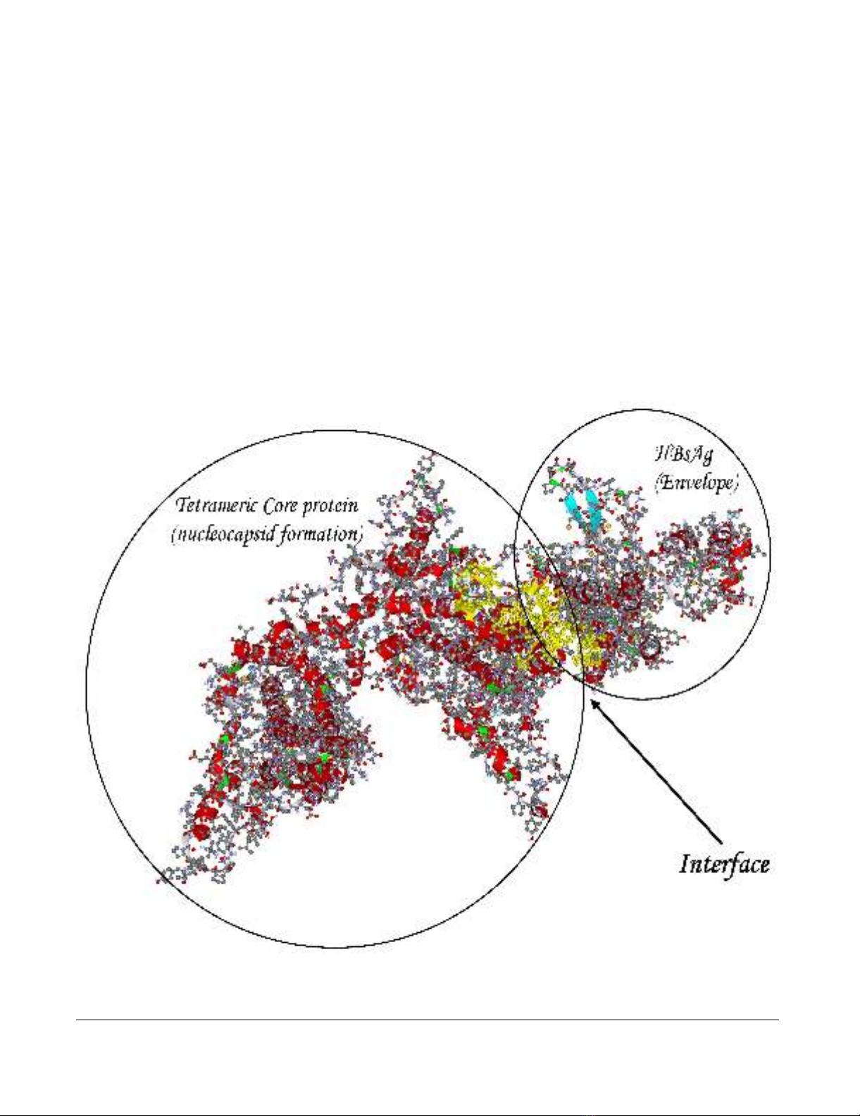
We have previously studied the amino acid composition
of interfaces between 3D-structured domains or proteins
of HBV [25] by means of computational alanine replace-
ment scanning [26]. The docking procedure [27] of mon-
omeric HBsAg with tetrameric core protein (PBD entry
1qgt) followed by ALASCAN-directed selection among the
alternative structures resulted in the complex with a yel-
low-colored interface region as shown in Fig 3. A PDB for-
matted data file carrying the coordinates of the complex is
provided online as Additional File 1. The corresponding
output of the ALASCAN server shows that the central part
of core protein (amino acid residues 67–96), the N-termi-
nal half of the a-determinant region (96–122) and the C-
terminal part of surface protein (169–195) participate in
the interface between core and surface proteins (Table 1)
in order to promote the formation of an infectious virus
particle. In d4 DNA, these regions display the d4-charac-
teristic feature of enhanced sequence divergence. Not all
of these nucleotide substitutions translate into amino acid
replacements. Replacements typical for d4 HBV are G74V,
I80A and Y100C in core and P111S, T123P, T125I, L175S
and M197T in surface protein, respectively. These results
indicate the evolutionary loss of the ability for S/C inter-
face formation during the development from a "wild
type" genotype D ancestor to the occult d4 phenotype. It
Model of the core/surface protein interactionFigure 3
Model of the core/surface protein interaction. A 3D-modeled complex of tetrameric core protein with HBsAg mono-
mer shows the yellow-colored amino acid residues comprising the interface between the two proteins.


![Báo cáo seminar chuyên ngành Công nghệ hóa học và thực phẩm [Mới nhất]](https://cdn.tailieu.vn/images/document/thumbnail/2025/20250711/hienkelvinzoi@gmail.com/135x160/47051752458701.jpg)


