
RESEARC H Open Access
The efficacy of preoperative PET/CT for prediction
of curability in surgery for locally advanced
gastric carcinoma
Hoon Hur
1
, Sung Hoon Kim
2
, Wook Kim
3
, Kyo Young Song
3
, Cho Hyun Park
3
, Hae Myung Jeon
3*
Abstract
Background: The benefits of preoperative
18
FDG-PET/CT for gastric cancer remain uncertain. The aim of this study
was to investigate the effects of preoperative
18
FDG-PET/CT on the surgical strategy for locally advanced gastric
cancer retrospectively.
Methods: From January 2007 to November 2008,
18
FDG-PET/CT was performed in 142 patients who had been
diagnosed with advanced gastric cancer by computed tomography or gastrofiberscope findings.
Results: Detection rates were 88.7% (126/142) for primary tumors and 24.6% (35/142) for local lymph nodes (LN).
Nine patients with metastatic lesions underwent induction chemotherapy without operation. Of 133 patients
subjected to operation, positive FDG uptake in primary tumors (p= 0.047) and local lymph nodes (p< 0.001) was
related to non-curable operations. The mean standard uptake value (SUV) of primary tumors of patients who
underwent non-curable operations was significantly higher than that of patients with curable operations (p= 0.001).
When the SUV was greater than 5 and FDG uptake of LN was positive, non-curable operations were predicted with a
sensitivity of 35.2%, a specificity of 91.0% and an accuracy of 76.7%.
Conclusions: High SUV of the primary tumor and positive FDG uptake in local lymph nodes at PET/CT could
predict non-curative resection in locally advanced gastric cancer. Therefore, information from preoperative PET/CT
can help physician decisions regarding other modalities without laparotomy.
Background
Preoperative imaging studies are used to evaluate clini-
cal and surgical factors of malignant tumors, including
resectability and identification of metastatic lesions that
contraindicate resection. Although the presence of loco-
regional disease in imaging studies will direct the surgi-
cal oncologist toward exploration with the intention of
complete resection, the ability of these studies to
exclude non-curability in surgery remains controversial.
In gastric cancer, the primary aim of surgery is curabil-
ity, i.e., elimination of macroscopic and microscopic rem-
nants of the malignant tumor by resection of the
stomach and proper lymphadenectomy [1]. Since non-
curative treatment is a definite poor prognostic factor for
patients who undergo surgery for gastric cancer [2,3],
other modalities may be needed in order to increase their
survival. However, it is not easy to preoperatively diag-
nose non-curability by conventional non-invasive ima-
ging methods such as computed tomography (CT),
endoscopic ultrasound (EUS) and magnetic resonance
imaging (MRI) without laparotomy or laparoscopic sta-
ging under general anesthesia.
Positron emission tomography (PET) imaging using the
radiolabeled glucose analog
18
fluorodeoxyglucose (FDG)
can present biologic images according to glucose meta-
bolism. PET imaging can be combined with anatomic
imaging such as conventional CT scanning in order to
increase diagnostic accuracy [4]. Although the National
Comprehensive Cancer Network (NCCN) recently
announced that preoperative PET/CT for gastric cancer
patients can be recommended as an option of preopera-
tive staging [5], the benefits of PET/CT remain uncertain.
Therefore, we analyzed information from preoperative
PET/CT for patients with locally advanced gastric
* Correspondence: hmjeon@catholic.ac.kr
3
Department of Surgery, The Catholic University of Korea, College of
Medicine, Seoul, Korea
Full list of author information is available at the end of the article
Hur et al.World Journal of Surgical Oncology 2010, 8:86
http://www.wjso.com/content/8/1/86 WORLD JOURNAL OF
SURGICAL ONCOLOGY
© 2010 Hur et al; licensee BioMed Central Ltd. This is an Open Access article distributed under the terms of the Creative Commons
Attribution License (http://creativecommons.org/licenses/by/2.0), which permits unrestricted use, distribution, and reproduction in
any medium, provided the original work is properly cited.

cancer and compared it with the surgical results, retro-
spectively. Uptake of FDG in the primary tumor or local
lymph node and the standardized uptake value (SUV)
were investigated for their potential in preoperative pre-
diction of non-curative surgery. Thus, the aim of this
study was to investigate the effects of preoperative PET/
CT on the surgical strategy in gastric cancer patients.
Methods
Patient selection and study
From January 2007 to November 2008, our institution
performed whole body
18
F-FDG PET/CT scans for 142
consecutive patients about three days before surgery.
These patients had been pathologically diagnosed with
gastric adenocarcinoma by endoscopic biopsy and sus-
pected of having advanced gastric cancer by endoscopic
findings or conventional enhanced CT scans. They
underwent careful physical examinations and other ima-
ging studies such as bone scans and chest radiography in
order to exclude distant metastasis. We obtained written
informed consent from the patients for preoperative
PET/CT, and then collected their preoperative staging
data and surgical results for this retrospective study.
PET/CT imaging
Before PET/CT scanning, all patients fasted for at least
6 hours. Patients were confirmed to have blood sugar
levels below 130 mg/mL and rested for approximately
45 minutes before receiving an intravenous injection of
440 MBq of 18F-FDG. Scanning began 60 minutes later.
A combined PET/CT in-line system (Biograph LSD,
Siemens, Knoxville, TN) was used for all data collection.
CT scanning was performed from the orbitomeatal line
to the upper thigh (30 mA; 130 kV; 5 mm-thick sec-
tions) prior to PET. PET was then immediately con-
ducted over the same body region with 6-8 bed
positions, with 2 min acquisition time per bed position.
Interpretation of PET/CT
PET/CT images were reviewed at a workstation with
fusion software (Syngo, Siemens, Knoxville, TN) by a
nuclear medicine physician who was given information
about the clinical findings in the patient. The images
were analyzed for the site and amount of positive FDG
uptake; FDG uptake was defined as qualitatively positive
when focal uptake was higher than normal background
FDG activity in the primary tumor, local lymph node
and metastatic lesions. FDG uptake in the bowel was
regarded as positive when there was wall thickening of
the same bowel at CT scan. The FDG uptake activity
within each lesion was corrected by the administered
dose and the patient weight to produce a maximum
standardized uptake value (SUV). For this study, we
only evaluated the SUV to primary tumors.
Conventional CT scan
Conventional abdominal enhanced CT scanning (Light-
Speed VCT, GE Healthcare, Milwaukee, WI) was per-
formed after intravenous administration of contrast
agents, with 5- to 10-mm slice thickness from the dia-
phragm to the symphysis pubis. The image was also
reviewed by a radiologist who was provided with patient
information. Non-curable operation was defined on CT
scans when suspicious findings met the criterion of
metastatic or non-resectable primary tumors in the sur-
gical strategy.
Treatment Plan
In our institution, we have the following treatment strat-
egy for gastric cancer: patients who have metastatic
lesions in either PET/CT or CT are started on induction
chemotherapy with or without pathologic confirmation.
Metastatic lesions of gastric cancer include liver and ret-
roperitoneal lymph nodes or seeding into the perito-
neum. A non-resectable primary tumor is indicated by
pancreatic or duodenal invasion requiring pancreatico-
duodenectomy, or invasion into the root of the meso-
colon. Cases with only one modality of PET/CT and CT
showing metastatic or non-resectable primary tumors
undergo additional imaging studies such as magnetic
resonance image (MRI) and ultrasound (US). Patients
with suspicious metastatic lesions in the imaging study
are subjected to surgical staging.
Surgery
If the patient had suspicious metastatic lesions or a non-
resectable primary tumor in the imaging studies, we first
performed a minilaparotomy in order to confirm metas-
tasis or the possibility of resectability. The abdominal
incision was extended in cases with resectability in the
surgical findings, and then surgery was performed by
conventional open gastrectomy with over D1 plus beta
lymphadenectomy with the intention of curability. Non-
curable operation was defined when we performed open
and close bypass surgery without tumor resection due
to metastatic lesions in other organs, the peritoneum
and retroperitoneal lymph, or when non-resectable pri-
mary tumors were found during surgery. In addition,
palliative resection of primary tumors in which micro-
scopic (R1) or macroscopic (R2) tumors remained was
also included in the category of non-curative operation.
Statistical analysis
Statistical analysis was performed with the statistical pack-
age for social sciences (SPSS) version 13.0. A Chi-square
test was performed in order to evaluate differences of
FDG uptake rates in primary tumors or local lymph nodes
according to the clinico-pathological factors. The SUVs of
curable and non-curable operations were compared by an
Hur et al.World Journal of Surgical Oncology 2010, 8:86
http://www.wjso.com/content/8/1/86
Page 2 of 7

independent t-test. The extent to which the SUV differed
between a curable and non-curable operation was assessed
using receiver operator characteristics (ROC) plots. We
plotted ROC curves for SUV to predict non-curable opera-
tion, and then calculated sensitivity, specificity, accuracy
and the positive predictive value at different SUV cutoffs
(5, 7 and 9) as well as positive uptake of local lymph
nodes.
Results
In 142 enrolled patients, the FDG uptake rate of primary
tumors was 88.7% (126/142) and that of local lymph
nodes was 24.6% (32/142). The mean SUV of primary
cancers was 5.7 (range, 1.89-19.06). In 2 patients, other
simultaneous malignancies (thyroid cancer and rectal
cancer) that the other imaging study could not detect
were incidentally found. We performed combined
operations for those patients.
Nine patients who had metastatic lesions or non-
resectable primary tumors in either PET/CT or con-
ventional CT scan were not operated on. The PET/CT
findings of these patients are listed in Table 1, showing
that all patients had positive FDG uptake in the pri-
mary tumor. We performed operations on the remain-
ing 133 patients and then evaluated the possibility of
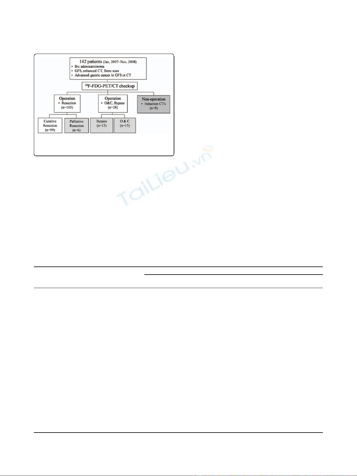
curative surgery (Fig 1).
The clinico-pathological characteristics of the 133
patients who underwent surgery are presented in Table
2. The rates of FDG uptake in the primary tumor and
local lymph nodes were compared according to age,
gender, diabetic mellitus, tumor size, tumor location,
histology and curability of operations. Except for non-
curative operation (97.1% vs. 84.8%, p= 0.047), no fac-
tors were significantly correlated with the FDG uptake
rate in the primary tumor. Patients with large tumor
sizes showed relatively high uptake rates in the primary
tumor (92.6% vs. 83.1%, p= 0.090). The FDG uptake
rate of local lymph nodes was significantly higher in
patients who underwent non-curative operations (44.1%
vs. 14.1%, p< 0.001).
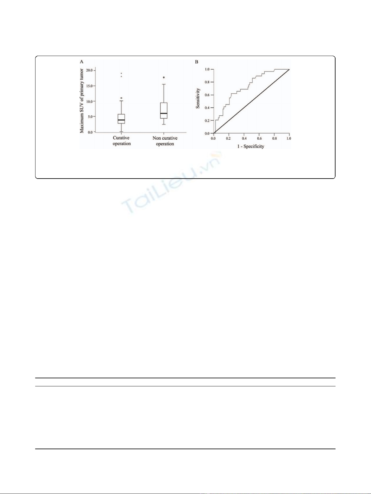
The mean maximum SUV of primary tumors in
patients with non-curative operations was 7.3 ± 4.5
(mean ± S.D.) and that of patients with curative opera-
tions was 4.4 ± 3.5 (mean ± S.D.). The difference in
SUV between the two groups was significant (p =
0.001), and a box plot of the SUVs in both groups is
presented in Fig 2A.
An ROC curve of the maximum SUV was plotted in
order to predict non-curative operations, and an area
under the curve of 0.730 (p< 0.001; 0.629 < 95% C.I. <
0.831) was obtained (Fig 2B). We calculated diagnostic
indices (sensitivity, specificity, accuracy and positive pre-
dictive value) at various SUV cutoffs for primary tumor
and lymph node FDG uptake, and then compared these
results with predictions from conventional enhanced CT
scanning. When the maximum SUV was greater than 5
and the FDG uptake of lymph node was positive, non-
curative operation was predicted with a sensitivity of
35.2%, a specificity of 91.0%, an accuracy of 76.7% and a
positive predictive value of 57.1%. These values are
higher than those obtained using other SUV cutoffs for
primary tumors or even with conventional enhanced CT
scanning (sensitivity of 17.6%, specificity of 87.9%, accu-
racy of 69.9% and a positive predictive value of 33.3%)
(Table 3).
Discussion
For patients with locally advanced gastric cancer, the
preoperative prediction of curability is important
because it can prevent unnecessary laparotomies and
Table 1 Study results of patients who underwent induction chemotherapy without operation
No CT finding PET/CT finding Additional study
Primary
SUV
Local LN
SUV
Other uptake
1 Lung metastasis 2.97 2.97 Lung, Bone Spine MRI
2 Peritoneal seeding
Liver metastasis
6.81 5.44 Mesentery
3 Peritoneal seeding
Esophagus invasion
7.89 0 Distal Esophagus
4 Peritoneal seeding 3.91 3.34 Peritoneum
5 Peritoneal seeding 3.73 8.15 Retroperitoneal LN
Lt. supraclaviclar LN
6 Liver metastasis 10.73 0 Liver Sono, Liver MRI
7 Liver metastasis 7.26 12.18 Liver Sono
8 Peritoneal seeding 2.4 0 T-colon, Omentum,
Retroperitoneal LN
9 Liver metastasis 11.9 0 Liver
PET, positron emission tomography, CT, computed tomography, LN, lymph nodes, SUV, standardized uptake value, MRI, magnetic resonance imaging.
Hur et al.World Journal of Surgical Oncology 2010, 8:86
http://www.wjso.com/content/8/1/86
Page 3 of 7
direct physicians toward treatment with other modalities
such as neoadjuvant chemotherapy. Conventional
enhanced CT scans are one of the most important ima-
ging methods for preoperative prediction of curability.
Therefore, patients diagnosed with definite metastatic
lesions (cM1) by CT scan might be treated systemically
without surgery. However, the treatment strategy for
patients with locally advanced gastric cancer and with-
out definite cM1 lesions has often been decided based
on surgical findings following laparotomy or laparo-
scopic staging [6]. Our results in patients with locally
advanced gastric cancer show that preoperative
18
F-FDG
PET/CT could provide objective information for deci-
sions regarding treatment strategies such as laparoscopic
staging and neoadjuvant chemotherapy.
At present, several studies have reported that FDG-
PET is the most sensitive non-invasive imaging strategy
for detecting distant metastasis [7,8]. Therefore, our
study was also designed that patients with suspected
metastatic lesions on CT scanning accompanied by FDG
uptake were started on induction chemotherapy without
operation. Previous studies reported that FDG-PET, and
not PET/CT, was more sensitive than CT scanning for
detecting primary tumors in advanced disease, but infer-
ior to CT for detecting intra-abdominal lymph node
metastasis [8,9]. In addition, recent studies showed that
FDG-PET had lower sensitivity for detection of lymph
nodes metastasis, and even had no definite role as preo-
perative imaging in gastric cancer [10,11]. Moreover,
studies validating the use of PET/CT in gastric carci-
noma are lacking thus far, and most physicians cannot
confirm whether adding CT information to FDG-PET
will improve diagnostic accuracy. Due to these reasons,
the current aims of preoperative PET/CT in most cen-
ters that perform operations for gastric cancer patients,
including our institution, are as follows: 1) to confirm
metastasis by contrast-enhanced CT scan; 2) to investi-
gate metastatic lesions that are not detected by contrast-
Figure 1 Treatment strategies for patients diagnosed with
gastric adenocarcinoma. GFS = gastrofiberscopy, CT = computed
tomography, O&C = open and closure, CTx = chemotherapy.
Table 2 Preoperative and operative findings of PET/CT in patients who underwent operation (n = 133)
n FDG uptake in primary tumor FDG uptake in local LN
Yes (%)
(n = 117)
No (%)
(n = 16)
p-value Yes (%)
(n = 29)
No (%)
(n = 104)
p-value
Age(years) <60 53 46(86.8) 7(12.1) 0.734 11(20.8) 42(79.2) 0.811
≥60 80 71(88.8) 9(10.7) 18(22.5) 62(77.5)
Gender Male 92 82(89.1) 10(10.9) 0.570 22(23.9) 70(76.1) 0.378
Female 41 35(85.4) 6(14.6) 7(17.1) 34(82.9)
DM Positive 18 16(88.9) 2(11.1) 1.000 5(27.8) 13(72.2) 0.543
Negative 115 101(87.8) 14(12.2) 24(20.9) 91(79.1)
Size(cm) <5 65 54(83.1) 11(16.9) 0.090 10(15.4) 55(84.6) 0.080
≥5 68 63(92.6) 5(7.4) 19(27.9) 49(72.1)
Location Upper 22 20(90.9) 2(9.1) 1.000 5(22.7) 17(77.3) 0.909
Middle and lower 111 97(87.4) 14(12.6) 24(21.6) 87(78.4)
Histology Tubular carcinoma 108 95(88.0) 13(12.0) 1.000 25(23.1) 83(76.9) 0.435
Signet ring/mucinous 25 22(88.0) 3(12.0) 4(16.0) 21(84.0)
Curability Curative operation 99 84(84.8) 15(15.2) 0.047 14(14.1) 85(85.9) <0.001
Non-curative operation 34 33(97.1) 1(2.9) 15(44.1) 19(55.9)
PET, positron emission tomography, CT, computed tomography, FDG, fluorodeoxyglucose, LN, lymph nodes, DM, diabetes mellitus.
Hur et al.World Journal of Surgical Oncology 2010, 8:86
http://www.wjso.com/content/8/1/86
Page 4 of 7
enhanced CT scan; 3) to evaluate other hidden simulta-
neous malignancies that are asymptomatic and unde-
tectable by CT scanning. Contrary to above usage of
PET-CT in gastric cancer, we focused on the prediction
of surgical finding through the result of preoperative
PET-CT. The results of our study suggested that treat-
ment strategy of gastric cancer could be decided accord-
ing to finding of FDG-PET CT.
With respect to preoperative PET/CT as a tool for sur-
gical strategy decisions, the present study uncovered sev-
eral relevant results. Using the semi-quantitative feature
of FDG-PET/CT, the degree of FDG uptake of the pri-
mary tumor and the SUV was analyzed for prediction of
curability. The mean SUV of the primary tumor in
patients who underwent non-curative surgery was signifi-
cantly higher than that of patients with curative surgery.
Therefore, the SUV of the primary tumor might be a pre-
dictive factor for non-curative surgery; this is supported
by the results of the ROC curve. When we defined a
mean primary tumor SUV of greater than 5.0 and posi-
tive uptake of FDG in perigastric lymph nodes as cutoff
values for prediction of non-curative resection, the sensi-
tivity, specificity and accuracy were higher than those of
enhanced CT scanning. Therefore, we find that FDG-
PET/CT may be a tool for decisions concerning laparo-
scopic staging or neoadjuvant chemotherapy.
SUV values are common indices of tracer uptake in
studies with PET, and can be calculated from the radio-
activity of tumors following injection of fluorine
18
F-FDG
according to body weight and physical decay [12]. The
possibility of applying the SUV to preoperative PET/CT
as a predictor for curability is explained by the following.
TheSUVmayrepresentthegrowthrateofmalignant
tumors. Several reports have described that glucose utili-
zation is higher in rapidly growing tumors than in less
aggressive neoplasia [13,14]. In our study, the mean SUV
was correlated with curability of advanced gastric cancer.
Diagnostic laparoscopy for the staging of gastric cancer
has the benefit for diagnosis of radiographically occult
metastatic disease. However, laparoscopic staging requires
general anesthesia and many studies have reported that
most patients who undergo laparoscopic staging also have
to undergo laparotomy [15-17]. In addition, animal studies
have shown that pneumoperitoneum due to laparoscopic
examination could impair immunity and promote tumor
growth [18-20]. Therefore, the routine use of laparoscopic
Figure 2 Maximum SUV of primary tumor related to curative or non-curative operation. A: Box plot of maximum SUV of primary tumor in
patients with curative or non-curative surgery; the mean values were significantly different between the two groups in an independent t-test
(p< 0.001). B: Receiver operator characteristics (ROC) curve of maximum SUV of primary tumor for predicting non-curative operation. The area
under the curve was 0.730 (p< 0.001, 0.629 <95% C.I. <0.831).
Table 3 Prediction of non-curative operation in patients who underwent operation (n = 133)
n Sensitivity Specificity Accuracy Positive predictive value
Enhanced CT Scan
(Suspicious
non-curability)
18 0.176 0.879 0.699 0.333
Tumor SUV > 5 54 0.676 0.687 0.684 0.426
Tumor SUV > 7 24 0.353 0.879 0.744 0.500
Tumor SUV > 9 17 0.265 0.919 0.752 0.530
Local LN
SUV uptake (+)
29 0.441 0.859 0.752 0.517
SUV > 5 and LN (+) 21 0.352 0.910 0.767 0.571
CT, computed tomography, SUV, standardized uptake value, LN, lymph nodes.
Hur et al.World Journal of Surgical Oncology 2010, 8:86
http://www.wjso.com/content/8/1/86
Page 5 of 7