JOURNAL OF
Veterinary
Science
J. Vet. Sci.
(2006),
7
(3), 233–239
Unique featur es of bovine lymphocytes exposed to a staphylococcal
enterotoxin
Yong Ho Park
1
, Sang Un Lee
2,†
, Witold A. Fer ens
2
, Sparrow Samuels
2
, W illiam C. Davis
3
, Lawrence K. Fox
3
,
Jong Sam Ahn
4
, Keun Seok Seo
2
, Byoung Sun Chang
1,5
, Sun Young Hwang
1
, Gregory A. Bohach
2,
*
1
Department of Microbiology, College of Veterinary Medicine, Seoul National University, Seoul 151-742, Kor ea
2
Departmen t of Microbiology, Mo lecu lar Biolog y and Bioc hemi stry, Un ive rsity o f Id aho, Mos cow, Idaho 83 844, USA
3
Departmen t of Veterina ry Mi crobiology and Path olog y, Washington State Universi ty, Pullman, WA 99164, US A
4
Department of Pathology and Microbiology, University of Nebraska Medical Center, Omaha, NE 68198, USA
5
Animal Hea lth Rese arch, LG Life Sc ien ces L td., Daej eon 305- 380, K orea
We previously demonstrated that stimulation of bovine
peripheral blood mononuclear cells (PBMCs) with
staphylococcal enterotoxin C (SEC), led to an inversion of
the CD4
+
:CD8
+
T cell rat io and genera tion of an at ypical
CD8
+
T cell subpopulation expre ss ing CD26. In the pre se nt
study, we examined T cell apoptosis and proliferation
profiles of PBMC subpopulations in cultures stimulated
with SEC. Unlike when stimulated with concanavalin A,
nucleic acid synthesis in bovine PBMC cultures stimulated
with SEC was low during the first fo ur days but incr e ased
greatly on day 5. In contrast, nucleic acid synthesis in
human PBMC cultures stimulated with SEC increased
continuously. To investigate the mechanism of delayed
bovine T cell proliferation, various cell phenotypes were
monitored. The inversion of the bovine CD4
+
:CD8
+
T cell
ratio in PBMC cul tur es stimulat ed by SEC was asso ciated
with higher proliferation and lower apoptosis of CD8
+
T
cells compared to CD4
+
T cells. The mRNA levels for
interleukin (IL)-4 and IL-13 were sustained over 4 days
but IL-12 mRNA levels dropped to background on day 2.
These data suggest that SEC induces a prolonged Th-2-
biased microenvironment, and together with the inversion
of the bovine CD4
+
:CD8
+
T cell ratios in bovine PBMC
cultur es with SEC, may in part expl ain the ina bility of the
mammary immune system to establish an effective
response to
S taphylococcus aureus
infections.
Key words:
bovine, enterotoxin, mastitis,
Staphylococcus
aureus
, superant igen
Introduction
Staphylococcus aureus
is a major cause of contagious
bovine intramam mary infection (IMI). This infection is often
subclinical or chronic and results in significant economic
losses in addition to being a potential human health threat.
Staphylococcal IMI can be refractory to therapy, suggesting
the influence of immunosuppression or a suboptimal immune
response to this pathogen [1].
S. aur eus
can produce over 30
extracellular proteins with enzymatic, immunomodulatory,
and/or toxic properties [15]. The virulence of bovine
S.
aureus
strains has been correlated with constitutive and
inducible factors that promote adhesion to the epithelium,
formation of a capsule or pseudocapsule, and secretion of
toxins [28]. However, a complete understanding of the
virulence factors necessary for causing mastitis or other
diseases has not been achieved.
Many bovine str ains of
S. aureus
associated with m astitis
produce staphylococcal enterotoxins (SEs) including
staphylococcal enterotoxin C (SEC) [17]. The SEs and toxic
shock syndrome toxin-1 belong to a family of pyrogenic
toxins is known as superantigen (SAg) [4]. The molecular
interactions of SAgs with the T cell receptor and major
histocompatibility complex (MHC) class II molecules lead
to oligoclonal activation of large numbers of T cells [31],
resulting in proliferation [8], anergy [16], and apoptosis [5,7].
SAg may disproportionately affect diff erent subpopulations o f
T cells [16] and reduce the CD4
+
:CD8
+
T cell ratio by
inducing CD8
+
T- cell-mediated suppr ession of proliferation
of CD4
+
T cell [23].
We recently demonstrated that, SEC induces aberrant
activation of a CD8
+
T cell subpopulation e xpre ss ing C D26
and a corresponding inversion of the CD4
+
:CD8
+
T cell ratio
[11,18]. In addition, staphylococcal infections were shown
previously to induce immunosuppressive CD8
+
T cells
in
vivo
[9,25], although it is unclear whether SAg moderated
†
Current address: Division of Infectious Diseases, Department of
Biomedical Sciences, Cummings School of Veterinary Medicine, Tufts
University, 200 Westboro Rd., North Gr afton, MA 0 15 36 , USA
*Corresponding author
Tel: +1-208-885-6666; Fax: +1-208-885-6518
E-mail: gbohach@u ida ho .ed u
234 Yong Ho Park
et al.
the effect in those prior studies. To further characterize the
responses of bovine peripheral blood mononuclear cell
(PBMC) stimulated by SAgs, this present study examined
bovine T cell prolifera tion, apoptos is , and c yt okine pr of iles,
associated with inversion of the CD4
+
:CD8
+
T cell ratio.
Materials and Methods
SEC toxin and monoclonal antibodies (mAbs)
SEC was purified from cultures of
S. aureus
RN4220,
harboring the recombinant
sec
structural gene from a bovine
mastitis
S. aureus
isolate RN3170 [20]. Cultures were grown
in medium containing beef heart broth and erythromycin (50
µ
g/ml). S EC was purified by ethanol precipitation fr om the
bacterial cultures, followed by preparative isoelectric focusing
with broad (PI 3-10) and narrow (PI 6-8) ranges of ampholytes
in succession as described previously [10].
The mAbs used in this study were obtained from the
Washington State University Monoclonal Antibody Center
(USA) and are s pecific for CD4 (mAbs CACT138 and IL-
A1 1A) or CD8 (mAbs 7C2B and CACT80A).
PBMC preparation
Bovine PBMCs were obtained fr om three purebr ed adult,
mid-lactated healthy Holstein-Frisian cows housed at
Washington State University Dairy Center (USA). Milk
samples were collected, screened for
S. aureus
using
standard culture methods, and confirmed to be culture-
negative. Hum an PBMCs were isola ted from venous blood
obtained by venip uncture from healthy human donors. Routin e
gradient cent rifugation m ethods de scribed pre viously [11,14]
were used to obtain enriched PBMCs from both sources.
Proliferation and apoptosis assays
3
[H]thymidine incorporation was used as an indicator to
monitor nucleic acid synthesis in PBMC cultures exposed to
SEC [26]. Bo vine or human PBM Cs w er e pla te d in t r ipl ic ate
in 96-well plates. Cultures were supplemented with SEC
(0.1
µ
g/ml) or concanavalin A (Con A; 5.0
µ
g/ml; Sigma,
USA). After incubating for various periods of time,
3
[H]thymidine (1.0
µ
Ci/well) was added and the cultures
were allowed to incubate for an additional 18 to 20 h before
harvesting.
In some experiments, ce ll prol iferati on and apop tosis le vels
in PBMC cultures were assessed simultaneously using
propidium iodide (PI) staining. Bovine PBMC suspensions
were adjusted to 2.0 × 10
6
cells per ml in full Dulbecco’s
Modified Eagle Medium (Gibco, USA) supplemented with
SEC (0.1
µ
g/ml) or Con A (5.0
µ
g/ml) and incubated in 6-
well plastic culture plates (5 ml/well). The cultures were
then incubated at 37
o
C in 5% CO
2
for up to 4 day s with no
change of medium. Cultures maintained for longer than 4
days were supplem ented w ith 4 ml of fresh m edium on day
4. Cells were harvested at varying time points, washed in
phosphate buffered saline (PBS), stained for surface markers
using anti-CD4 or -CD8 mAbs as described previously [1 1],
fixed with ice-cold 70% ethanol, and stored at
−
20
o
C until
final processing. After washing in PBS, the cells were
incubated in phosphate citrate buffer (192 ml of 0.2 M
Na
2
HPO
4
, 8 ml of 0.1 M citric acid, pH 7.8) at room
temperature for 5 min, washed again, and placed in a
solution of PI (20
µ
g/ml) and RNas e A (100
µ
g/ml; Sigm a,
USA) for 30 min. Cells were then analyzed using a
FACSCalibur flow cytometer operated with CellQuest
software (BD Biosciences, USA). T cells were considered
apoptotic if their PI fluorescence intensity was below baseline
levels (<2n). Proliferating T cells exhibited elevated (>2n)
PI fluorescence [3]. Specific subpopulations of cells were
enumerated using fixed attractor regions, with a cut-off
channel at 1.1 log. Activated cells, identified by their large
size, were detectable on plots of forward/right angle light
scatter. A cut off value of linear forward light scatter
(typically >550 channels), was set to differentiate small cells
(resting cells and cells in the initial stages of activation) from
larger blast cells (cells in the later stages of activation and
proliferation).
Analysis of cytokine gene expression
PBMC cultures were stim ulated with S EC (0.1
µ
g/ml) as
described above for the proliferation assays. Cells were
harvested at 24 h intervals for 7 days and analyzed for
cytokine expression. Reverse transcription-PCR (RT-PCR)
amplification of interferon (IFN)-
γ
, interleukin ( IL)-2, IL-4,
IL-12, IL-13 and glyceraldehydes-3-phosphate dehydrogenase
(GAPDH) mRNA was performed as previously described
[12,13]. Amplified RT-PCR products were resolved on 4%
NuSieve 3 : 1 agarose gels (FMC BioProducts, USA)
containing ethidium bromide. mRNA quantities in each
sample were determined by densitometric image analysis
(IS-1000 Digital Imaging System and Alpha-EASE 3.21
software; Alpha Innotech, USA). The normalized expression
index was calculated by dividing the quantity of cytokine
mRNA by the quantity of GAPDH mRNA.
Results
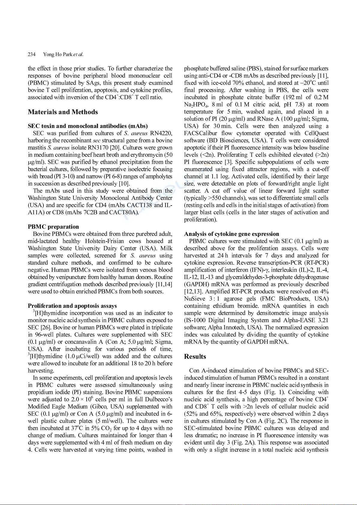
Con A-induced stimulation of bovine PBMCs and SEC-
induced stimulation of human PBMCs resulted in a constant
and nearly linear increase in PBMC nucleic acid synthesis in
cultures for the first 4-5 days (Fig. 1). Coinciding with
nucleic acid synthesis, a high percentage of bovine CD4
+
and CD8
+
T cells with >2n levels of cellular nucleic acid
(52% and 65%, respectively) were observed within 2 days
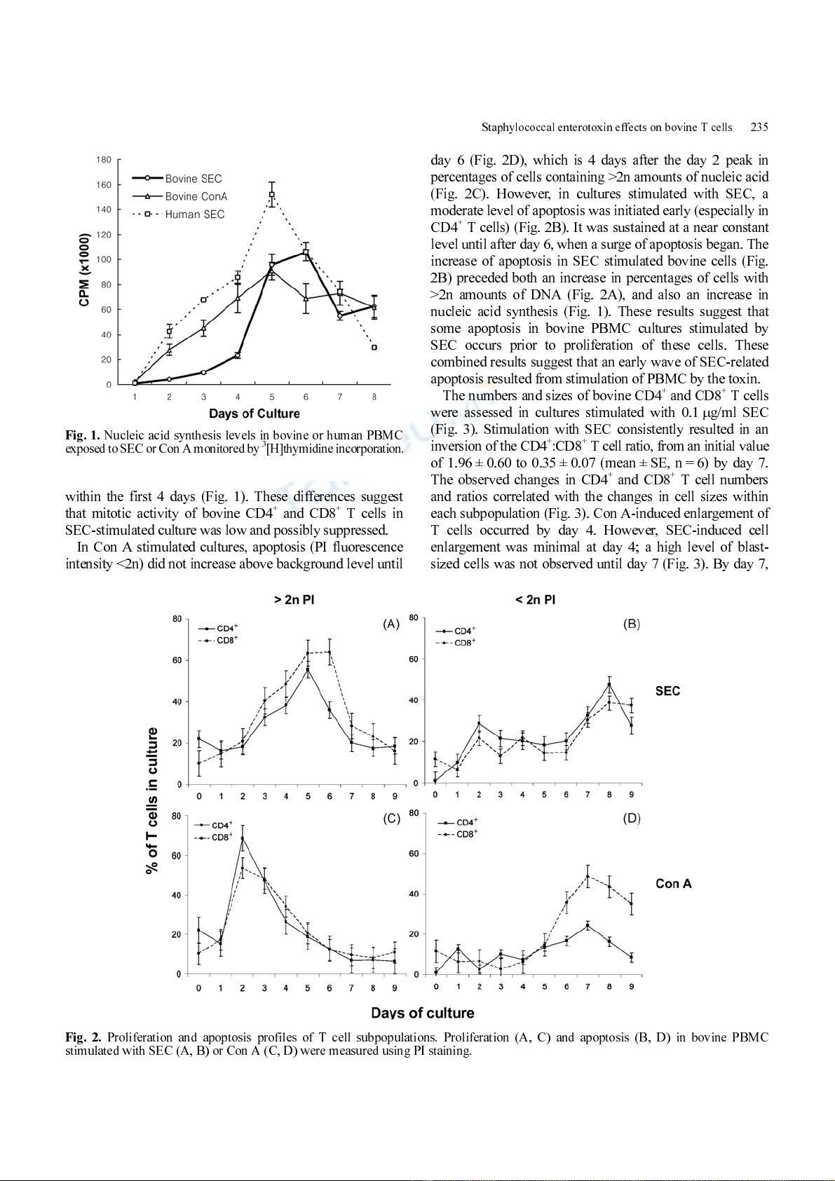
in cultures stim ulated by Con A (Fig. 2C). The r esponse in
SEC-stimulated bovine PBMC cultures was delayed and
less dramatic; no increase in PI fluorescence intensity was
evident until day 3 (Fig. 2A) . This response was ass ociated
with only a slight increase in a total nucleic acid synthesis
Staphylococcal enteroto xin effects on bovin e T cells 235
within the first 4 days (Fig. 1). These differences suggest
that mitotic activity of bovine CD4
+
and CD8
+
T cells in
SEC-stimulated culture was low and possibly suppressed.
In Con A stimulated cultures, apoptosis (PI fluorescence
intensity <2n) did not increase above background level until
day 6 (Fig. 2D), which is 4 days after the day 2 peak in
percentages of cells containing >2n amounts of nucleic acid
(Fig. 2C). However, in cultures stimulated with SEC, a
moderate level of apoptosis was initiated early (especially in
CD4
+
T cells) (Fig. 2B) . It was sustained at a near constant
level until after day 6, when a surge of apoptosis began. The
increase of apoptosis in SEC stimulated bovine cells (Fig.
2B) preceded both an increase in percentages of cells with
>2n amounts of DNA (Fig. 2A), and also an increase in
nucleic acid synthesis (Fig. 1). These results suggest that
some apoptosis in bovine PBMC cultures stimulated by
SEC occurs prior to proliferation of these cells. These
combined results sugge st that an early wave of SE C-r elated
apoptosis resulted from stimulation of PBMC by the toxin.
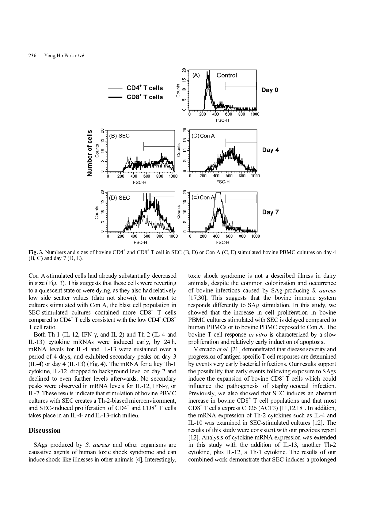
The number s and sizes of bovine CD 4
+
and CD8
+
T cells
were assessed in cultures stimulated with 0.1
µ
g/ml SEC
(Fig. 3). Stimulation with SEC consistently resulted in an
inversion of the CD4
+
:CD8
+
T cell ratio, from an initial value
of 1.96 ± 0.60 to 0.35 ± 0.07 (mean ± SE, n = 6) by day 7.
The observed changes in CD4
+
and CD8
+
T cell numbers
and ratios correlated with the changes in cell sizes within
each subpopulation (Fig. 3). Con A - induced enl argement of
T cells occurred by day 4. However, SEC-induced cell
enlargement was minimal at day 4; a high level of blast-
sized cells was not obs erved until day 7 (Fig. 3) . By day 7,
Fig. 1.
Nucleic acid synthesis levels in bovine or human PBMC
exposed to S EC or Con A monitored by
3
[H]thymidine incorporation
.
Fig. 2.
Proliferation and apoptosis profiles of T cell subpopulations. Proliferation (A, C) and apoptosis (B, D) in bovine PBMC
stimulated with SEC (A, B) or Con A (C, D) were measured using PI staining.
236 Yong Ho Park
et al.
Con A-stimulated cells had already substantially decreased
in size (Fig. 3). Thi s s uggests that t hese cells were reverting
to a quiescent state or were dying, as they also had relatively
low side scatter values (data not shown). In contrast to
cultures stim ulated with Con A, the blast cell population in
SEC-stimulated cultures contained more CD8
+
T cells
compared to CD4
+
T cells consistent with the lo w CD4
+
:CD8
+
T cell ratio.
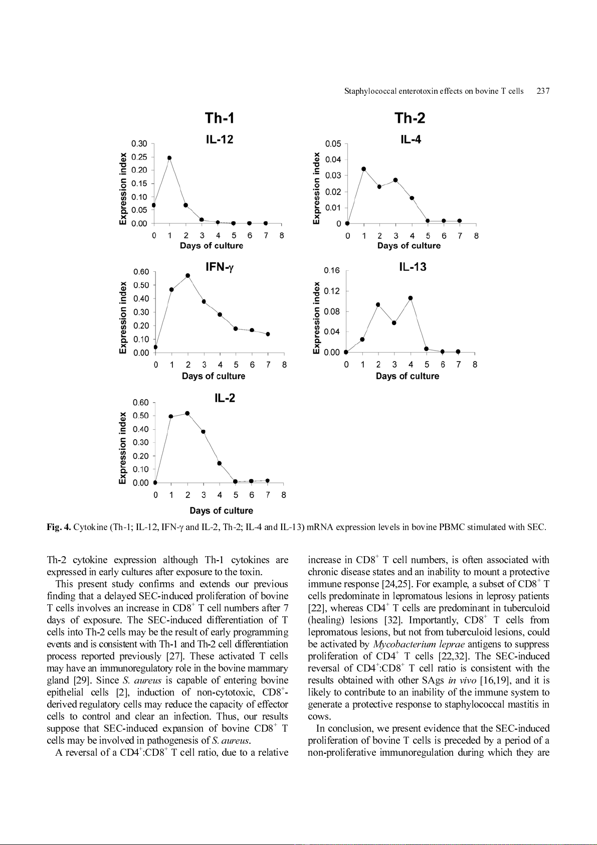
Both Th-1 (IL-12, IFN-
γ
, and IL-2) and Th-2 (IL-4 and
IL-13) cytokine mRNAs were induced early, by 24 h.
mRNA levels for IL-4 and IL-13 were sustained over a
period of 4 days, and exhibited secondary peaks on day 3
(IL-4) or day 4 (IL -13) (Fig. 4). The m RNA for a key Th- 1
cytokine, IL-12, dr opped to background level on day 2 and
declined to even further levels afterwards. No secondary
peaks were observed in mRNA levels for IL-12, IFN-
γ
, or
IL-2. These results indicate that stimulation of bovine PBMC
cultures with SEC creates a Th-2-biased microenvironment,
and SEC-induced proliferation of CD4
+
and CD8
+
T cells
takes place in an IL-4- and IL-13-rich milieu.
Discussion
SAgs produced by
S. aureus
and other organisms
are
causative agents of human toxic shock syndrome and can
induce shock- like i llnes ses in oth er a nim al s [4] . In ter esti ngly,
toxic shock syndrome is not a described illness in dairy
animals, despite the common colonization and occurrence
of bovine infections caused by SAg-producing
S. aureus
[17,30]. This suggests that the bovine immune system
responds differently to SAg stimulation. In this study, we
showed that the increase in cell proliferation in bovine
PBMC cultures stimulated with SEC is delayed compared to
human PBMCs or to bovine PBMC exposed to Con A. The
bovine T cell response
in vitro
is characterized by a slow
proliferation and relatively early induction of apoptosis.
Mercado
et al.
[21] demonstrated that disease severity and
progression of anti gen-specific T cell response s are determined
by events very early bacterial infections. Our results support
the possibility that early events followi ng exposur e to SA gs
induce the expansion of bovine CD8
+
T cells which could
influence the pathogenesis of staphylococcal infection.
Previously, we also showed that SEC induces an aberrant
increase in bovine CD8
+
T cell populations and that most
CD8
+
T cells express CD26 (ACT3) [1 1,12,18]. In addition,
the mRNA expression of Th-2 cytokines such as IL-4 and
IL-10 was examined in SEC-stimulated cultures [12]. The
results of this study were consistent with our previous report
[12]. Analysis of cytokine m RNA expre ssion was extended
in this study with the addition of IL-13, another Th-2
cytokine, plus IL-12, a Th-1 cytokine. The results of our
combined work dem onstrate that SEC induces a prolonged
Fig. 3.
Numbers and sizes of bovine CD4
+
and CD8
+
T cell in SEC (B, D) or Con A (C, E) stimulated bovine PBMC cultures on day 4
(B, C) and day 7 (D, E).
Staphylococcal enteroto xin effects on bovin e T cells 237
Th-2 cytokine expression although Th-1 cytokines are
expressed in early cultures after exposure to the toxin.
This present study confirms and extends our previous
finding that a delay ed SEC-induced proliferation of bovine
T cells involves an increas e in CD8
+
T cell numbers after 7
days of exposure. The SEC-induced differentiation of T
cells into Th-2 cells may be the result of early programming
events and is consist ent with Th -1 and Th-2 ce ll d ifferentiation
process reported previously [27]. These activated T cells
may ha ve an imm uno r egu lat or y r ole in the bov ine mam mary
gland [29]. Since
S. aureus
is capable of entering bovine
epithelial cells [2], induction of non-cytotoxic, CD8
+
-
derived regulatory cell s may reduce the capacity of effector
cells to control and clear an infection. Thus, our results
suppose that SEC-induced expansion of bovine CD8
+
T
cells may be involved in pathogenesis of
S. aur eus
.
A reversal of a CD4
+
:CD8
+
T cell ratio, due to a relative
increase in CD8
+
T cell numbers, is often associated with
chronic disease state s and an i nability to m ount a prot ective
imm une r espons e [24,25]. For example, a subset of CD8
+
T
cells predom inate in lepr om atous le sions in le prosy pati ents
[22], whereas CD4
+
T cells are predominant in tuberculoid
(healing) lesions [32]. Importantly, CD8
+
T cells from
lepromatous lesions, but not from tuberculoid lesions, could
be activated by
Mycobacterium leprae
antigens to suppress
proliferation of CD4
+
T cells [22,32]. The SEC-induced
reversal of CD4
+
:CD8
+
T cell ratio is consistent with the
results obtained with other SAgs
in vivo
[16,19], and it is
likely to contribute to an inability of the im mune system to
generate a protecti ve response to staphyloc occal mastitis in
cows.
In conclusion, we present evidence that the S EC-induced
proliferation of bovine T cells is preceded by a period of a
non-proliferative immunoregulation during which they are
Fig. 4.
Cytokine (Th -1; IL -12 , IFN -
γ
and IL-2, Th-2; IL-4 and IL-13) mRNA expression levels in bovine PBMC stimulated with SEC.