
RESEARCH Open Access
Characterization of the HIV-1 RNA associated
proteome identifies Matrin 3 as a nuclear
cofactor of Rev function
Anna Kula
1
, Jessica Guerra
1,3
, Anna Knezevich
1
, Danijela Kleva
1
, Michael P Myers
2
and Alessandro Marcello
1*
Abstract
Background: Central to the fully competent replication cycle of the human immunodeficiency virus type 1 (HIV-1)
is the nuclear export of unspliced and partially spliced RNAs mediated by the Rev posttranscriptional activator and
the Rev response element (RRE).
Results: Here, we introduce a novel method to explore the proteome associated with the nuclear HIV-1 RNAs. At
the core of the method is the generation of cell lines harboring an integrated provirus carrying RNA binding sites
for the MS2 bacteriophage protein. Flag-tagged MS2 is then used for affinity purification of the viral RNA. By this
approach we found that the viral RNA is associated with the host nuclear matrix component MATR3 (Matrin 3) and
that its modulation affected Rev activity. Knockdown of MATR3 suppressed Rev/RRE function in the export of
unspliced HIV-1 RNAs. However, MATR3 was able to associate with Rev only through the presence of RRE-
containing viral RNA.
Conclusions: In this work, we exploited a novel proteomic method to identify MATR3 as a cellular cofactor of Rev
activity. MATR3 binds viral RNA and is required for the Rev/RRE mediated nuclear export of unspliced HIV-1 RNAs.
Introduction
Viruses have evolved to optimize their replication poten-
tial in the host cell. For this purpose, viruses take advan-
tage of the molecular strategies of the infected host and,
therefore, represent invaluable tools to identify novel
cellular mechanisms that modulate gene expression [1].
The primary viral transcription product is utilized in
unspliced and alternatively spliced forms to direct the
synthesis of all human immunodeficiency virus (HIV-1)
proteins. Although nuclear export of pre-mRNA is
restricted in mammalian cells, HIV-1 has evolved the
viral Rev protein to overcome this restriction for viral
transcripts [2,3], recently reviewed in [4]. Rev promotes
the export of unspliced and partially spliced RNAs from
the nucleus through the association with an RNA ele-
ment called the Rev response element (RRE) that is pre-
sent in the env gene [5-7]. In the cytoplasm, the RRE-
containing HIV-1 transcripts serve as templates for the
expression of viral structural proteins, and the full-length
unspliced forms serve as genomic RNAs that are pack-
aged into viral particles. In order to fulfill its function,
Rev requires the assistance of several cellular cofactors
(reviewed in [8]). Rev interacts with a nucleocytoplasmic
transport receptor, Exportin 1 (CRM1), to facilitate the
export of viral pre-mRNAs [9]. Rev also engages the
activity of cellular RNA helicases [10] and capping
enzymes [11] that are required for the correct nuclear
export of Rev interacting viral RNAs.
The nucleus is a complex organelle where chromo-
somes occupy discrete territories and specific functions
are carried out in sub-nuclear compartments [12-15].
Transcription, for example, has been proposed to occur
in ‘factories’where genes and the RNA polymerase com-
plex transiently assemble [16,17]. Once integrated, the
HIV-1 provirus behaves like a cellular gene, occupying a
specific sub-nuclear position and takes advantage of the
cellular machinery for transcription and pre-mRNA pro-
cessing [18-21]. Control of HIV-1 gene expression is cri-
tical for the establishment of post-integrative latency
and the maintenance of a reservoir of infected cells
* Correspondence: marcello@icgeb.org
1
Laboratory of Molecular Virology, International Centre for Genetic
Engineering and Biotechnology (ICGEB), Padriciano, 99, 34012 Trieste, Italy
Full list of author information is available at the end of the article
Kula et al.Retrovirology 2011, 8:60
http://www.retrovirology.com/content/8/1/60
© 2011 Kula et al; licensee BioMed Central Ltd. This is an Open Access article distributed under the terms of the Creative Commons
Attribution License (http://creativecommons.org/licenses/by/2.0), which permits unrestricted use, distribution, and reproduction in
any medium, provided the original work is properly cited.

during antiretroviral therapy [22]. Beyond transcriptional
control, processing of the RNA may also concur in the
establishment of a latent phenotype [23].
The spatial positioning of chromatin within the
nucleus is maintained by a scaffold of filamentous pro-
teins generally known as the nuclear matrix [24].
Although the exact function of the nuclear matrix is still
debated [25], several of its components have been impli-
cated in nuclear processes that include DNA replication,
repair, transcription, RNA processing and transport
[26-28]. Matrin3 (MATR3) is a highly conserved compo-
nent of the nuclear matrix [29-31]. MATR3 is a 125 kDa
protein that contains a bipartite nuclear localization sig-
nal (NLS), two zinc finger domains, and two canonical
RNA recognition motifs (RRM) [32]. Little is known
about the function of MATR3. A missense mutation in
the MATR3 gene has been linked to a type of progres-
sive autosomal-dominant myopathy [33]. MATR3,
together with the polypyrimidine tract-binding protein
associated splicing factor (PSF) and p54
nrb
, has been
implicated in the retention of hyperedited RNA [34].
Recently, MATR3 has also been involved in the DNA
damage response [35]. Hence, MATR3 may be at the
crossroad of several nuclear processes, serving as a plat-
form for the dynamic assembly of functional zones of
chromatin in the cell nucleus in a so-called ‘functional
neighborhood’[36].
In the present work, we developed a novel proteomic
approach for the identification of host factors involved
in nuclear steps of HIV-1 RNA metabolism. In our pro-
teomic screen, we identified MATR3, and we provide
evidence that it binds viral RNA and is required for
Rev- activity.
Results
Generation and characterization of cell lines expressing
tagged HIV-1 RNAs
The MS2 phage coat protein is a well-described tool for
RNA tagging [37]. Modified MS2 homodimers bind with
high affinity to a short RNA stem loop that can be engi-
neered in multimers in the RNA of interest for various
purposes. On one hand, MS2 fused to the green fluores-
cent protein (GFP) has been used to visualize mRNAs
in living cells allowing for the kinetic analysis of mRNA
biogenesis and trafficking [38-40]. Alternatively, MS2
fused to the maltose binding protein (MBP) has been
used to purify the spliceosome by affinity chromatogra-
phy of cellular extracts [41]. Recently, to visualize and
analyze the biogenesis of HIV-1 mRNA, we inserted
twenty-four MS2 binding sites in the 3’UTR of an HIV
vector and demonstrated that this system fully recapitu-
lates early steps of HIV-1 transcription [42,43].
In this work, we aimed to develop an MS2-based
approach to identify novel host factors associated with
HIV-1 RNA. To this end we took advantage of two
HIV-1 derived vectors called HIV_Exo_24 × MS2
(HIVexo) and HIV_Intro_24 × MS2 (HIVintro),
described earlier [42-45], which carry the MS2 tag either
intheexonicorintheintronicpartoftheviral
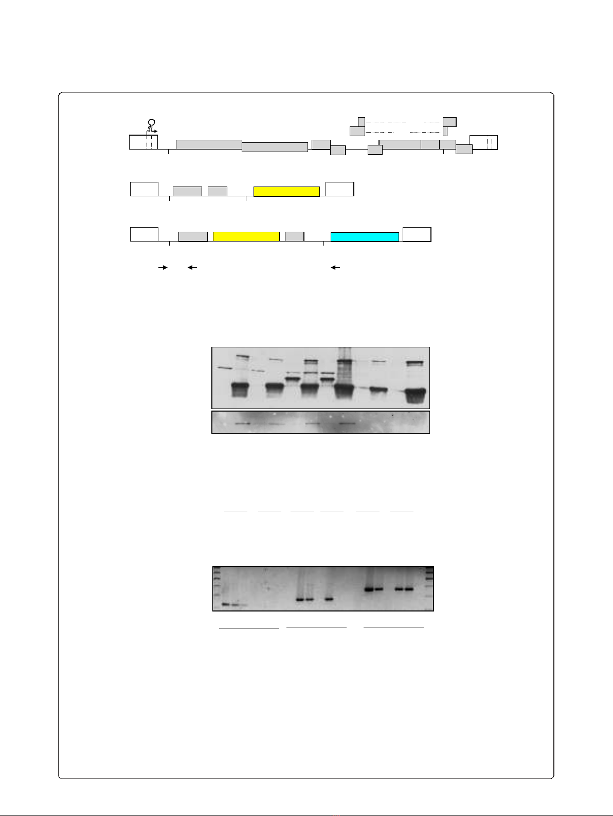
sequence, respectively (Figure 1A andAdditional File 1).
These HIV-1 reporter vectors contain the cis acting
sequences required for viral gene expression and down-
stream steps in replication: the 5’LTR, the Tat respon-
sive region TAR, the major splice donor (SD1), the
packaging signal ψ,aportionofthegag gene, the Rev
responsive region RRE, the splice acceptor SA7 flanked
by its regulatory sequences (ESE and ESS3), and the 3’
LTR that drives 3’-end formation (Figure 1A). The
HIVintro vector carries additionally the reporter gene
coding for the cyan fluorescent protein fused with per-
oxisome localization signal (ECFPskl). Moreover, place-
ment of the 24xMS2 tag inside the intron of the
HIVintro vector increases the probability of purifying
proteins involved in early nuclear steps of HIV-1 RNA
processing [44]. To demonstrate that it was feasible to
pull-down proteins associated with viral RNA via flag-
tagged MS2, we transfected 293T cells with HIVintro,
together with a construct expressing the Tat trans-acti-
vator fused to CFP and a construct expressing a flag-
tagged MS2nls. Total cell extracts were immunoprecipi-
tated with anti-flag antibodies and blotted against GFP
or flag. As shown in Figure 1B, Tat-mediated viral
expression is indicated by the presence of reporter
CFPskl in the lysates (lanes 5 and 7). Importantly, Tat-
CFP is immunoprecipitated when pHIVintro is present,
but the interaction is lost in the presence of RNase
(compare lane 6 and 8) demonstrating that HIV-1 RNAs
carrying both the TAR and the MS2 repeats are
required to pull down Tat-CFP.
Next, two U2OS cell lines carrying stable arrays of
either HIVexo or HIVintro were selected that show
robust trans-activation by Tat and other stimuli known
to induce transcription of integrated HIV-1 [42,43]. To
demonstrate that our strategy was able to distinguish
between the unspliced and spliced viral RNAs in the
pull-down, U2OS HIVintro and U2OS HIVexo cells
were transfected with plasmids expressing Tat-CFP and
flag-MS2nls. Cell lysates were immunoprecipitated with
anti-flag antibodies, extensively washed and used as
templates for RT-PCR using primers that are able to
distinguish unspliced (A+B, 372 bp) and spliced (A+C,
280 bp) RNAs. As shown in Figure 1C, only the spliced
RNA of HIVexo (lane 11), but not of HIVintro (lane
12), was immunoprecipitated, whereas both unspliced
RNAs could be detected (lanes 17, 18). The absence of
the spliced product in the pull-down from HIVintro is
explained by the loss of the MS2 tag after splicing and
demonstrates the specificity of the MS2-based RNA
Kula et al.Retrovirology 2011, 8:60
http://www.retrovirology.com/content/8/1/60
Page 2 of 15
A
B
Ψ
SD1 SA7
gag RRE
LTR LTR
24×MS2 repeats
gag
Ψ
SD1 SA7
LTR
polyA
pol RRE
vif vpr vpu
tat
rev
TAR
LTR
nef
HIVexo
24×MS2 repeats
Ψ
SD1 SA7
RRE LTR LTR
HIVintro
A B C
ECFPskl-IRES-TK
gag
200bp
300bp
400bp
WL
U2OS wt
U2OSHIVexo
U2OSHIVintro
+
+
+
IP
+
+
+
+
+
+
+
+
+
+
+
+
+
+
+
WL IP WL IP
β-actin spliced
(A+C, 280bp)
unspliced
(A+B, 372bp)
RNase
flag-MS2
HIVintro
Tat-CFP +
+
+
+
+
+
+
+
+
+
+
+
+
+
+
+
+
+
+
+
+
+
+
-IgH
-IgL
Tat-CFP-
ECFPskl -
flag-MS2 -
WL IP WL IP WL IP WL IP WL IP WL IP
C
1 2 3 4 5 6 7 8 9 10 11 12
1 2 3 4 5 6 7 8 9 10 11 12 13 14 15 16 17 18
env
Figure 1 Detection and identification of HIV-1 RNA associated factors. A) Description of the HIV-1 constructs. Above an outline of the full-
length viral genome, below the two constructs used in this work: HIVexo (carrying the MS2 binding sites after the SA7 splice site) and HIVintro
(carrying the MS2 repeats in the intron). Black arrows indicate the RT-PCR primers listed in Table 2. The scheme is not drawn to scale. B)
Pulldown of HIV-1 RNA and associated Tat. 293T cells expressing the indicated constructs were lysed and immunoprecipitated with anti-flag
beads. Immunoblots with anti-GFP antibodies show Tat-CFP (lanes 1, 3, 5 7) and ECFPskl (lanes 5 and 7) expressed by the HIVintro construct. Tat
could be immunoprecipitated only when the HIV-1 RNA is present and the association is disrupted by RNase treatment (compare lanes 6 and 8).
IgH and IgL are the heavy and light chains of the immunoglobulins used in the immunoprecipitation. IP and WL stand for immunoprecipitation
and whole cell’s lysate, respectively. C) MS2-dependent pulldown of specific HIV-1 RNAs. U2OS clones and U2OS wt cells expressing Tat-CFP and
flag-MS2nls were lysed and immunoprecipitated with anti-flag beads. RNA was extracted from immunoprecipitations and the RNA reverse-
transcribed and PCR amplified with primers for b-actin mRNA (lanes 1-6), as well as with primers that differentiate spliced (lanes 7-12) and
unspliced (lanes 13-18) forms of the HIV-1 RNAs which are outlined in Figure 1A.
Kula et al.Retrovirology 2011, 8:60
http://www.retrovirology.com/content/8/1/60
Page 3 of 15

affinity purification. Moreover, detection of unspliced
HIV RNA in both IPs reinforces the notion that a cer-
tain proportion of this product is maintained during
transcription of HIV-1. All together these observations
show that the MS2-based strategy can be successfully
used for the purification of factors interacting with viral
transcripts.
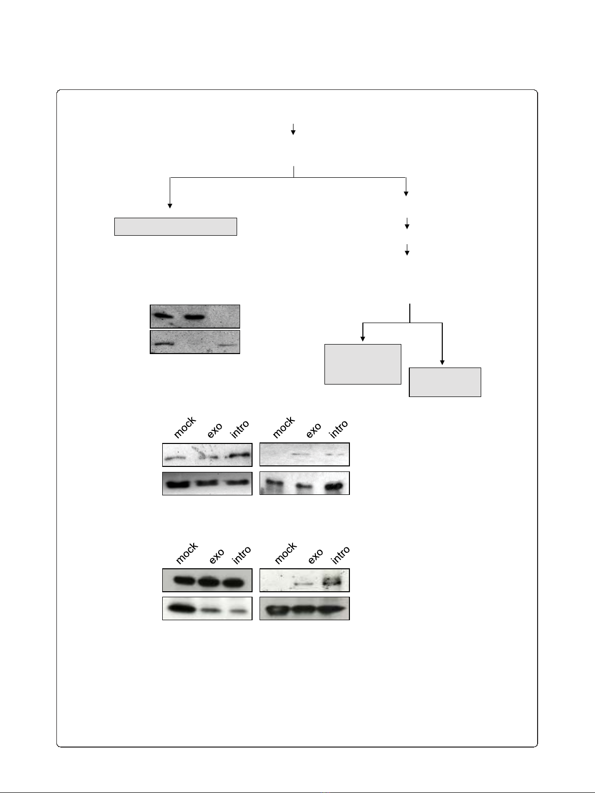
Identification of proteins associated with HIV-1 RNA
As we described above, we used the MS2 tagging for the
purpose of HIV-1 RNA affinity purification. Next, to
identify nuclear factors associated with viral RNA, we
proceeded as follows: U2OS HIVexo and U2OS HIVin-
tro stable cell lines together with wild type U2OS were
transfected with vectors expressing Tat-CFP and flag-
MS2nls proteins. Since we were interested in the identi-
fication of factors involved in nuclear HIV-1 RNA meta-
bolism, we subjected the cells to biochemical
fractionation for the extraction of the nucleoplasmic
fraction (NF) (Figure 2A). Indeed, the procedure
resulted in clean preparation of NF as controlled by
immunoblotting with nuclear (tubulin) and cytoplasmic
(RecQ) markers as shown in Figure 2B. The nuclear
fraction was further subjected to flag-immunoprecipita-
tion. IPs were extensively washed in the presence of
nonspecific competitors as described in Materials and
Methods, and the specificity of pulldown was assessed
by immunoblotting as shown in Figure 2C. Lastly, IPs
were subjected to mass spectrometry analysis as
described in details in Materials and Methods. We were
interested in proteins that associated with both HIVexo
and HIVintro RNAs because they represent hits
obtained from two totally independent procedures. The
combined results of two immunoprecipitations led to
the identification of 32 proteins that were specific for
the stable cell lines carrying the virus (Table 1). Indeed,
most of the identified proteins have been characterized
in RNA binding and/or regulation. Proteins such as
BAT1, FUS and hnRNPs have been already found in
large-scale proteomic analysis of the human spliceosome
[46,47]. BAT2 and CAPRIN1 were shown to associate
with pre-mRNA, although their role in pre-mRNA pro-
cessing is yet to be demonstrated [48,49]. Interestingly,
many of the identified proteins have been already shown
to be involved in various steps of HIV-1 RNA metabo-
lism. DBPA and RPL3 were shown to interact with the
TAR while ILF3 interacts with both - the TAR and the
RRE [50-52]. DDX3X, SFPQ and Upf1 were shown to
regulate Rev-dependent unspliced and partially spliced
viral transcripts while PTB was shown to regulate Rev-
independent, multiply spliced HIV-1 RNA [10,23,53,54].
MOV10 belongs to a family of Upf1-like RNA helicases,
and it has been shown to inhibit viral replication at mul-
tiple stages although its activity on viral RNA is yet to
be discovered [55,56]. Interestingly, in both screens we
identified the nuclear matrix protein MATR3 as a strong
candidate according to the number of non-redundant
peptides sequenced (the log(e) score was -44.4 for U2OS
HIVintro and -38.2 for U2OS HIVexo). MATR3 is of
particularinterestbecauseverylittleisknownaboutits
nuclear function, and it has never been described in the
context of HIV-1 replication. Although MATR3 con-
tains two canonical RNA recognition motifs (RRM), its
RNA target is unknown. Intriguingly, MATR3 was
shown to interact with the SFPQ/p54
nrb
complex which
triggers the nuclear retention of A to I hyperedited
RNA [34]. Therefore, we were stimulated to further
investigate the possible MATR3 interaction with HIV-1
RNA.
To confirm that MATR3 specifically co-immunopreci-
pitates with viral RNA, we transfected U2OS HIVexo
and U2OS HIVintro stable cell lines and wild type
U2OS with flag-MS2nls and Tat. Cells were lysed, and
the resulting cell extract was subjected to immunopreci-
pitation with anti-flag antibodies. Resulting pulldowns
were immunoblotted with MATR3 and flag antibodies.
AsshowninFigure2D,MATR3isdetectedonflag-
MS2 pulldown only in cells expressing the HIV vectors,
both HIVexo and HIVintro, and not in mock cells con-
firming that MATR3 interacts with HIV-1 RNA.
Our preliminary observations suggest that MATR3 is a
novel HIV RNA-binding factor. Therefore, we decided
to further investigate the functional meaning of this
interaction.
MATR3 is required for Rev activity
To investigate the functional role of MATR3 in HIV-1
replication, we measured the effect of RNAi-mediated
knockdown on a full-length HIV-1 molecular clone car-
rying the luciferase reporter gene in nef (pNL4.3R-E-
luc).AsshowninFigure3A,luciferaseactivitythat
depends on the Rev-independent nef transcript was not
affected by MATR3 knockdown. However, gag expres-
sion that is dependent on Rev-mediated export of RRE
containing RNAs was greatly affected (Figure 3B). These
findings suggest that MATR3 acts at a post-transcrip-
tional level on gag mRNA.
In order to confirm that the identified cellular factor
impacts the activity of Rev, we knocked down MATR3
by siRNAs in the context of ectopic Rev expression
along with Tat and the HIV-1 derived vector vHY-
IRES-TK described in [57] and in Additional File 1. As
shown in Figure 4A, efficient knockdown of MATR3
was obtained in the presence and absence of Rev. Next
we examined the levels of unspliced viral RNA by RT-
PCR.AsshowninFigure4B,inthepresenceofRev,
the level of unspliced viral RNA was increased due to
Rev activity (compare lane 3 and 4). Interestingly, the
Kula et al.Retrovirology 2011, 8:60
http://www.retrovirology.com/content/8/1/60
Page 4 of 15
B
C
CF NF
- α-tubulin
- RecQ
Tat-CFP
WL
Flag-MS2
input IP
Transfect U2OS cell clones with flag-MS2 and Tat-CFP
24h later harvest cells, pellet, wash with PBS, resuspend in buffer A
Pellet 5 2000 rpm
Resuspend buffer B
Incubate 4 °C for 30
snap-freeze/thaw 3x
pellet high speed 15
supernatant
nucleoplasmic
fraction (NF)
pellet
supernatant
cytoplasmic fraction (CF)
Nuclear
insoluble
fraction (NP)
pellet
A
MATR3
Flag-MS2
input IP
D
Figure 2 Immunoprecipitation of HIV-1 RNA from nucleoplasmic fractions. A) Biochemical fractionation for the proteomic analysis. Nuclear
extraction scheme showing the various phases of the protocol used to produce the nucleoplasmic fraction. B) Control of nuclear extraction in
U2OS cells. The fractions obtained by the protocol outlined in Figure 2A were loaded on a gel for immunoblotting against a-tubulin (upper
panel) that shows up only in the cytoplasmic fraction (CF) and against the nuclear protein RecQ (bottom panel) that was present only in the
nucleoplasmic fraction (NF). C) Control of HIV-1 RNA associated factor Tat in the NF. Nuclear extracts from U2OS cells (mock), U2OS HIV_Exo_24
× MS2 (exo) or U2OS HIV_Intro_24 × MS2 (intro) were immunoprecipitated for HIV-1 RNA as described above, loaded on SDS-PAGE and blotted
against GFP to detect the RNA-bound Tat-CFP protein (IP). Immunoblots for the nuclear extracts against GFP and flag-MS2nls (input) are shown.
D) Pulldown of HIV-1 RNA and endogenous MATR3. Whole cell extracts from U2OS cells (mock), U2OS HIV_Exo_24 × MS2 (exo) or U2OS
HIV_Intro_24 × MS2 (intro) were immunoprecipitated for HIV-1 RNA as described above, loaded on SDS-PAGE and blotted against MATR3 to
detect the RNA-bound endogenous protein (IP). Immunoblots for the whole cell extracts against MATR3 and flag-MS2nls (input) are shown.
Kula et al.Retrovirology 2011, 8:60
http://www.retrovirology.com/content/8/1/60
Page 5 of 15


![PET/CT trong ung thư phổi: Báo cáo [Năm]](https://cdn.tailieu.vn/images/document/thumbnail/2024/20240705/sanhobien01/135x160/8121720150427.jpg)


