
BioMed Central
Page 1 of 5
(page number not for citation purposes)
Journal of Medical Case Reports
Open Access
Case report
Long-term remission of myopic choroidal neovascular membrane
after treatment with ranibizumab: a case report
Neruban Kumaran*, Dawn A Sim and Adnan Tufail
Address: Department of Medical Retina, Moorfields Eye Hospital, London, UK
Email: Neruban Kumaran* - neruban@doctors.org.uk; Dawn A Sim - dawnsim@doctors.org.uk; Adnan Tufail - adnan.tufail@moorfields.nhs.uk
* Corresponding author
Abstract
Introduction: Myopia has become a big public health problem in certain parts of the world. Sight-
threatening complications like choroidal neovascularisation membranes occur in up to 10% of
pathological myopia, and natural history studies show a trend towards progressive visual loss.
There are long-term financial and quality-of-life implications in this group of patients, and treatment
strategies should aim for long-term preservation of vision.
Case presentation: A 56-year-old Caucasian woman presented with a best-corrected visual
acuity of 6/6-1 in her right eye and 6/24 in her left. Fundal examination revealed pathological myopia
in both eyes and an elevated lesion associated with pre-retinal haemorrhage in the left macula.
Ocular coherence tomography and fundus fluorescein angiogram confirmed a subfoveal classic
choroidal neovascularisation membrane. The patient decided to proceed with intravitreal
ranibizumab (0.5 mg) therapy. One month after treatment, best-corrected visual acuity improved
to 6/12 in her left eye, with complete resolution subretinal fluid on ocular coherence tomography.
After three months, best-corrected visual acuity further improved to 6/9, which was maintained up
to 16 months post-treatment.
Conclusion: We suggest intravitreal ranibizumab as an alternative treatment for long-term
remission of myopic choroidal neovascular membrane. It also suggests that myopic choroidal
neovascularisation membranes may require fewer treatments to achieve sustained remission.
Furthermore, this could serve as a feasible long-term management option if used in conjunction
with ocular coherence tomography.
Introduction
In certain parts of the world, myopia has reached epi-
demic proportions and is now a major public health prob-
lem [1]. The prevalence of high and pathological myopia
appears to be rising in Asia and other parts of the world.
This has a large public health impact because of the asso-
ciated increase in potentially blinding ocular complica-
tions. High myopia or myopia with increased risks of
ocular morbidity can be defined as a spherical equivalent
of at least -6 OD. The resulting ocular pathology is usually
due to excessive elongation of the eyeball and associated
with pathological changes in the fundus [2].
Myopia accompanied by degenerative changes in the
sclera, choroid, retinal pigment epithelium and associated
compromises in visual function have also been termed
'degenerative', 'malignant' and 'pathological' [3].
Published: 28 October 2009
Journal of Medical Case Reports 2009, 3:84 doi:10.1186/1752-1947-3-84
Received: 28 October 2009
Accepted: 28 October 2009
This article is available from: http://www.jmedicalcasereports.com/content/3/1/84
© 2009 Kumaran et al; licensee BioMed Central Ltd.
This is an Open Access article distributed under the terms of the Creative Commons Attribution License (http://creativecommons.org/licenses/by/2.0),
which permits unrestricted use, distribution, and reproduction in any medium, provided the original work is properly cited.
Journal of Medical Case Reports 2009, 3:84 http://www.jmedicalcasereports.com/content/3/1/84
Page 2 of 5
(page number not for citation purposes)
Many complications and associations have been noted
with such 'pathological' myopia. Evidence from both
clinic and population-based studies suggest that high and
low myopia in European and Afro-Caribbean populations
[4,5] may be associated with cataract (posterior subcapsu-
lar, nuclear and occasionally, cortical cataract), the lead-
ing cause of blindness in the world [6].
Myopic eyes are known to have longer axial lengths and
vitreous chamber depths compared to emmetropic eyes.
Eyes with longer axial lengths tend to have higher cup-disc
ratios, increased optic nerve fibre layer defects and possi-
bly greater deformity of the lamina cribrosa, leading to
high susceptibility to glaucomatous optic disc changes
[7]. Such elongation may lead to mechanical stretching
and thinning of the choroid and retinal pigment epithe-
lium and other vascular degenerative changes. These
changes include choroidal neovascularisation, macular
holes, chorioretinal atrophy, Fuchs' spots, lacquer cracks,
lattice degeneration and retinal breaks. Here, we describe
the presentation, follow-up and management of a myopic
patient who presented with a choroidal neovascular mem-
brane (CNVM), as a result of choroidal neovascularisation
(CNV).
Case presentation
A 56-year-old Caucasian woman with high-myopia (-
6.00) presented with a one month history of sudden,
painless distortion of vision in her left eye. She noted that
reading had been more difficult for the last two weeks.
Previous documented best corrected visual acuity (seven
years ago) was 6/5 in the right eye and 6/6 in the left eye.
Previous ocular history of note was macular change sec-
ondary to myopia, diagnosed by her optician eleven years
previously. Both of her parents were myopic, but her med-
ical history was otherwise unremarkable.
On examination, best-corrected visual acuities were 6/6-1
in the right and 6/36, improving to 6/24 with pinhole, in
the left eye. The left eye was noticed to have an Adie
(tonic) pupil. Both anterior segments were deep and quiet
and the intraocular pressure was 16 mmHg in each eye.
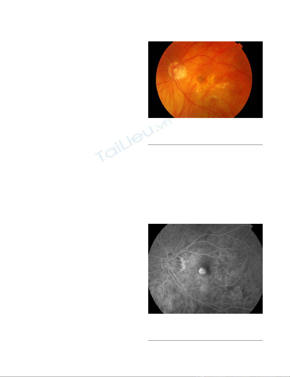
Examination of the left fundus revealed a myopic, tilted
disc and staphyloma and an elevated grayish lesion asso-
ciated with small pre-retinal haemorrhage. The vitreous
was quiet and retinal vessels were of normal calibre (Fig-
ure 1). Ocular coherence tomography (OCT) showed no
sub-retinal fluid but did reveal a choriodal neovascular
membrane (CNVM). Fundus fluoroscein angiogram
(FFA) showed a classic subfoveal CNVM, with early, well-
defined hyperfluorescence (Figure 2) and late leakage.
Therefore, the drop in the vision of her left eye was attrib-
uted to the development of a CNVM. Myopic changes
were seen in the right eye but it was otherwise unremark-
able.
After considering discussion of various treatment options,
the patient decided to proceed with 0.5 mg of intravitreal
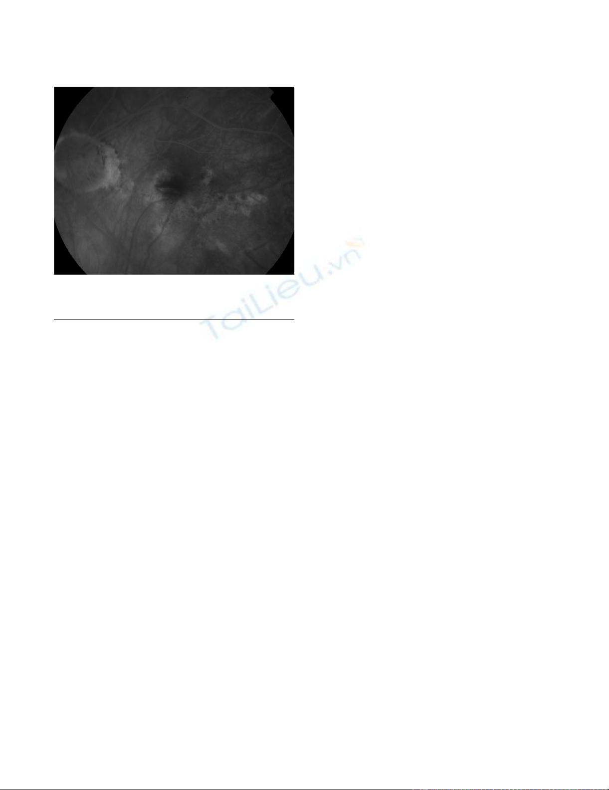
ranizumab (Lucentis). One month following intravitreal
injection into the left eye, her visual acuity improved from
6/32 to 6/12. OCT and FFA showed no subretinal fluid
and furthermore regression of CNVM complex (Figure 3).
However, the patient still complained of distortion and
that the images are still smaller and darker in the left eye
compared to the right eye.
Colour fundus photo of the left eye with myopic macular degeneration, atrophy and an elevated greyish lesion with associated pre-retinal haemorrhageFigure 1
Colour fundus photo of the left eye with myopic mac-
ular degeneration, atrophy and an elevated greyish
lesion with associated pre-retinal haemorrhage.
Early phase fundus fluoroscein angiogram showing choroidal neovascular membrane with well defined hyperfluorescenceFigure 2
Early phase fundus fluoroscein angiogram showing
choroidal neovascular membrane with well defined
hyperfluorescence.
Journal of Medical Case Reports 2009, 3:84 http://www.jmedicalcasereports.com/content/3/1/84
Page 3 of 5
(page number not for citation purposes)
Three months post-treatment, her visual acuity in the left
eye was 6/18, improving to 6/12 with pinhole. She
noticed further improvement in left reading vision. The
patient re-presented four and a half months post-treat-
ment with new distortion in the left eye. FFA showed a
small area of new leakage away from the centre of vision.
It was decided to withhold further treatment at that time
as her visual acuity was 6/12, improving to 6/9 with pin-
hole, which remained stable for 16 months. The patient
agreed to monitor for any changes with an Amsler chart.
VA and OCT findings in her subsequent follow up
appointments were stable up to one year after treatment
with Lucentis.
Discussion
Choroidal neovascular membrane is one of the leading
causes of severe visual loss. Usually a manifestation in the
elderly, it is often associated with age-related macular
degeneration. In this case, however, it is as a cause of the
myopia of the patient.
It appears the balance between antiangiogenic factors
(e.g., pigment epithelium derived factor) and angiogenic
factors (e.g. vascular endothelial growth factor or VEGF)
determines the growth of CNV and VEGF has been tempo-
rally and spatially correlated with the development of
CNV [8].
The main treatment options for CNV are photodynamic
therapy, surgery and anti-vascular endothelial growth fac-
tor (anti-VEGF) treatment.
VEGF was isolated in 1989 [9] and VEGF-A is now known
to promote growth of vascular endothelial cells from
arteries, veins and lymphatics and is needed as a survival
factor for vascular endothelial cells [10]. Eventually, in
2005 VEGF-A, a known mediator of tumour angiogenesis,
was documented to have a key role in the development of
the choroid vasculature. Examples of VEGF inhibitors
include pegaptanib (Macugen), ranibizumab (Lucentis)
and bevacizumab (Avastin).
The use in myopic CNVM of intravitreal bevacizumab
(Avastin), a cheaper and closely related alternative to
ranibizumab, has been reported in both retrospective
[11,12] and prospective studies [13,14], with the majority
of patients achieving CNVM remission and improvement
in visual acuity. Currently, bevacizumab is the mainstay of
management both as a mono-therapy and as an adjuvant
to PDT. Furthermore, although bevacizumab appears to
be a safe and effective treatment for myopic CNVM, fol-
low-up periods have been relatively short, ranging from
35 days to seven months and long-term outcome is
unknown.
In 2008, Silva et al. conducted a retrospective, non-rand-
omized interventional case series study on the short term
efficacy and safety of intravitreal ranibizumab for myopic
CNV. A significant mean improvement in VA was noted at
one, three and six months, with a significant reduction in
mean central retinal thickness, as seen on OCT [15]. In
addition, in 2009 a prospective study of 31 newly diag-
nosed patients showed a similar improvement in VA, in
non-AMD related CNV with a mean follow up of 13.4
months [16]. Treatment of myopic CNVM with intravit-
real ranibizumab with a 16-month-follow-up has not pre-
viously been reported in the literature. The prohibitive
cost of ranibizumab has led to widespread use of bevaci-
zumab.
Our patient had treatment with ranibizumab (Lucentis).
Ranibizumab was developed due to questions over the
ability of intravitreally injected molecules to penetrate
across the retinal layers and reach the choroid.
The safety and efficacy of ranibizumab in the treatment of
neovascular AMD have been evaluated in two large phase
III, multicenter, randomized, double-masked, controlled
pivotal trials, including different neovascular AMD
patient populations.
The MARINA trial randomized 716 subjects in the United
States with CNV to one of three treatment arms: monthly
placebo injections, monthly intravitreal injections of 0.3
mg of ranibizumab, or monthly intravitreal injections of
0.5 mg of ranibizumab.
The ANCHOR trial randomized 423 subjects in the
United States, Europe, and Australia who had CNV to one
of three treatment arms: verteporfin photodynamic ther-
Fundus fluoroscein angiogram showing regression of the choroidal neovascular membrane complexFigure 3
Fundus fluoroscein angiogram showing regression of
the choroidal neovascular membrane complex.

Journal of Medical Case Reports 2009, 3:84 http://www.jmedicalcasereports.com/content/3/1/84
Page 4 of 5
(page number not for citation purposes)
apy with monthly placebo ocular injections, monthly
intravitreal injections of 0.3 mg of ranibizumab with a
placebo photodynamic therapy procedure, and monthly
intravitreal injections of 0.5 mg of ranibizumab with a
placebo photodynamic therapy procedure.
Analyses of these two phase III studies (ANCHOR and
MARINA trials) indicate that ranibizumab results not only
in a slowing down of vision loss but also a clinically
meaningful vision gain at the primary 12-month assess-
ment in a significant proportion of patients. In the case of
the MARINA study these benefits were also observed
through the final 24-month assessment [17].
In 2007 an open-label single centre prospective study
called the prospective OCT imaging of patients with neo-
vascular AMD Treated with intra-ocular Lucentis
(PrONTO) was designed to investigate the role of OCT in
guiding retreatment decisions for a variable dosing regi-
men in patients with choroidal neovascularisation (CNV)
secondary to AMD. The aim of the study was to find out if
an OCT-guided treatment regimen could be used to main-
tain improvements in visual acuity over two years after
three consecutive monthly doses of Lucentis (500 μg)
[18].
The results showed rapid improvements in visual acuity
and OCT measurements. After 12 and 24 months, out-
comes in the study were similar to the MARINA and
ANCHOR phase III study results. It is worth noting that
the mean frequency of dosing reduced by more than half.
Based on these results, OCT appears to be a useful tool for
guiding retreatment decisions such as the frequency of
treatment of patients with CNV. However a prospective,
randomized clinical trial is needed to confirm these
results [18].
Conclusion
In conclusion, we presented a patient with myopic CNVM
whose vision improved and stabilized at 6/6 after one
treatment of ranibizumab (Lucentis). Furthermore, a
review of major trials that have been done on CNV show
that ranibizumab in CNV not only reduces loss of vision
but in fact results in visual gain. In addition, the PrONTO
trial shows that the frequency of treatment should be
guided by investigations such as OCT and the treatment
tailored to the individual findings in the patient (such as
an increase in central retinal thickness of 100 μm or more
on OCT.) This is further supported by studies showing the
use of intravitreous anti-VEGF resulting in long term
remission of other Type 2 CNVM.
Due to the relative rarity of myopic CNVM, there is lack of
evidence for intravitreal anti-VEGF treatment. Treatment
of CNVM should therefore be individualized and the
chance of spontaneous resolution discussed with patients.
This case report presents intravitreal ranibizumab as a rea-
sonable treatment option, and shows that the frequency
of treatment can be modulated according to OCT find-
ings.
Abbreviations
CNV: choroidal neovascularisation; CNVM: choroidal
neovascular membrane; OCT ocular coherence tomogra-
phy; FFA: fundus fluoroscein angiogram; VEGF: vascular
endothelial growth factor.
Competing interests
The authors declare that they have no competing interests.
Authors' contributions
DS reviewed the patient in clinic. AT and DS structured the
management plan and followed up the patient. NK and
DS reviewed the article for intellectual content while NK
carried out a literature review. NK and DS read and
approved the final script.
Consent
Written informed consent was obtained from the patient
for publication of this case report and any accompanying
images. A copy of the written consent is available for
review by the journal's Editor-in-Chief.
References
1. Grosvenor T: Why is there an epidemic of myopia? Clin Exp
Optom 2003, 86:273-275.
2. Goldschmidt E: Ocular morbidity in myopia. Acta Ophthalmol
Suppl 1988, 185:86-87.
3. Duke-Elder S: Pathological refractive errors. System of Ophthal-
mology. St. Louis, Mosby 1970, V:.
4. Leske MC, Wu SY, Nemesure B, Hennis A, Barbados Eye Studies
Group: Risk factors for incident nuclear opacities. Ophthalmol-
ogy 2002, 109:1303-1308.
5. Saw Seang-Mei, Gazzard Gus, Shih-Yen Edwin Chan, Chua Wei-Han:
Myopia and associated pathological complications. Ophthal-
mic & physiological optics: the journal of the British College of Ophthalmic
Opticians (Optometrists) 2005, 25(5):381-391.
6. Resnikoff S, Pascolini D, Etya'ale D, Kocur I, Pararajsegaram R,
Pokarel G, Mariotti S: Global data on visual impairment in the
year 2002. Bull World Health Organ 2004, 82:844-851.
7. Fong DS, Epstein DL, Allingham RR: Glaucoma and myopia: are
they related? Int Ophthalmol Clin 1990, 30:215-218.
8. Bhatt NS, Diamond JG, Jalali S, Das T: Choroidal neovascular
membrane. Indian J Ophthalmol 1998, 46(2):67-80.
9. Ferrara N, Henzel WJ: Pituitary follicular cells secrete a novel
heparin-binding growth factor specific for vascular endothe-
lial cells. Biochem Biophys Res Commun 1989, 161:851-858.
10. Ferrara N, Damico L, Shams N, Lowman H, Kim R: Development
of Ranibizumab, and anti-vascularendothelial growth factor
antigen binding fragment, as therapy for neovascular age-
related macular degeneration. Retina 2006, 26:859-870.
11. Yamamoto I, Rogers AH, Reichel E, Yates PA, Duker JS: Intravitreal
bevacizumab (Avastin) as treatment for subfoveal choroidal
neovascularisation secondary to pathological myopia. Br J
Ophthalmol 2007, 91:157-160.
12. Sakaguchi H, Ikuno Y, Gomi F, Kamei M, Sawa M, Tsujikawa M,
Oshima Y, Kusaka S, Tano Y: Intravitreal injection of bevacizu-
mab for choroidal neovascularisation associated with patho-
logical myopia. Br J Ophthalmol 2007, 91:161-165.

Publish with Bio Med Central and every
scientist can read your work free of charge
"BioMed Central will be the most significant development for
disseminating the results of biomedical research in our lifetime."
Sir Paul Nurse, Cancer Research UK
Your research papers will be:
available free of charge to the entire biomedical community
peer reviewed and published immediately upon acceptance
cited in PubMed and archived on PubMed Central
yours — you keep the copyright
Submit your manuscript here:
http://www.biomedcentral.com/info/publishing_adv.asp
BioMedcentral
Journal of Medical Case Reports 2009, 3:84 http://www.jmedicalcasereports.com/content/3/1/84
Page 5 of 5
(page number not for citation purposes)
13. Chan WM, Lai TY, Liu DT, Lam DS: Intravitreal bevacizumab
(Avastin) for myopic choroidal neovascularization: six-
month results of a prospective pilot study. Ophthalmology 2007,
114(12):2190-2196.
14. Ruiz-Moreno JM, Gomez-Ulla F, Montero JA, Ares S, Lopez-Lopez F,
Rodriguez M, Fernandez M: Intravitreal bevacizumab to treat
subfoveal choroidal neovascularization in highly myopic
eyes:1 year outcome. Br J Ophthalmol 2009, 93:448-451.
15. Silva RM, Ruiz-Moreno JM, Nascimento J, Carneiro A, Rosa P, Bar-
bosaa A, Carvalheira F, Abreu JR, Cunha-Vaz JG: Short-term effi-
cacy and safety of intravitreal ranibizumab for myopic
choroidal neovascularization. Retina 2008, 28(8):1117-1123.
16. Konstantinidis L, Mantel I, Pournaras JA, Zografos L, Ambresin A:
Intravitreal ranibizumab (Lucentis) for the treatment of
myopic choroidal neovascularization. Graefes Arch Clin Exp Oph-
thalmol 2009, 247(3):311-318.
17. Rosenfeld PJ, Brown DM, Heier JS, Boyer DS, Kaiser PK, Chung CY,
Kim RY, MARINA Study Group: Ranibizumab for neovascular
age-related macular degeneration: 2-year results of the
MARINA study. N Engl J Med 2006, 355(14):1419-1431.
18. Grossniklaus HE, Green WR: All About PrONTO: Study
Yielded Good Results in AMD With Treatment Guided by
OCT MAY/JUNE. Retina Today 2007:41-48.


![PET/CT trong ung thư phổi: Báo cáo [Năm]](https://cdn.tailieu.vn/images/document/thumbnail/2024/20240705/sanhobien01/135x160/8121720150427.jpg)


