
BioMed Central
Page 1 of 6
(page number not for citation purposes)
Journal of Medical Case Reports
Open Access
Case report
Rapidly progressing, fatal and acute promyelocytic leukaemia that
initially manifested as a painful third molar: a case report
Juan A Suárez-Cuenca*1,2,3, José L Arellano-Sánchez1,2, Aldo
A Scherling-Ocampo1,2, Gerardo Sánchez-Hernández2,
David Pérez-Guevara4 and Juan R Chalapud-Revelo5
Address: 1Department of Internal Medicine, Ticomán General Hospital, SSDF Mexico City, Mexico, 2Xoco General Hospital, SSDF Av México
Coyoacán s/n, esq Bruno Traven, Mexico City, Mexico, 3Cellular Biology Department (Postgraduate Program in Biomedical Science), Institute of
Cellular Physiology, Circuito Exterior s/n Ciudad Universitaria, Mexico City, Mexico, 4Maxillofacial Surgery Department (Division of Research and
Postgraduate Studies), Faculty of Dentistry, Av Institutos s/n Ciudad Universitaria, Mexico City, Mexico and 5Haematology Department, National
Cancer Institute, Av San Fernando, Mexico City, Mexico
Email: Juan A Suárez-Cuenca* - jsuarez@ifc.unam.mx; José L Arellano-Sánchez - md_arlos79@hotmail.com; Aldo A Scherling-
Ocampo - aldosterona1@hotmail.com; Gerardo Sánchez-Hernández - gmtp@prodigy.net.mx; David Pérez-Guevara - dvdprz@hotmail.com;
Juan R Chalapud-Revelo - injuan@hotmail.com
* Corresponding author
Abstract
Introduction: Acute promyelocytic leukaemia, an uncommon and devastating subtype of leukaemia, is
highly prevalent in Latin American populations. The disease may be detected by a dentist since oral signs
are often the initial manifestation. However, despite several cases describing oral manifestations of acute
promyelocytic leukaemia and genetic analysis, reports of acute promyelocytic leukaemia in Hispanic
populations are scarce. The identification of third molar pain as an initial clinical manifestation is also
uncommon. This is the first known case involving these particular features.
Case presentation: A 24-year-old Latin American man without relevant antecedents consulted a dentist
for pain in his third molar. After two dental extractions, the patient experienced increased pain, poor
healing, jaw enlargement and bleeding. A physical examination later revealed that the patient had pallor,
jaw enlargement, ecchymoses and gingival haemorrhage. Laboratory findings showed pancytopaenia,
delayed coagulation times, hypoalbuminaemia and elevated lactate dehydrogenase. Splenomegaly was
detected on ultrasonography. Peripheral blood and bone marrow analyses revealed a hypercellular
infiltrate of atypical promyelocytic cells. Cytogenetic analysis showing genetic translocation t(15;17)
further confirmed acute promyelocytic leukaemia. Despite early chemotherapy, the patient died within
one week due to intracranial bleeding secondary to disseminated intravascular coagulation.
Conclusion: The description of this unusual presentation of acute promyelocytic leukaemia, the
diagnostic difficulties and the fatal outcome are particularly directed toward dental surgery practitioners
to emphasise the importance of clinical assessment and preoperative evaluation as a minimal clinically-
oriented routine. This case may also be of particular interest to haematologists, since the patient's
cytogenetic analysis, clinical course and therapeutic response are well documented.
Published: 3 November 2009
Journal of Medical Case Reports 2009, 3:102 doi:10.1186/1752-1947-3-102
Received: 18 October 2008
Accepted: 3 November 2009
This article is available from: http://www.jmedicalcasereports.com/content/3/1/102
© 2009 Suárez-Cuenca et al; licensee BioMed Central Ltd.
This is an Open Access article distributed under the terms of the Creative Commons Attribution License (http://creativecommons.org/licenses/by/2.0),
which permits unrestricted use, distribution, and reproduction in any medium, provided the original work is properly cited.

Journal of Medical Case Reports 2009, 3:102 http://www.jmedicalcasereports.com/content/3/1/102
Page 2 of 6
(page number not for citation purposes)
Introduction
Up to 65% of patients with acute leukaemia consult a den-
tist due to oral manifestations, or the disease is detected
from suggestive findings during periodontal and/or phys-
ical examination. According to reports, the most common
findings in the oral cavity include gingival enlargement,
local abnormal colour or gingival haemorrhage,
petechiae, ecchymoses, mucosal ulceration, paresthesia
and/or oral infections [1]. These clinical manifestations
are consequences of gingival infiltration and abnormal
proliferative neoplastic white blood cells that affect the
normal production of erythrocytes, leukocytes and plate-
lets. Toothache as an initial clinical manifestation of acute
leukaemia without accompanying oral or systemic mani-
festation is very uncommon [2,3].
Acute promyelocytic leukaemia (M3-APL) is a malignant
subtype of acute myeloid leukaemia (AML), comprising
approximately 8 to 13% of reported cases of leukaemia.
Prevalence is especially high among Hispanic populations
and among younger patients [4]. In most cases, chromo-
somal abnormality from genetic translocation t(15;17)
that leads to leukaemic transformation can be seen by
cytogenetics [5]. Oral symptoms in M3-APL are similar to
those found in other leukaemias. Takagi et al. [6]
described oral manifestations in 16 patients with sponta-
neous gingival bleeding, post-oral surgery bleeding and
gingival swelling as the most common symptoms of M3-
APL. Half of these patients consulted a dentist during an
early stage of the disease.
The clinical outcome of M3-APL is characterised by bleed-
ing disorders secondary to disseminated intravascular
coagulation (DIC). This may account for the worst fea-
tures associated with leukaemia since it causes a fulmi-
nant disorder that primarily affects young people. The
effects are devastating on an individual's life and causes
death for a large number of patients during the initial
phases of treatment. However, M3-APL is the most treata-
ble of AMLs if early diagnosis and treatment are per-
formed [7].
Case presentation
A 24-year-old, obese, Latin American man with a history
of measles, scarlet fever, and appendectomy, and taking
no medications, consulted a private odontological care
centre because of two days of toothache on the right side
of his mouth. Evaluated as a common painful third molar,
surgical extraction was attempted, but resulted in partial
extraction due to the dentist's inexperience. The patient
experienced mild haemorrhage and pain which was con-
trolled by conventional haemostatic measures and anal-
gesia. A prophylactic antibiotic was prescribed to the
patient, and a later appointment was scheduled for the
extraction of the residual tooth.
On the following day, the patient continued to have mild
haemorrhaging and local swelling. The same supportive
measures were prescribed since these were considered
normal outcomes of a traumatic procedure. On the third
day after surgery, persistent pain accompanied by malaise,
moderate haemorrhaging and progressive local swelling
prompted hospital care, where management included
conventional haemostatic measures, analgesic, one-day
hospital surveillance and early discharge. No laboratory
analysis was ordered.
The next day, the patient complained of mild local pain,
accompanied by trismus, minor local bleeding and clots,
ecchymoses and mild jaw enlargement. Surgical extrac-
tion of the tooth residues and wound closure were per-
formed, without apparent hemorrhagic complications.
The dentist assumed that the patient had a coagulation
disorder, based on such slow healing, and recommended
a blood test and medical evaluation at the hospital
because the private odontological clinic lacked the neces-
sary laboratory resources. That same day, the patient expe-
rienced exacerbated pain, poor response to analgesia and
continued local bleeding, which required emergency hos-
pital care and a stay in the internal medicine department.
Physical examination revealed that the patient had pallor,
right-sided jaw enlargement, ecchymoses (Figure 1, panel
A) and an oral cavity with active gingival haemorrhage
from surgery (Figure 1, panel B). There was no gingival
enlargement, local abnormal colour, or clinical hepat-
osplenomegaly and/or lymphadenopathy.
The patient's complete blood count (CBC) (Table 1)
showed pancytopaenia with differential count of neu-
trophils 0.489 × 10⁹/L (31.5%), lymphocytes 0.924 × 10⁹/
L (59.5%), monocytes 0.122 × 10⁹/L (7.84%), eosi-
nophils 0.006 × 10⁹/L (0.38%), and basophils 0.012 ×
109/L (0.76%). The patient had normocytic normochro-
mic anaemia (Hb 107 g/L, MCV 84fL, MCH 29 pg and
MCHC 351 g/L), thrombocytopenia (platelets 6.3 × 10⁹/
L) and abnormal coagulation times (prothrombin time
(PT) 16 s with control sample 11 s, INR 1.43, activated
partial tissue thromboplastin (aPTT) 39 s with control
sample 32 s). Additional laboratory data revealed dyslipi-
daemia, hypoalbuminaemia and high levels of lactate
dehydrogenase. An abdominal ultrasound revealed mild
splenomegaly.
During a short stay of three days in the internal medicine
department, a peripheral blood smear revealed a 6% blast
cell content with Auer rods and promyelocytic cells. The
patient was referred to the National Cancer Institute,
where M3-APL was diagnosed based on bone marrow
samples, with hypercellularity and 80% neoplastic pro-
myelocytes (Figure 2). The findings were subsequently
Journal of Medical Case Reports 2009, 3:102 http://www.jmedicalcasereports.com/content/3/1/102
Page 3 of 6
(page number not for citation purposes)
confirmed by fluorescence in situ hybridisation (FISH) for
chromosomal translocation t(15;17) (Figure 3).
Supportive measures were established based on the
patient's stay at the internal medicine department. The
patient experienced transfusion-associated fever that
occurred after he was transfused with erythrocytes and
platelets. Thrombocytopenia was refractory to platelet
transfusion and he maintained platelet counts between
6.3 × 10⁹/L and 30 × 109/L. The patient further developed
neutropaenia and fever, and the protocol for a high risk of
opportunistic infection included isolation and antibiotics
(ceftriaxone, metronidazole and fluconazole). At the
National Cancer Institute, specific treatment was started
immediately after diagnosis. His induction therapy
included ATRA (all-trans retinoic acid) and daunorubicin.
Laboratory analyses showed platelets at 8 × 10⁹/L, delayed
coagulation times and a fibrinogen level of 43 mg/dl (nor-
mal range 159 to 317). Further supportive measures
included cryoprecipitates in addition to fresh frozen
plasma (15 mg/kg q.i.d.) when the patient's fibrinogen
level was lower than 100 mg/dl, and platelet transfusion
when his platelet count was below 50 × 109/L. His CBC
and coagulation parameters (PT, aPTT, thrombin time
and fibrinogen levels) were monitored daily.
Despite early specific treatment, the patient suffered gen-
eralised seizures secondary to intracranial bleeding caused
by DIC, which was confirmed by delayed coagulation
times, low serum fibrinogen, high fibrin degradation
products and D-dimer. The patient died after four days of
hemorrhagic complications.
Discussion
Oral manifestations of many haematological diseases are
clinically similar to locally-occurring lesions. For this rea-
son, a specific diagnosis of blood dyscrasia is difficult, if
not impossible, to establish on the basis of oral findings
alone. Commonly, the dentist is the first professional in
contact with the patient and has the best chance to detect
special cases by means of an accurate medical history, a
recording of systemic disease-oriented information and
an identification of signs that are relevant to the patient's
current problem.
Toothache is a common symptom in oral medicine and
possible sources include local diseases like tooth infec-
tion, decay, nerve irritation, repetitive motions and any
injury that bruises the tooth. Non-dental causes include
acute ulceration of the gingiva or soft tissues, pericoroni-
tis, dry socket, trigeminal neuralgia, sinusitis, otitis media,
mastoiditis, temporomandibular joint pain radiation,
and, occasionally, systemic diseases. In this case, third
molar pain was identified and treated by surgical extrac-
tion. However, a few special considerations may be appro-
priate. During routine practice of dental extraction,
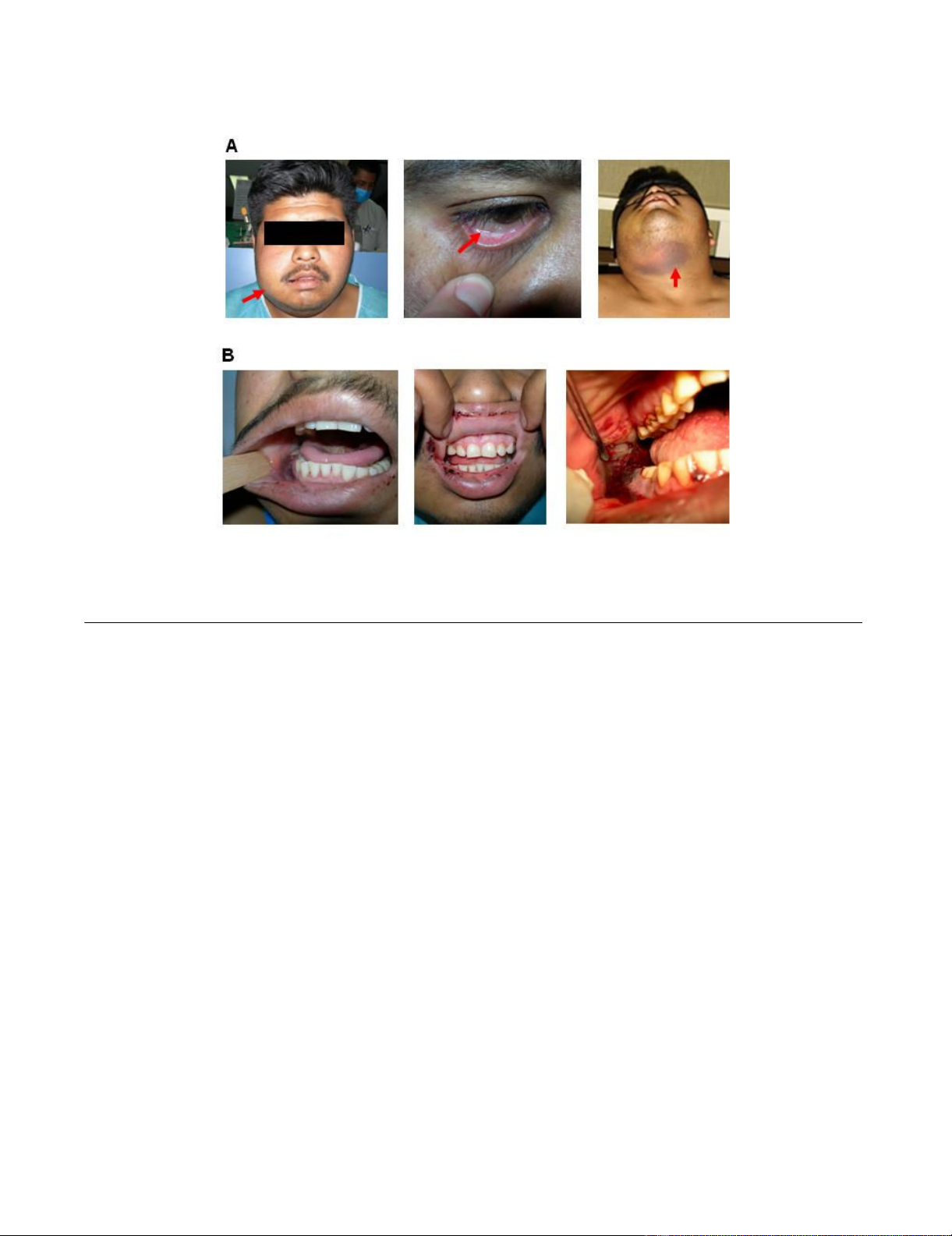
Clinical appearance of the patient after second dental procedureFigure 1
Clinical appearance of the patient after second dental procedure. Upon hospital admission, the patient exhibited jaw
enlargement, conjunctival pallor and ecchymoses (panel A, left, middle and right, respectively). Oral cavity examination showed
gingival damage with active gingival haemorrhage from surgery (panel B).

Journal of Medical Case Reports 2009, 3:102 http://www.jmedicalcasereports.com/content/3/1/102
Page 4 of 6
(page number not for citation purposes)
preoperative assessment commonly includes a brief clini-
cal history with details about the outcome of previous
dental procedures, including the duration of bleeding
after extraction and treatment, previous hemorrhagic epi-
sodes, outcomes from other surgical procedures or inju-
ries, family history, drugs or medications, identification or
hospital cards and physical findings. Laboratory blood
analyses are only performed if the abovementioned items
are suspicious. The lack of suggestive medical and/or fam-
ily history or symptoms in the present case made the diag-
nosis of this haematological disease unlikely.
Nevertheless, the physical examination and outcomes
revealed the manifestations of the underlying disease: a)
patient appearance with pallor guided to anaemia, b)
poor tissue healing was secondary to leucopoenia, and c)
excessive bleeding after extraction is commonly an acute
leukaemia-associated symptom due to thrombocytopenia
and delayed coagulation times [1,8]. In this case, the
excessive bleeding was underestimated. If CBC and coag-
ulation tests had been ordered by the dentist or during the
one-day hospital surveillance, it would have led to an ear-
lier diagnosis and treatment that could have possibly lead
to a better outcome. However, in the absence of support-
ing data, hemorrhagic manifestations are easily misinter-
preted as a consequence of an imperfect surgical
technique and local damage instead of a haematological
disease. In the case of thrombocytopenia-related gingival
bleeding, local treatment is recommended. Once bleeding
is controlled, an early reference to a haematologist or
internist should occur if there is any suspicion of a possi-
ble haematological disease.
Table 1: Complete blood count and blood chemistry
Complete Blood Count
Test Value
White Blood Cells 1.5 × 10⁹/L
Red Blood Cells 3.6 × 10¹²/L
Platelets 6.3 × 10⁹/L
Haemoglobin 107.0 g/L
Haematocrit 0.30
Glucose 6.9 mmol/L
Blood Biochemistry
Test Value
BUN 12.8 mmol/L
Creatinine 159.1 μmol/L
Uric acid 422.31 μmol/L
Calcium 2.14 mmol/
Magnesium 1.03 mmol/L
Cholesterol 3.59 mmol/L
C-HDL 0.21 mmol/L
C-LDL 1.91 mmol/L
Triglycerides 3.21 mmol/L
Total bilirubin 9.06 μmol/L
Indirect bilirubin 6.33 μmol/L
Total protein 86 g/L
Globulin 30 mg/L
Albumin 27 g/L
Aspartate aminotransferase 30 U/L
Alanine aminotransferase 31 U/L
Alkaline phosphatase 75 U/L
Gamma-glutamyl transferase 82 U/L
Lactate dehydrogenase (diluted) 742 U/L
BUN, Blood Urea Nitrogen; C-HDL, High density lipoprotein
cholesterol; C-LDL, Low density lipoprotein cholesterol.
Histological features of acute promyelocytic leukaemia (M3 subtype from FAB)Figure 2
Histological features of acute promyelocytic leukae-
mia (M3 subtype from FAB). Bone marrow aspirate
shows neoplastic promyelocytes, with abnormally coarse and
numerous azurophilic granules (1000×).
Journal of Medical Case Reports 2009, 3:102 http://www.jmedicalcasereports.com/content/3/1/102
Page 5 of 6
(page number not for citation purposes)
The patient was referred to our hospital without any diag-
nostic consideration or laboratory data. Upon admission
to the hospital, the patient's CBC indicated that a haema-
tological disease was a possibility. Pancytopaenia clearly
supported a haematological disorder, while normocytic
normochromic anaemia, thrombocytopenia, mild
splenomegaly, and abnormally elevated levels of LDH
reasonably ruled out other causes of pancytopaenia and
suggested bone marrow involvement with a neoplastic
origin as a likely cause. A diagnostic protocol and initial
supportive measures were rapidly established at the inter-
nal medicine department. In collaboration with the
National Cancer Institute, the diagnosis of M3-APL was
made based on the identification of abnormally excessive
haematopoietic cells, namely promyelocytes, in the
peripheral blood and bone marrow. Chromosomal trans-
location t(15;17) was revealed by FISH to further confirm
the diagnosis.
M3-APL is considered a special variant for its particular
biology, severe manifestations, haematological complica-
tions and rapid fatal course. Conversely, M3-APL is cur-
rently the AML with the highest rate of favourable
response to therapy, especially at an early stage. Morpho-
logically, the bone marrow is effaced by heavily granu-
lated cells with folded and twisted nuclei. In most cases,
reciprocal translocation t(15;17) of the PML gene on
chromosome 15q22 and the RARα gene on chromosome
17, can be seen cytogenetically [5]. Its demonstration is
important because the molecular rearrangement is crucial
for leukaemogenesis and treatment. Furthermore, demon-
stration of t(15;17) or PML/RARα gene fusion is a manda-
tory requirement for confirming the diagnosis of M3-APL
and has relevant prognostic implications [9,10]. Cyto-
chemistry and immunophenotyping provide additional
characteristics of M3-APL, and, although not essential for
diagnosis, yield to desirable analyses in most cases.
M3-APL frequently affects younger, obese and Latin Amer-
ican populations [4,11]. Patients typically display symp-
toms associated with cytopaenias. Oral symptoms of M3-
APL are similar to those found in other leukaemias,
including spontaneous gingival bleeding, post-oral sur-
gery bleeding and gingival swelling [6]. The outcome is
characterised by bleeding disorders, which are the main
causes of death within the first ten days after onset [12].
Most of these features were true for this case (young His-
panic man with post-extraction toothache, pallor, gingival
haemorrhage, ecchymoses, jaw enlargement, pancytopae-
nia, evidence of neoplastic haematopoietic cells and fatal
hemorrhagic complications). Moreover, life-threatening
coagulopathy accompanying M3-APL is relatively com-
mon (80% of cases at the time of diagnosis) and is
ascribed to either primary fibrinolysis or DIC, resulting
from abnormal promyelocytes lysis and release of proco-
agulant mediators [13,14].
Early recognition of M3-APL is important because current
therapy of ATRA combined with chemotherapy or arsenic
trioxide regimens results in 70 to 80% of patient survival
and disease-free outcome after five years. Despite the
favourable response rate, patients with M3-APL and
bleeding disorders, especially with intracranial bleeding,
have an unfavourable prognosis. Peri-induction mortality
is significant, reaching around 50%, according to litera-
ture reports [13,15].
Conclusion
Periodontal signs are commonly the first clinical manifes-
tations of AML, including M3-APL. This case emphasises
the importance of performing a detailed periodontal his-
tory and also obtaining relevant medical information,
adequate interpretation of physical findings and follow-
ing established protocols. Furthermore, it highlights the
relevance of a systematic clinical assessment by dental sur-
geons by ordering pertinent laboratory tests and making
early references to haematologists and/or internists if any
suspicions arise from the preoperative assessment.
Abbreviations
M3-APL: Acute Promyelocytic Leukaemia; AML: Acute
Myeloid Leukaemia; DIC: disseminated intravascular
coagulation; CBC: Complete Blood Count; aPTT: Partial
Tissue Thromboplastin; FISH: Fluorescence in situ hybrid-
isation; ATRA: All-trans Retinoic Acid.
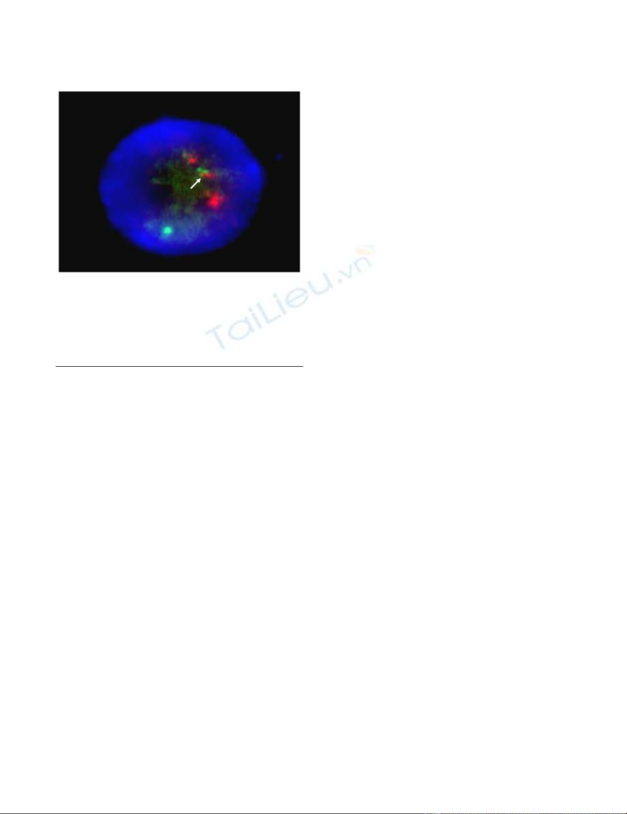
Cytogenetic analysis reveals chromosome translocation t(15;17)Figure 3
Cytogenetic analysis reveals chromosome transloca-
tion t(15;17). Fluorescence in situ hybridisation (FISH) of
abnormal promyelocytic cells with a translocation of chro-
mosome 15 and 17 (nucleus in blue, PML gene in red, RARα
in green and arrow shows fused signals at translocation 15
and 17).


![PET/CT trong ung thư phổi: Báo cáo [Năm]](https://cdn.tailieu.vn/images/document/thumbnail/2024/20240705/sanhobien01/135x160/8121720150427.jpg)


