
RESEARC H Open Access
Structural and functional characterization of
human apolipoprotein E 72-166 peptides in
both aqueous and lipid environments
Yi-Hui Hsieh, Chi-Yuan Chou
*
Abstract
Backgrounds: There are three apolipoprotein E (apoE) isoforms involved in human lipid homeostasis. In the
present study, truncated apoE2-, apoE3- and apoE4-(72-166) peptides that are tailored to lack domain interactions
are expressed and elucidated the structural and functional consequences.
Methods & Results: Circular dichroism analyses indicated that their secondary structure is still well organized.
Analytical ultracentrifugation analyses demonstrated that apoE-(72-166) produces more complicated species in PBS.
All three isoforms were significantly dissociated in the presence of dihexanoylphosphatidylcholine.
Dimyristoylphosphatidylcholine turbidity clearance assay showed that apoE4-(72-166) maintains the highest lipid-
binding capacity. Finally, only apoE4-(72-166) still maintained significant LDL receptor binding ability.
Conclusions: Overall, apoE4-(72-166) peptides displayed a higher lipid-binding and comparable receptor-binding
ability as to full-length apoE. These findings provide the explanation of diverged functionality of truncated apoE
isoforms.
Introduction
Human apolipoprotein E (apoE)
1
comprises 299 amino
acids and there are three isoforms, apoE2, apoE3, and
apoE4, encoded by the ε2, ε3, and ε4 genes, respectively.
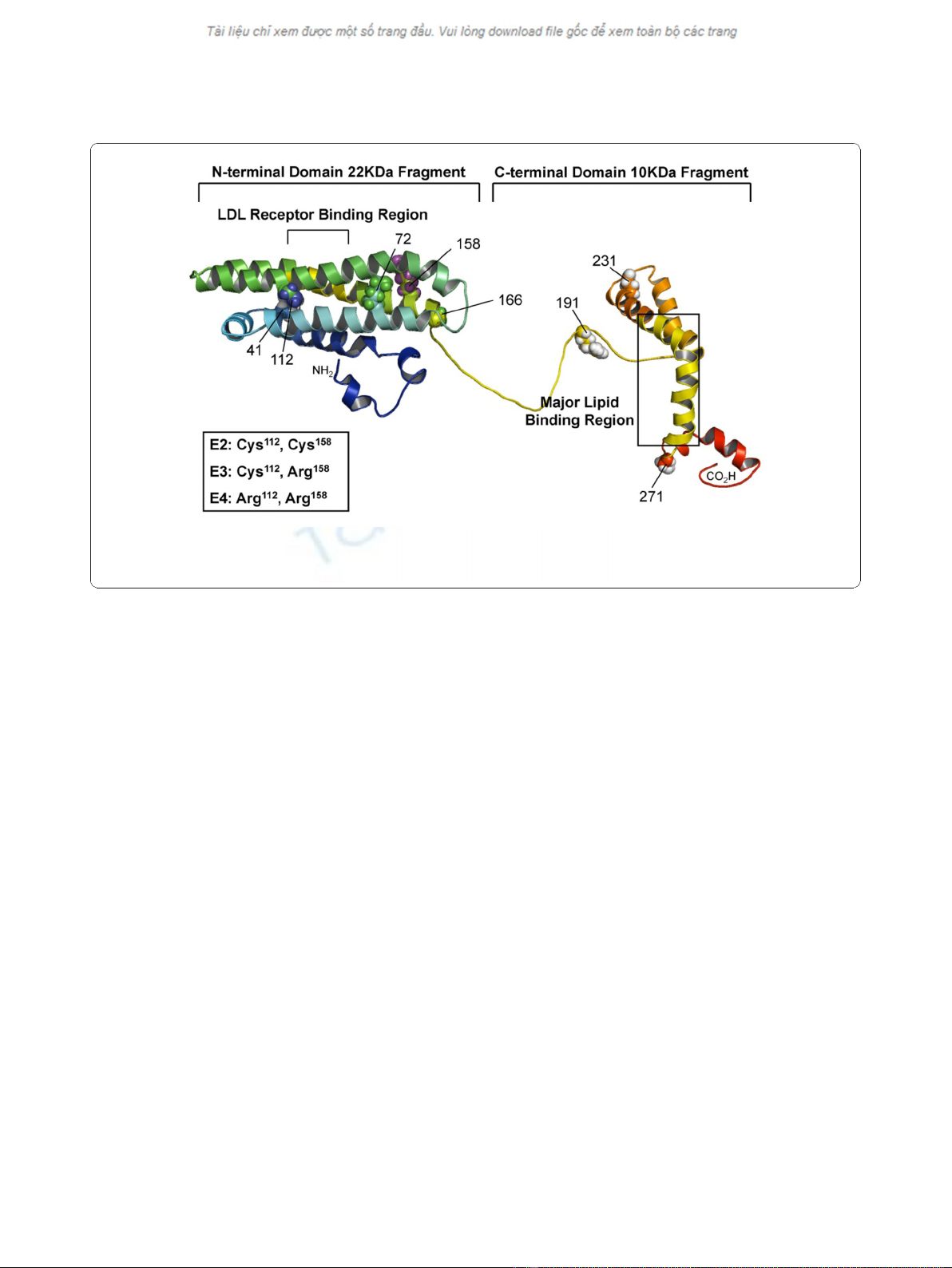
These isoforms differ from each other only at residues
112 and 158 i.e. Cys112 and Arg158 in apoE3, a cysteine
at both positions in apoE2, and an arginine at both posi-
tions in apoE4 [1]. The amino-terminal (NT) domain of
apoE contains four amphipathic a-helices and has
pronounced kinks in the helices near the end of the
four-helix bundle that correlates with the lipid binding
ability (Figure 1) [2,3]. The residues between 140-150 in
the fourth a-helix, comprising many basic amino acids,
has been identified as the low-density lipoprotein recep-
tor (LDLR) binding region [4], with the lipid binding
regionshowntobeinthecarboxyl-terminal(CT)
domain [5,6]. The lipid association is required for high
affinity binding of apoE to the LDLR because of the
increased exposure of basic region on the fourth a-helix
after interacting with lipids [7].
ApoE is involved in facilitating the transportation of
plasma chylomicron remnant to the liver through either
the remnant receptor or LDLR [8,9]. Owing to distinct
domain interactions, apoE2 and apoE3 bind preferen-
tially to small lipoproteins such as high-density lipopro-
tein (HDL), whereas apoE4 has a higher affinity to
very-low-density lipoprotein (VLDL) [6,10]. Different to
apoE3, apoE4 is prone to raise the plasma LDL to high
levels and cause high oxidative stress that can facilitate
atherosclerosis progression [11,12], whilst apoE2 is asso-
ciated with type III hyperlipoproteinemia [13]. The ε4
allele is also associated with familial late-onset and
sporadic Alzheimer’s disease (AD) [14,15]. ApoE4 has
been found to interact with beta-amyloid peptides (Ab)
and induce neurofibrillary tangle (NFT) formation
[16,17]. It preferentially undergoes proteolysis to yield
NT- and CT-truncated that interact with cytoskeletal
components to form NFT-like inclusions in neuronal
cells [16]. To understand the pathogenesis of different
isofomic apoE, most studies are focused on the delinea-
tion of the structure and function characterization of
the full-length apoE, varied length CT, or a “four
a-helix bundle”NT domain [18-21].
* Correspondence: cychou@ym.edu.tw
Department of Life Sciences and Institute of Genome Sciences, National
Yang-Ming University, Taipei 112, Taiwan
Hsieh and Chou Journal of Biomedical Science 2011, 18:4
http://www.jbiomedsci.com/content/18/1/4
© 2011 Hsieh and Chou; licensee BioMed Central Ltd. This is an Open Access article distributed under the terms of the Creative
Commons Attribution License (http://creativecommons.org/licenses/by/2.0), which permits unrestricted use, distribution, and
reproduction in any medium, provided the original work is properly cited.
In the present studies, we attempted to clarify the
structural and functional consequences of NT- and
CT-truncated apoE peptides, i.e. apoE-(72-166). This
truncation still maintains the LDLR binding region, and
removes the first two a-helices and the complete CT
domain. The aim is to create a shorter but still functional
apoE for potential therapeutic approach. Analytical ultra-
centrifugation was used to elucidate the quaternary struc-
tural properties of the three apoE-(72-166) isoforms. In
the presence of lipid, the degree of apoE-(72-166) disso-
ciation and extended conformation was significantly
elevated. The functional assays conclude that apoE-(72-
166) peptides still maintain comparable LDLR and higher
lipid binding ability as to full-length apoE, particularly
apoE4-(72-166). These findings suggest a crucial role of
shorter NT-domain in the biological function of apoE
and provide the basis for the explanation of diverged
functionality of truncated apoE isoforms.
Materials and methods
Plasmids
The construction of pET-apoE2, apoE3, apoE4, apoE3-
(72-166), and apoE4-(72-166) vectors were described
previously [22]. The apoE2-(72-166) DNA fragment was
amplified by PCR, and the forward primer was 5’-AAA-
CATATGAAGGCCTACAAATCGGA, whereas the
reverse primer was 5’-AACTCGAGGGCCCCGGCCT.
The NdeI-XhoI digested apoE2-(72-166) cDNA was then
ligated to the 5.2-kb NdeI-XhoI pET-29a(+) fragment.
Expression and Purification of ApoE Proteins
Protein induction and purification procedures have been
described previously [22,23]. Typical yields of the apoE-
(72-166) proteins were 5-10 mg after purification from 1
liter of E. coli culture medium. The purity of all recombi-
nant proteins was estimated by SDS-PAGE to be > 95%
and the molecular mass of the apoE-(72-166) proteins
was 12 kDa. The purified proteins were buffer-changed
to phosphate buffered saline (PBS) (pH7.3) using Amicon
Ultra-4 10-kDa centrifugal filter (Millipore).
Preparation of Micelle Solution
Dihexanoylphosphatidylcholine (DHPC) has a critical
micelle concentration of 16 mM, at which micelle mono-
mers are formed containing 19 to 40 molecules based on
ultracentrifugation, NMR, and small angle neutron scat-
tering, respectively [24-26]. We used several concentra-
tions of DHPC (5, 50, and 100 mM) to establish an
appropriate lipid environment containing submicelles or
micelles. In current studies, all experiments related to
DHPC were executed at 20°C for the same lipid state.
Circular Dichroism Spectroscopy
Circular dichroism (CD) spectra of the apoE-(72-166)
peptides using a JASCO J-810 spectropolarimeter
(Tokyo, Japan) showed measurements from 250 nm to
190 nm at 20°C in PBS (pH 7.3) with or without 50 mM
DHPC. The protein concentration was 0.5 mg/ml. In
wavelength scanning, the width of the cuvette was 0.1
Figure 1 Structure of human apoE proteins. The model structure illustrating the full-length apoE with NT and CT domains. The structure was
modified from apoE299_20K (S. Y. Sheu, unpublished data). The polymorphic sites (residues 112 and 158) that distinguished the three isoforms
are highlighted. The picture was produced with PyMOL [46].
Hsieh and Chou Journal of Biomedical Science 2011, 18:4
http://www.jbiomedsci.com/content/18/1/4
Page 2 of 9
mm. The far-UV CD spectrum data were analyzed with
the CDSSTR program [27,28]. In this analysis, the
a-helix, b-sheet, and random coil were split. To estimate
the goodness-of-fit, the normalized root mean square
deviation (NRMSD) was calculated.
Unfolding of the ApoE-(72-166) Proteins in Guanidinium
Chloride
ApoE-(72-166) proteins (0.1 mg/ml) with or without 50
mM DHPC were unfolded with different concentrations
ofGdnClinPBS(pH7.3)at4°Covernighttoreach
equilibrium. The unfolding of the proteins was moni-
tored by measuring the CD signal of 222 nm at 20°C
and the width of the cuvette was 1 mm. The unfolding
data were analyzed using thermodynamic models by
global fitting of the measurements to the two-state
unfolding model [29] as follows:
yyye
e
obs NU
GmGdnCl
RT
G
HONU NU
HON
=+•
+
−−
[]
⎛
⎝
⎜
⎜
⎞
⎠
⎟
⎟
−
→→
→
Δ
Δ
()
()
2
2
1
UU NU
mGdnCl
RT
−
[]
⎛
⎝
⎜
⎜
⎞
⎠
⎟
⎟
→
(1)
where y
obs
is the observed biophysical signal; y
N
and
y
U
are the calculated signals of the native and unfolded
states, respectively. GdnCl is the GdnCl concentration,
and ΔGHON U()
2→isthefreeenergychangeforthe
N®Uprocess. m
N®U
is the sensitivity of the unfolding
process to a denaturant concentration.
Sedimentation Velocity
Sedimentation velocity (SV) experiments were per-
formed with an XL-A analytical ultracentrifuge (Beck-
man, Fullerton, CA) as described previously [23]. All
studies were performed at 20°C with a rotor speed of
42,000 rpm in PBS (pH 7.3) with or without DHPC.
The protein concentration was 0.5 mg/ml. Multiple
scans at different time periods were then fitted to a con-
tinuous c(s) distribution model using the SEDFIT
program as described previously [30,31]. All continuous
size distributions were calculated using a confidence
level of p= 0.95, a best fitted average anhydrous friction
ratio (f
r
), a resolution value N of 200, and sedimentation
coefficients between 0 and 20 S. For the data fitting of
apoE-(72-166) in PBS and 5 mM DHPC, the partial spe-
cific volume was set to 0.73 for proteins species. Differ-
ently, for those in 50 and 100 mM DHPC, the value was
set to 0.86 because the influence of DHPC micelle.
Previous studies have suggested that DHPC’s partial spe-
cific volume is 0.99 ml/g [32]. According to our calcula-
tion, higher partial specific volume will lower the best
fitted average f
r
, while the c(s) distribution will not have
any difference.
Sedimentation Equilibrium
Sedimentation equilibrium (SE) experiments were per-
formed with six-channel epon charcoal-filled center-
pieces as described previously [22]. The cells were then
mounted into an An-60 Ti rotor and centrifuged at
10,000 rpm, 15,000 rpm, and 20,000 rpm, respectively,
each for 18 h at 20°C. Ten A
280 nm
measurements with
a time interval of 8-10 min were performed for each dif-
ferent rotor speed to check the equilibrium state. The
SV and SE spectrum of each apoE-(72-166) protein
under the same environments were combined and then
fitted to a global discrete species model using
SEDPHAT program as described previously [22,33].
DMPC Turbidity Clearance Assay
The preparation of DMPC (Sigma, St Louis, MO) multi-
lamellar vesicles (mLV) has been described previously
[22,34-36]. ApoE (250 μg) was added to DMPC mLV
solution (0.5 mg/ml) in a quartz cuvette which had been
preincubated at 24°C in a Perkin-Elmer Lambda 35
spectrophotometer with water circulated temperature
control. Vesicle solubilization was monitored as a
decrease in the absorbance at 325 nm. The time course
of the clearance measurements were fitted by nonlinear
regression to the biexponential decay equation,
YAe Be C
kt kt
=⋅ +⋅ +
−⋅ −⋅
12 (2)
where Y is the absorbance at 325 nm and k,k
1
or k
2
are the rate constants for different kinetic phases of the
solution clearance. Aand Bare the changes in turbidity
for different phases (pool sizes), tis the time, and Cis
the remaining turbidity at the completion of the
reaction.
In vitro VLDL Binding Assay
ApoE proteins were incubated with apoE(-) mice serum
at 37°C. The molar ratio of apoE and VLDL was 1:1 for
the apoE and 5:1 for the apoE-(72-166) proteins. After a
4 h incubation, the apoE-VLDL particles and free apoE
were separated by NaBr density ultracentrifugation
(Optima L-90K ultracentrifuge, Beckman). At first, the
density of serum was corrected to 1.211 g/ml by adding
NaBr. The serum solution was then loaded into 10-ml
ultracentrifuge bottles (polycarbonate, Beckman, Fuller-
ton, CA) and centrifugation was performed for 24 h
with a rotor (Beckman 70.1 Ti) speed of 44,000 rpm at
4°C. After centrifugation, the lipoproteins (HDL, LDL,
and VLDL) float on the solution surface and can be
recovered by pipetting. The binding of apoE-VLDL was
then confirmed by lipoprotein electrophoresis (hydragel
lipo + Lp(a) K20, Sebia) at 50 V, a current of 25 mA,
and a power setting of 5 W for 3 h. The LDL, VLDL,
and HDL molecules were separated by their charge and
Hsieh and Chou Journal of Biomedical Science 2011, 18:4
http://www.jbiomedsci.com/content/18/1/4
Page 3 of 9

the VLDL band was shifted with the binding of apoE
proteins.
LDLR Binding Assay
The detailed procedures for the LDLR binding assay
have been described previously [22,37,38]. Briefly,
human hepatoblastoma cells (HepG2) were incubated in
DMEM with 10% fetal bovine serum at 37°C followed
by incubation with DMEM containing
3
H-LDL and
different receptor binding competitors (apoE proteins)
at 4°C for 2 h. After washing, cells were released, lysed,
and the radioactivity was determined using a liquid
scintillation counter (Beckman, Fullerton, CA).
Results and Discussions
Secondary Structures of the apoE-(72-166) peptides is
well organized and a-helical dominant
Based on the far-UV CD measurements we made,
apoE2-, apoE3-, and apoE4-(72-166) peptides main-
tained 49, 48, and 53% a-helical structure in PBS; and
47, 49, and 45% in DHPC micellar solution, respectively
(Additional file 1: Figure S1A, B, and Table S1). The
structure of apoE-(72-166) peptides was estimated to be
a-helix dominant in both aqueous and DHPC micellar
solution, although the content of a-helix was lower than
the value from the solved crystal structure of NT
domain (residues 23-166, pdb code: 1LPE), which is 74%
[39]. The shorter length of our peptides and lower pro-
tein concentration used in CD may be the reason. Over-
all, the content of a-helix in all three isoforms did not
change too much in the two environments, while the
content of b-strand increased by 8-10% in DHPC micel-
lar solution. Consequently, their random coil decreased
by 1-11%. These data indicated that in the aqueous or
DHPC micellar solution, the secondary structure of
apoE-(72-166)waswellorganizedanddidnotshow
very significant isoformic difference.
The secondary structure of apoE-(72-166) was more
stable in the solution containing DHPC micelles
To delineate the structural stability of the apoE-(72-166)
peptides with or without DHPC, the GdnCl denaturation
experiments were executed. The denaturation of the
three apoE-(72-166) proteins followed a two-state transi-
tion (Additional file 1: Figure S1C, D). Our experimental
data was then fitted using equation 1 to calculate the
change of free energy, m value, and [GdnCl]
0.5
(Table 1).
InthepresenceofDHPCmicelle,themvalueofthe
three isoforms showed a significant decrease, while
ΔG
HON U()
2
→
didnot.Itresultedinthe[GdnCl]
0.5
of the
three isoform increased by 0.8-0.86 M, respectively, com-
paring to those in PBS. These differences suggested that
the secondary structure of apoE-(72-166) was more
stable in the solution containing DHPC micelles. Recent
studies for apolipoprotein C-II amyloid fibrils have
shown similar phenomenon that phospholipid interac-
tions can stabilize regular secondary structure formations
and molecular-level polymorphisms [40].
Similar to full-length apoE proteins in a lipid-free
solution [20], the differences between the apoE-72-166
protein isoforms in terms of structural stability was in
the order of apoE2 > apoE3 > apoE4. Previous structural
studies indicated that Cys112 of apoE3 is partially
buried between helices 2 and 3, while Arg112 of apoE4
could be easily accommodated by filling the solvent
region surrounding the helix pair [39]. This variation
may cause apoE4 more unstable. By the way, it further
suggests that the structure of apoE4-(72-166) is more
easily opened and exposed more hydrophobic residues.
Indeed, by 1-anilino-8-naphthalenesulfonic acid titration
analysis (our unpublished data), the apoE4-(72-166)
shows the highest hydrophobic exposure, which can
further explain the highest ability of DMPC turbidity
clearance of apoE4-(72-166) (see below). Differently but
not surprisingly, apoE-(72-166) displayed a two-state
transition, whereas full-length apoE showed a three-state
unfolding process. We also found that the [GdnCl]
0.5
values for apoE2-, and apoE3-(72-166) were about
1.1-1.4 M, very close to the [GdnCl]
0.5,N-I
of full-length
apoE2 and apoE3. However, the [GdnCl]
0.5
of apoE4-
(72-166) was only 0.6 M, which was lower than the
[GdnCl]
0.5,N-I
measurement of full-length apoE4 (0.9 M).
Remarkably, the relatively unstable apoE4-(72-166) frag-
ment still possessed a 53 % a-helical structure. More
Table 1 Guanidine hydrochloride denaturation of apoE-(72-166) proteins with and without DHPC
Buffer Protein ΔGHON U()
2→
a
(kcal mol
-1
) m (kcal mol
-1
M
-1
) [GdnCl]
0.5
(M)
PBS apoE2-(72-166) 1.93 ± 0.14 1.37 ± 0.09 1.40 ± 0.14
apoE3-(72-166) 1.71 ± 0.18 1.51 ± 0.13 1.13 ± 0.15
apoE4-(72-166) 1.52 ± 0.20 2.45 ± 0.27 0.62 ± 0.11
PBS + 50 mM DHPC apoE2-(72-166) 1.89 ± 0.24 0.84 ± 0.11 2.25 ± 0.41
apoE3-(72-166) 2.18 ± 0.23 1.13 ± 0.11 1.93 ± 0.28
apoE4-(72-166) 1.30 ± 0.26 0.88 ± 0.15 1.48 ± 0.39
a
The denaturation data were analyzed by the two-state unfolding model (eq. 1). The R
sqr
of each result was from 0.975 to 0.997.
Hsieh and Chou Journal of Biomedical Science 2011, 18:4
http://www.jbiomedsci.com/content/18/1/4
Page 4 of 9
detailed structural analysis may be required to explain
the reciprocal low structural stability and high a-helical
content of apoE4-(72-166) in aqueous environment.
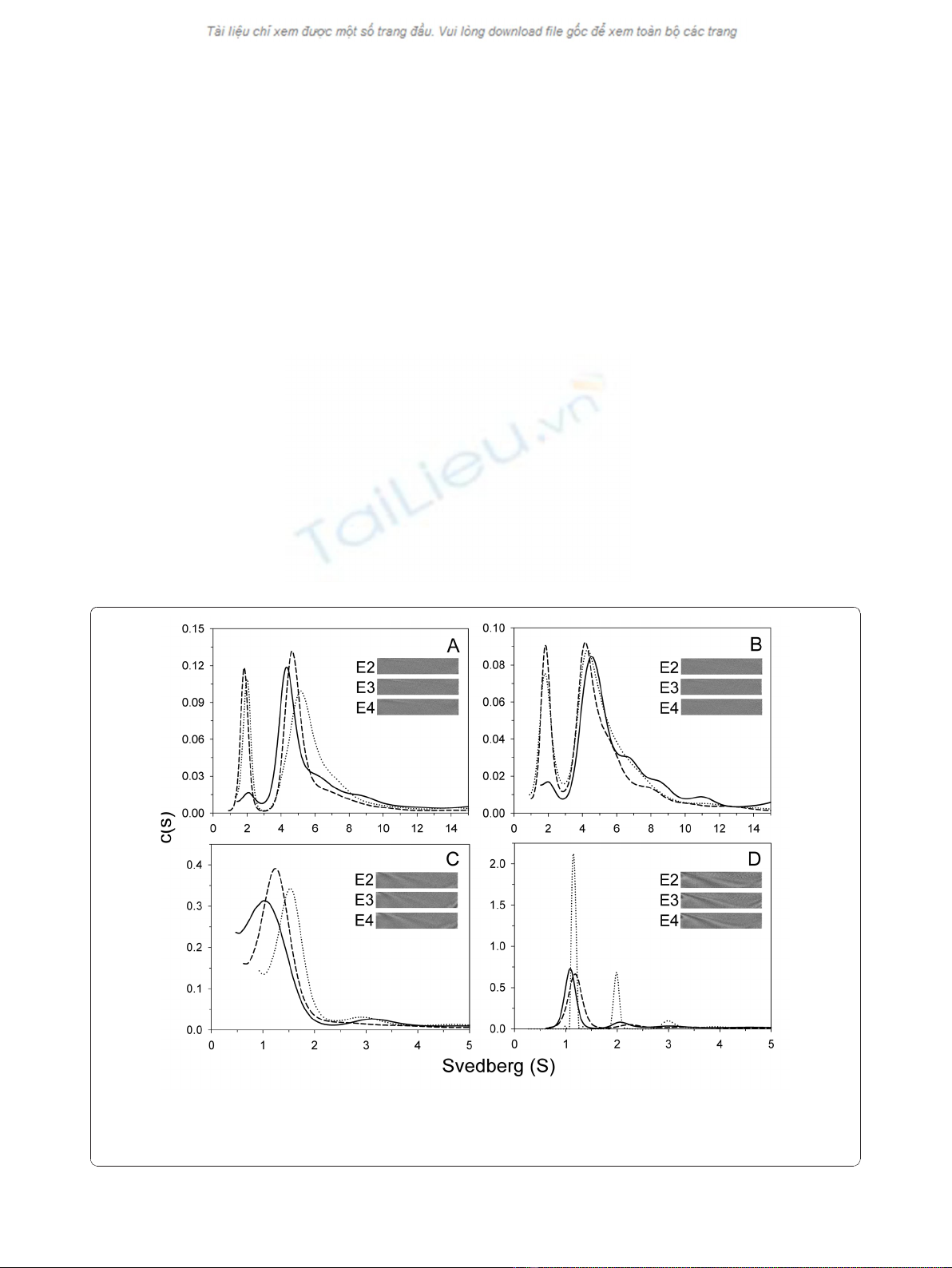
Our SV experiments and c(s) distribution analysis
demonstrate a different species distribution of
apoE-(72-166) in aqueous and lipid environments
In PBS, apoE-(72-166) proteins showed a distribution
pattern of two major species (Figure 2A). The first of
these showed a sedimentation coefficient distribution of
20 % for apoE2-(72-166) and 23 % for apoE3-(72-166) at
s = 2.0, but only 6 % for the same species of apoE4-(72-
166). The second major species was a broad peak at
s=3.5to6.5,withatotaloccupancyof46%for
apoE2-(72-166), 55 % for apoE3-(72-166), and 59 % for
apoE4-(72-166). This region may be the result of a con-
tribution by multi-oligomers. Besides, there were 22-35
% distribution belonged to large aggregated forms. In
the 5 mM DHPC submicellar solution, the small species
(s = 2) of the three apoE-(72-166) increased by 1.3 to
4 % (Figure 2B), whereas the major species at s = 3.5-
6.5 decreased by 2 to 8 %. It suggested that submicellar
DHPC can induce the dissociation of apoE-(72-166)
peptides but not very significantly. In 50 mM DHPC, 76
to 82 % of the apoE-(72-166) proteins dissociated to a
species at s = 1.2-1.5 (Figure 2C). Finally, whilst apoE2-
(72-166) maintained a two species distribution (s = 1.1
and 2.0) in 100 mM DHPC, its apoE3 and apoE4 coun-
terparts maintained a single major species at s = 1.1
(Figure 2D). Furthermore, by c(s) distribution analysis
we found that the average f
r
of apoE-(72-166) in PBS
was around 1.3-1.5, but in 5-50 mM DHPC was around
1.7-1.8, which increased to 1.7-2.1 in 100 mM DHPC
(partial specific volume at 0.86). These differences indi-
cated that when the DHPC concentration increases,
apoE-(72-166) not only displays a dissociation tendency,
but also adopts a more elongated conformation.
The mass variation of the apoE-(72-166) in PBS and in
DHPC was analyzed by global discrete species model
To further clarify the mass variation of the three apoE-
(72-166) peptides in PBS and also in the presence of
DHPC, SE experiments were performed. The SE and SV
data were combined and globally fitted to a multiple
discrete species model using SEDPHAT. Figure 3
showed the best-fit results of apoE3-(72-166) in PBS.
Figure 2 c(s) distribution of apoE-(72-166) proteins in PBS with or without DHPC. The sedimentation velocity data was fitted with the
SEDFIT program using the continuous c(s) distribution model [30]. The fitted curves for apoE2-, apoE3-, and apoE4-(72-166) are shown as dotted,
dash, and solid lines, respectively. Panels A-D: proteins were in PBS, and with 5 mM, 50 mM, or 100 mM DHPC, respectively. Insets, grayscale of
the residual bit map showing the quality of data fitting.
Hsieh and Chou Journal of Biomedical Science 2011, 18:4
http://www.jbiomedsci.com/content/18/1/4
Page 5 of 9


![PET/CT trong ung thư phổi: Báo cáo [Năm]](https://cdn.tailieu.vn/images/document/thumbnail/2024/20240705/sanhobien01/135x160/8121720150427.jpg)


