
Can Tho Journal of Medicine and Pharmacy 9(6) (2023)
136
EX VIVO PERMEATION STUDY OF NANOSTRUCTURED DOSAGE
FORM CONTAINING MANGO SEED KERNEL EXTRACT USING
FRANZ CELL
Than Dang Tuyet Minh, Binh Thi Anh Thu, Le Tra My, Nguyen Huu Nhan,
Le Thi Thanh Yen, Nguyen Ngoc Nha Thao*
Can Tho University of Medicine and Pharmacy
*Corresponding author: nnnthao@ctump.edu.vn
Received: 13/8/2023
Reviewed:07/9/2023
Accepted: 05/10/2023
ABSTRACT
Background: mango seeds (Mangiferin indica L.) have the ability to inhibit P. acnes, S.
aureus, and E. coli bacteria and inhibit inflammation, potentially for transdermal therapeutic
dosage forms. The nano-emulsion dosage forms were prepared based on the SNEDDS system with
nano-oil droplets containing mango seed extract. These dosage forms contributed to carrying the
active ingredient deeply into the impact site, bringing the highest efficiency, but this needs to be
proven. Thus, it is necessary to study the process to evaluate the permeability of these formulas to
prove their availability improvement. Objectives: To assess the transdermal permeability and active
substance release of nanostructured dosage forms by a validated procedure. Methods: an ex vivo
experiment was designed by adding a sample to diffuse through Franz cells. In addition, to ensure
the quality and effectiveness of preparations containing mango seeds, developing and validating a
process for quantifying the total polyphenols in the preparation are extremely necessary. Total
polyphenols were quantified by color complexing with Folin-Ciocalteu reagents, with maximum
absorption at 765 nm. Results: the procedure had been validated according to the International
Conference on Harmonization (ICH) on the criteria of specificity, system compatibility with RSD =
1.02%, linearity built on the concentration range of 10–50 µg/ml with R2 = 0.998, accuracy,
precision with %recovery in the range of 97.73% to 102.56%. The results showed that more than
300 mg/g of polyphenol was released after 6 hours from the tested nanostructured dosage form,
about 4.3 times as many as the total amount of polyphenols in the comparative cream. Conclusions:
the quantification of polyphenols diffused through Franz cells helps evaluate the quality of the
preparation. The procedure had been validated according to the International Conference on
Harmonization (ICH) and could be applied to evaluate nanostructured dosage forms containing
mango seed kernel.
Keywords: mango seed kernel extract, Franz cell, polyphenols, nanostructured dosage forms.
I. INTRODUCTION
Mango’s scientific name is Mangifera indica L (M. indica) in the family Anacardiaceae.
Some studies in the world indicate that mango seeds have antioxidant effects applied to
protect food [1]; in addition, there are anti-inflammatory, antibacterial, and anti-fungal effects
applied to the digestive system, skin disease treatment, etc. [2], [3], [4], [5], [6]. According to
a study by Ha Cao Thien et al. (2022), mango seed kernel extract (MSKE) has significantly
better antibacterial and anti-inflammatory activity than mango seed coat extract [7]. On the
other hand, there has been no further research on biological activity or the application of these
activities to make preparations in Vietnam. In addition, we noticed that nowadays, a common
trend among consumers is using cosmetics made from natural medicinal herbs because of

Can Tho Journal of Medicine and Pharmacy 9(6) (2023)
137
their proximity, safety, and high efficiency. Therefore, MSKE's effects can be applied and
developed into therapeutic dosage forms through the skin.
The self-nanoemulsifying drug delivery system (SNEDDS) is a form of pre-emulsion,
usually formulated in anhydrous forms to form tablets to improve the bioavailability of the drug
when taken orally. SNEDDS is a homogeneous mixture of oil and surfactant or co-surfactant.
When entering the stomach, it will slowly come into contact with the aqueous phase of gastric
digestive juice. Under the contractile action of the stomach, it will form a nanometer-sized oil
and droplet-sized emulsion [8]. For cosmetic products used on the skin, SNEDDS is prepared
to enhance the permeability of therapeutic dosage forms through the skin.
To evaluate the efficacy of these dosage forms, it is essential to develop an ex vivo
Franz static permeation cell experiment to assess transdermal permeability and release of
the active ingredient in the formulation containing MSKE.
This study aims to develop and validate the process of quantifying the total
polyphenols loaded in the preparation by UV-Vis spectroscopy. The process is validated
with the following criteria: specificity, system compatibility test, linearity, precision, and
accuracy. This validated procedure is then applied to quantify the total amount of
polyphenols in creams diffusing through Franz cells.
II. MATERIALS AND METHODS
2.1. Materials, chemicals, and instruments
Materials, chemicals, and reagents: gallic acid (98%) was supplied by Sigma
Aldrich, sodium hydroxide (NaOH), potassium monobasic phosphate (KH2PO4), and
sodium carbonate (Na2CO3) were mixed for day use, and Folin-Ciocalteu reagent was
purchased from Nanjing Duly Biotech. Nanostructured dosage forms of MSKE were
prepared using the SNEDDS technique.
Instruments: instruments used in the present study consist of UV/Vis V-730
spectrophotometer (Kern-Germany) instruments, an analytical balance (Kern AES –
Germany) with an accuracy of 0.0001 g, a magnetic stirrer, a laboratory water bath
(Membert – Germany), and a Franz cell (Vietnam).
2.2 Methods
2.2.1. Method validation
Franz static permeation cells had a receptor compartment volume of 13.45 mL and
an area for permeation of 2 cm2. The solution in the receptor compartment was a phosphate
buffer at pH 7.4. Permeation membranes were taken from the young white porcin ears’
epidermal skin. After purchased, they were cleaned with distilled water and stored
immediately in a NaCl 0.9% solution at 4–8 oC. Hairs, subcutaneous fat tissues, and blood
vessels were removed using scissors to obtain a full-thickness skin of about 500 µm, an area
of 2x2 [10], [11]. Receptor compartment solution: phosphate buffer pH 7.4 was maintained
at 37 ± 0.5 °C. The receptor compartment solution and permeation membranes temperature
was maintained at 37 ± 0.5 °C. Agitation was maintained at 400 rpm. Before applying the
preparation to the permeation membrane surface in the donor compartment, the membranes
were stabilized at this condition for about 30 minutes.
Determination of the maximum wavelength of total polyphenols: the gallic acid standard
solution was diluted employing phosphate buffer pH 7.4 to gain a gallic acid solution

Can Tho Journal of Medicine and Pharmacy 9(6) (2023)
138
concentration of 30 μg/mL. Then, the solution was reacted with a Folin-Ciocalteu reagent. The
absorbance of the solution was scanned in the wavelength range of 450–900 nm using a UV/Vis
V-730 spectrophotometer with phosphate buffer pH 7.4 and Folin-Ciocalteu reagent as a blank.
Determination of total polyphenol concentration: the recovered solution was
reacted with Folin-Ciocalteu reagents to quantify the total polyphenol concentration. The
reaction samples were photometrically measured at the maximum absorption wavelength
(expected at 765 nm). Total polyphenol content is calculated according to gallic acid. For
the test sample: accurately aspirate 1,0 ml of the recovered solution into the test tube, add
5.0 ml of Folin-Ciocalteu reagent, and shake well for 2 minutes. Left at room temperature
for 10 minutes. Add 4,0 ml of sodium carbonate (Na2CO3) at 7.5%. Shake well for 2
minutes, cover tightly, and leave at room temperature for 60 minutes.
Validation of the method: appraisal according to the International Conference on
Harmonization (ICH) guidelines [9] includes the following indicators:
Specificity: scanning the spectrums of gallic acid standard solution, the cream, and
the placebo diffused through the Franz static permeation cells in 6 hours. All were reacted
with a Folin-Ciocalteu reagent.
System Suitability Test: preparation of a gallic acid standard solution with a 10 µg/ml
concentration from the original standard solution. Accurately aspirated 1.0 ml of this solution,
reacted with the Folin-Ciocalteu reagent, and measured absorption at 765 nm (n = 6).
Linearity: on a series of standard solutions with a concentration range of 10–50
µg/ml, we constructed a regression equation representing the linear dependence between
concentration and absorption. Requirements: R2 ≥ 0.99.
Precision: prepare samples on the intraday (n = 6) and interday (n = 3) days. Intraday
precision was evaluated by measuring the total amount of polyphenols in six samples on the
same day. Interday precision was evaluated by continuously measuring the total amount of
polyphenols in three days. All test samples were reacted with a Folin-Ciocalteu reagent and
measured at 765 nm. Calculation of results: From the sample's absorption, calculate the
concentration of total polyphenols in the sample. From the concentration of total
polyphenols in the sample, we measured the total amount of polyphenols in the preparation
that diffused through the Franz static permeation cells in 6 hours by the formula:
Qn = V x Cn. Where: V (ml): volume of the receptor compartment; Cn (g/ml): concentration
of sample received at tn; Qn (µg): the total amount of polyphenols in the preparation that
diffuses through the Franz static permeation cells.
Accuracy: 1.0 mL of reference solution of 16, 20, and 24 µg/ml was added to the test tubes
containing 1.0 mL of test sample taken from the receptor comparison solution in 6 hours (n = 9).
All samples were reacted with the Folin-Ciocalteu reagent, and absorption was measured at 765 nm.
The accuracy was determined based on a percentage of recovery (%Recovery) parameter.
2.2.2. Determination of the release of a nanostructured dosage form containing
mango seed kernel extract by the ex vivo Franz static permeation cells experiment
The release of the MSKE-loaded nanostructured dosage form was determined by
determining the total amount of polyphenols that diffused through the Franz static
permeation cells in the prepared formulation.
The test nano-emulsion cream composition was prepared with the formula: oil phase
including vitamin E, lanolin, stearyl alcohol, and cetyl alcohol; surfactant including tween,
Can Tho Journal of Medicine and Pharmacy 9(6) (2023)
139
span, and polyethylene glycol 400 (PEG 400) as co-surfactant; water phase including
glyceryl, MSKE, and distilled water. The ratio of nano-emulsion phases is 15:50:35. The
water phase was slowly administered with continuous stirring to adjust the nano-emulsion
volume. The MSKE concentration administered was 5% w/w. To compare the ability to
diffuse through the skin and release the active ingredient of nano-emulsion cream
containing MSKE simultaneously, we designed the experiment under the same conditions
as nano-emulsion cream. A comparative cream (normal cream) was prepared with the same
formula but a different preparation method for nano-emulsion cream. The method involved
heating the oil and water phases and then mixing the two phases vigorously and
homogeneously. 30mg of cream was placed on the membrane surface and continuously
contacted the surface for 6 hours in the donor compartment. After 6 hours, 2.0 mL of
solution was removed from the receptor compartment to recover and analyze.
2.2.3. Statistical Analysis
All experiments were conducted in triplicate, and the data were reported in terms of mean
± SD (standard deviation). A student’s t-test (independent sample t-test) was utilized for statistical
purposes to determine the total amount of polyphenol diffused through the ex vivo Franz static
permeation cells between two comparisons, with a p-value of <0.05 for significant comparisons.
III. RESULTS
3.1. Method validation
The maximum wavelength of gallic acid standard solution in phosphate buffer pH
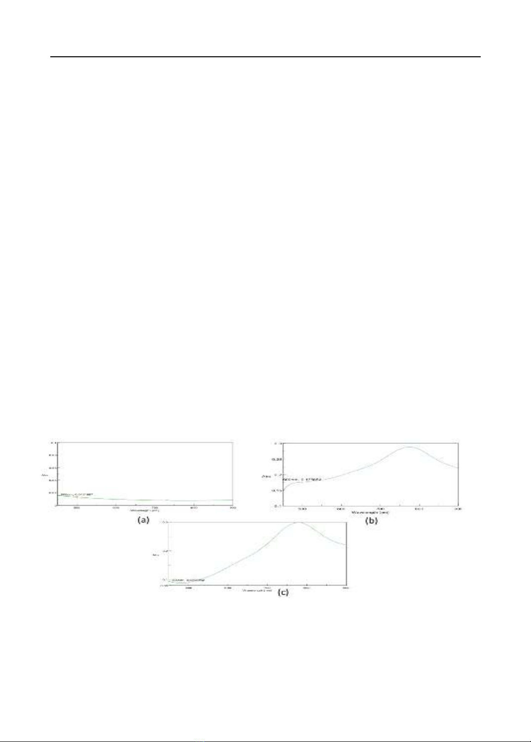
7.4 obtained from the absorbance scanning in the range of 450-900 nm is 765 nm (Figure
1c). Therefore, the maximum wavelength at 765 nm was determined as a quantification
wavelength for further total polyphenols assays in this study.
Specificity: results of the spectrogram (Figure 1) indicated that at the absorption
peak (765nm) of the cream sample, there was no absorption in the placebo sample. Thus,
the process achieves specificity.
Figure 1. UV spectrum of samples(a) Placebo in 6 hours reacted with Folin-Ciocalteu
reagents; (b) Cream in 6 hours reacted with Folin-Ciocalteu reagents; (c) Gallic acid
standard solution reacted with Folin-Ciocalteu reagents.
System suitability test: the measured absorption was 0.098 ± 0.001 (RSD = 1.02%). The
result showed a relative standard deviation (RSD) of absorption of the standard solution after
Can Tho Journal of Medicine and Pharmacy 9(6) (2023)
140
reacting with the Folin-Ciocalteu reagent of 2%. Thus, this analysis method is compatible with
the UV-Vis spectroscopy system.Figure 1. UV spectrum of samples(a) Placebo in 6 hours
reacted with Folin-Ciocalteu reagents; (b) Cream in 6 hours reacted with Folin-Ciocalteu
reagents; (c) Gallic acid standard solution reacted with Folin-Ciocalteu reagents
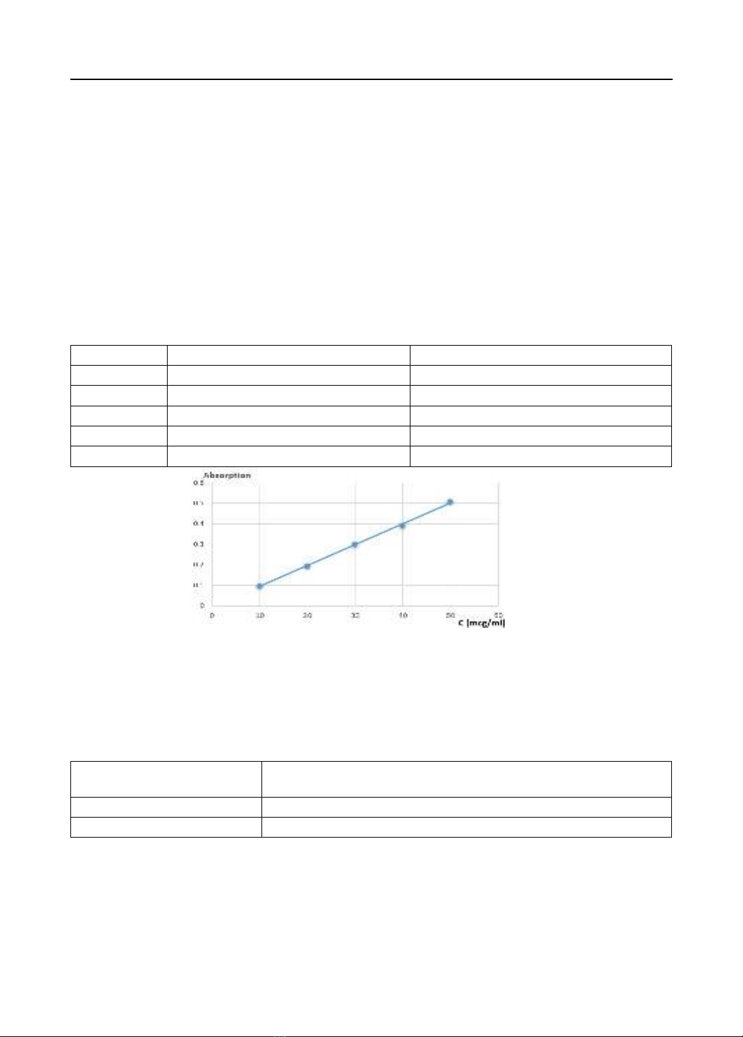
Linearity: accurately taking 1, 2, 3, 4, and 5 ml of the original standard solution into
10 ml flasks, filled to the line with phosphate buffer pH 7.4, These, solutions were shaken
thoroughly to achieve equivalent concentrations of 10, 20, 30, 40, and 50 µg/ml gallic acid.
Accurately aspirated 1.0 ml of the above solutions into test tubes and reacted with Folin-
Ciocalteu reagents. Measuring the absorption of these solutions at 765 nm. The correlation
between concentration and absorption of standard gallic acid is shown in Table 1 and Figure
2. Absorption into gallic acid concentration was linearly correlated with s correlation
coefficient (r) R2=0.998, and the linear regression equation was y = 0.0102x – 0.0056.
Table 1. Absorption of reference solution at concentrations (n = 3)
No.
Concentration (µg/ml)
Absorption (Mean ± SD)
1
10
0.098 ± 0.001
2
20
0.196 ± 0.003
3
30
0.302 ± 0.001
4
40
0.392 ± 0.001
5
50
0.508 ± 0.002
Figure 2. Plots of the calibration curve of the gallic acid standard
Precision: measuring the total amount of polyphenols that diffused through the
membrane in 6 hours based on the intraday (n=6) and the interday (n=3). The results showed
that the relative standard deviations (RSD) for both intraday and interday measurements were
<2% (Table 2). Thus, the method is highly accurate and meets the analysis requirements.
Table 2. Precision results
Precision
The total amount of polyphenols that diffused through the
membrane in 6 hours (µg)
Intraday (Mean ± SD)
300.54 ± 4.11* RSD= 1.37%
Interday (Mean ± SD)
295.62 ± 5.12* RSD= 1.73%
*Concentration was calculated from the linear regression equation.
Accuracy: based on the linear regression equation, calculate the recovery
concentration and determine the percentage of recovery (%Recovery). The quantitative
method exhibits high accuracy, with the recovery percentage ranging from 97.37% to
102.65%, averaging 100.12%, and showing an RSD of 1.57% (Table 3).


![Tài liệu học tập Chuyên đề tế bào [mới nhất]](https://cdn.tailieu.vn/images/document/thumbnail/2025/20250906/huutuan0/135x160/56151757299182.jpg)

![Câu hỏi ôn tập Sinh học tế bào [chuẩn nhất]](https://cdn.tailieu.vn/images/document/thumbnail/2025/20250709/kimphuong1001/135x160/771752031316.jpg)
![Lysosome là gì? - Nguyễn Huỳnh Thịnh [Giải thích A-Z]](https://cdn.tailieu.vn/images/document/thumbnail/2015/20151217/conheokiss/135x160/463746382.jpg)


![Tài liệu giảng dạy Sinh học và di truyền [mới nhất]](https://cdn.tailieu.vn/images/document/thumbnail/2026/20260323/hoatudang2026/135x160/42181774414220.jpg)

![Giáo trình Công nghệ vi sinh (Nghề Công nghệ sinh học TC/CĐ) - Trường Cao đẳng Đà Lạt [Mới nhất]](https://cdn.tailieu.vn/images/document/thumbnail/2026/20260224/hoacattuong2026/135x160/87621772161812.jpg)


![Giáo trình Vi sinh vật học môi trường Phần 1: [Thêm thông tin chi tiết nếu có để tối ưu SEO]](https://cdn.tailieu.vn/images/document/thumbnail/2025/20251015/khanhchi0906/135x160/45461768548101.jpg)


