7
Journal of Medicine and Pharmacy, Volume 12, No.07/2022
The role of Hepatitis B Core Antibody: Significance and Clinical
practice
Phan Thi Hang Giang1*, Le Ba Hua1, Phan Ngoc Dan Thanh1, Nguyen Thi Huyen1, Tran Long Anh1,
Tran Thi Bich Ngoc1, Truong Thi Bich Phuong1, Nguyen Thi Phuoc1, Phan Thi Minh Phuong1
(1) Department of Immunology-Pathophysiology, University of Medicine and Pharmacy, Hue university
Abstract
Hepatitis B infection remains a global health problem, with progression to acute-chronic hepatitis,
severe liver failure, and death making hepatitis B one of the most serious infections worldwide. The disease
is most widely transmitted from an infected mother to her baby, after exposure to infected blood or body
fluids or unsafe sexual contact. Pregnant women, adolescents, and all adults at high risk for chronic infection
are recommended to be screened for hepatitis B. Serological tests allow the distinction between acute and
chronic hepatitis. Meanwhile, the molecular tests performed provide detection and quantification of viral
DNA, genotyping, drug resistance, and pre-core/core mutation analysis to confirm infection and follow
monitoring disease progression in patients with chronic hepatitis B. Because, the current treatment is only
based on nucleotide analogs and pegylated interferons that save lives by decreasing liver cancer death, liver
transplant, slowing or reversing the progression of liver disease as well as the virus infectivity. In this review,
we clearly light the role of Hepatitis B Core Antibody, therefore clinicians understand the need to screen
for hepatitis B core antigen (Anti-HBc), proper interpretation of HBV biomarkers, and that “anti-HBc only
indicates HBV exposure, lifelong persistence of cccDNA with incomplete infection control, and potential risk
for reactivation.
Keywords: Hepatitis B virus (HBV), Hepatitis B Core Antibody (Anti-HBc/HbcAb).
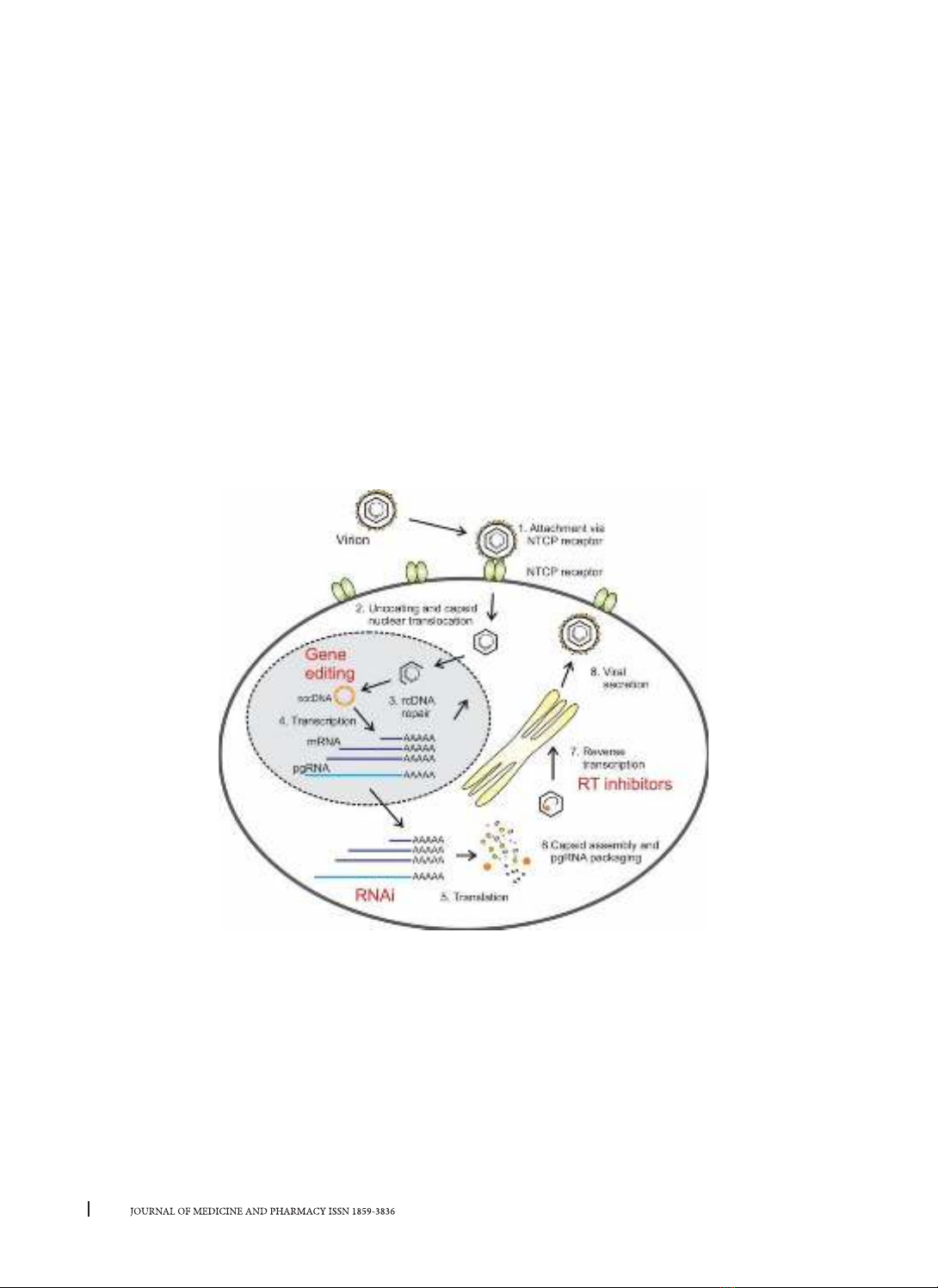
1. INTRODUCTION
Despite the availability of vaccines and robust
treatment strategies, infection with the Hepatitis
B virus remains a severe worldwide illness because
Hepatitis B virus (HBV) infection leads to acute-
chronic hepatitis, severe liver failure, and liver cancer
with high morbidity and mortality. An estimated 2
billion people worldwide have been infected with the
hepatitis B virus in the presence of the hepatitis B core
antigen (anti-HBc). In approximately 95% of adults,
exposure to HBV leads to an acute infection that
rapidly resolves without long-term consequences,
while the remaining 5% do not control viral infection,
leading to chronic [1, 2]. Over 292 million people
are living with chronic hepatitis B (CHB) worldwide
Global HBV with surface antigen (HBsAg) positivity
was estimated at 3.9% in 2016 [3]. Annually, 887,000
deaths yearly occur from HBV and related diseases,
mainly cirrhosis, and advanced cirrhosis. The risk and
progression of chronic infection are age-dependent
and occur mostly in immunocompromised
individuals. It is shown that acute HBV infection is
usually cleared in immunocompetent individuals,
but chronic HBV infection develops in about 90% of
infants, 30 - 50% of children aged five, and 5 - 10% of
adults [4].
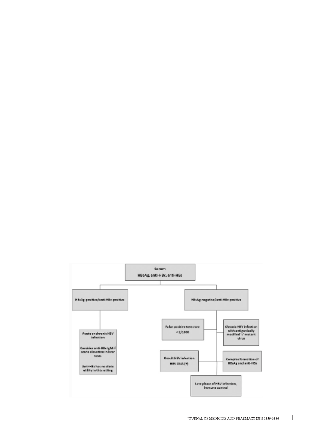
Occult Hepatitis B infection (OBI) was defined in
the group with undetectable HBsAg, defined as the
presence of HBV DNA in the liver of HBsAg-negative
individuals. OBI has been shown to occur both in the
absence and presence of anti-HBc and/or anti-HBs.
OBI rates have been reported to be more common
in patients at high risk for gastrointestinal infections
such as hepatitis C virus (HCV) and HIV infection
[5]. OBI is associated with severe liver injury and
hepatocellular carcinoma (HCC), and poses a risk
to individuals, particularly in transfusion infections,
HBV reactivation, chronic liver disease, and HCC [6].
Isolated anti-HBc (IAHBc) is a particular serotype
seen in immunocompromised patients. Isolated
anti-HBc is determined by negative anti-hepatitis
B antigen and positive anti-hepatitis B antibody
(whether in the form of IgM or IgG). It is especially
important to screen immunocompromised patients
for IAHB because HBV replication can be reactivated
with the potential for morbidity and mortality [7].
This review describes virological tests, including
serological and molecular techniques for the
diagnosis of HBV infection, and specifically updates
the role of Anti-HBc in clinical practice.
Corresponding author: Phan Thi Hang Giang, email: pthgiang@huemed-univ.edu.vn
Recieved: 14/10/2022; Accepted: 18/11/2022; Published: 30/12/2022
DOI: 10.34071/jmp.2022.7.1
8
Journal of Medicine and Pharmacy, Volume 12, No.07/2022
2. PATHOPHYSIOLOGY OF HBV INFECTION
The Hepatitis B virus belongs to the family
Hepadnaviridae. Virions also known as Dane
granules, are made by a capsid with an envelope
around it, which is entirely infectious. HBV contains
viral DNA and two enzymes DNA-polymerase and
protein kinase. The HBV genome is an open circular
DNA molecule with four open reading frames called
P, S, C, and X encode for viral proteins.
The HBV replication cycle initiates the
interaction between HBsAg and hepatocyte
surface proteins that help HBV attach to and enter
hepatocytes. This is followed by relaxed cyclic DNA
(rcDNA) which is changed to Covalently closed
circular DNA (cccDNA) and is required for the
transcription and production of new RNA and DNA,
which is then matured by the viral polymerase
(Figure 1). HBV DNA can exist in hepatocytes as
cccDNA, unlike rcDNA found in viruses. CccDNA
also contributes to core antigen (HBcAg) synthesis
in hepatocytes and the production of anti-HBc
antibodies, explaining their presence in acute,
chronic and resolved HBV infections Normally,
there are Four types of responses that have been
described in HBV infection. Firstly, there can be a
strong immune response leading to the complete
and fast elimination of HBV and infected cells,
leading to acute hepatitis and/or hepatocellular
necrosis. Secondly, the immune response can
be useful but not strong, often presenting an
asymptomatic infection to resolve progressively.
Thirdly, the immune response is inadequate, with
a state of tolerance resulting a part in considerable
prolonged HBV replication. Lastly, the immune the
response may be unstable, which is usually related
to the asymptomatic carrier of HBV [8, 9].
Figure 1. Hepatitis B virus life in hepatocytes [10].
3. LABORATORY DIAGNOSIS OF HEPATITIS B
VIRUS
In practice, the assessment of HBV infection
begins with the patient history, physical examination,
and assessment of liver function, through analysis
of various hepatitis serologic markers and/or their
combinations such as HBsAg, HBeAg, anti-HBs,
anti-HBc (IgM, IgG), anti-HBe, and biochemical
parameters, molecular assay [11]. The Hepatitis B
Foundation recommends screening all adults for HBV
with a triad serological marker that includes HBsAg,
anti-HBs, and total anti-HBc. To stage infection in
HBV-infected patients, the following should be
performed: 1) testing for HBsAg, HBeAg/anti-HBe,
HBV DNA; 2) liver blood tests including aspartate
aminotransferase (AST), alanine transaminase
(ALT), and 3) transient elastography (Fibroscan) as a
noninvasive test or needle biopsy of the liver as an
9
Journal of Medicine and Pharmacy, Volume 12, No.07/2022
invasive method for the presence of cirrhosis [12].
3.1. HBV serological markers
To assess the patient’s HBV status, HBsAg is always
tested along with anti-HBc and anti-HBs (Figure 2). A
positive HBsAg antigen indicates ongoing infection and
infection. Initially, the majority of infections are positive
for hepatitis B e antigen (HBeAg). However, with
chronic infection, HBeAg is often lost. Loss of HBeAg,
whether spontaneous or treatment-induced and
with or without anti-HBe seroconversion, represents
partial immune control of chronic HBV infection, often
resulting in a low replication phase [11]. In later stages
of the disease, loss of HBeAg is sometimes caused by
viral variants or mutations. It is interesting to note that
total anti-HBc is usually lifelong.
HBsAg: HBsAg is an envelope protein expressed
on the surface of infectious virions called Dane
particles. The detection of positive HBsAg in the
serum indicates the current HBV infection condition
[11]. The incubation stage for hepatitis is around
60–150 days after exposure to HBV, and HBsAg
appears in the blood between 1–10 weeks after
initial exposure to HBV [11]. During the immune
window, HBsAg can disappear rapidly without the
appearance of Anti-HBs, and IgM Anti-HBc is the only
evidence of infection during this period. If HBsAg
positivity persists after 6 months, it means the
progression of a chronic HBV infection. Quantitative
immunofluorescence analysis was performed to
assess the HBsAg level of chronic HBV patients and
is a useful marker for interferon-alfa-treated chronic
HBV patients with HBeAg-negative [12].
Anti-HBs/HBsAb: The presence of anti-HBs
in serum indicates convalescence and immunity
against HBV infection by an infected virus or by
immunized HBV vaccine. People with first-degree
relatives such as a parent-child or a partner who are
chronic carriers are recommended to be vaccinated
if their triple sera screening test is negative [12].
The titer of anti-HBs should be ≥10 mIU/ml for
protection [13].
HBeAg and anti-HBe: HBeAg stands for Hepatitis
B Envelope Antigen. The presence of HBeAg
correlated with active viral replication is indicative
of the infectivity of patients with HBV. Meanwhile,
the appearance of anti-HBe indicates low viral
replication and is strong evidence for infection
resolution [14]. These tests are commonly used to
determine the stage of chronic HBV infection [12].
Anti-HBc/HBcAb: Anti-HBc also known as
HBcAb is an antibody against the core antigen of the
hepatitis B virus, this antibody appears very early
and persists for life. Detection of anti-HBc antigens
confirms HBV exposure and indicates acute, chronic,
or resolved infection but not vaccine immunity [15].
The presence of IgM Anti-HBc, along with HBsAg
positivity, usually indicates an acute infection, which
usually lasts no more than six months [16]. Anti-
HBc-positive and Anti-HBs-negative individuals have
chronic infections and show a reduced risk of HBV
reactivation. However, there is also no clinical benefit
of vaccination for individuals who are only Anti-HBc
positive due to HBV exposure or who are anti-HBc
and anti-HBs positive due to immune control [17].
Figure 2. Interpretation of HBsAg, Anti-HBs and anti-HBc test results [12]
10
Journal of Medicine and Pharmacy, Volume 12, No.07/2022
3.2. Biochemical parameters
The severity of liver damage as inflammation and
fibrosis is assessed using biochemical parameters,
including AST (Aspartate aminotransferase)
and ALT (Alanine aminotransferase) which are
enzymes released from the liver in response to
damage and liver disease. Besides, There are other
biochemical parameters including gamma-glutamyl
transpeptidase (GGT), alkaline phosphatase (ALP),
bilirubin, serum albumin gamma globulin, and
prothrombin time (PT) for diagnosing liver disease
[12]. But, in case biochemical and HBV markers are
not sure, then invasive and noninvasive methods
are chosen to determine the stage of liver injury
[12]. Because liver biopsy is an invasive, expensive,
and hurtful technique compared to the other
techniques, many non-invasive techniques are safe
to identify the stage of the presence of fibrosis in
chronic HBV patients. Therefore, the APRI index
is calculated according to the formula = [AST/AST
upper limit of normal (ULN) × 100/platelet count]
[18] of The WHO recommends estimating the stage
of liver fibrosis [11]. It has been recommended that
40 IU/ml as the AST ULN value should be used in
the APRI formula. In addition, ALT levels must also
be evaluated in chronic HBV patients as it is relative
to the severe disease. According to the WHO
guidelines, the ULN ALT levels are below 30 U/l for
men and 19 U/l for women [11].
3.3. Molecular assays
Molecular diagnostic methods are used for
HBV DNA quantification, genotype, determining
mutations of drug resistance, and analysis of
pre-core/core mutation [19]. Currently, various
molecular assays are recommended by FDA (2019)
for the diagnosis of HBV infection such as the
UltraQual HBV PCR technique, COBAS AmpliScreen
HBV technique, Procleix Ultrio test, Procleix
Ultrio Plus technique, and COBAS TaqScreen MPX
technique, nucleic acids amplification tests (NATs).
HBV DNA quantification: HBV DNA by NATs is
recommended to evaluate the infectivity of HBsAg-
positive individuals and HBsAg-positive pregnancy
to prevent transmission risk and lead to a conclusion
of whether to treat HBV disease. And HBV DNA
quantification with molecular assays allows early
detection of risk people with HBV acute disease
before HBsAg emerges and excludes OBI [20]. The
methods of HBV DNA are also used to follow the
treatment response in chronic HBV infection patients
[11]. The HBV DNA concentration reflects the disease
evaluation, the long-term following of chronic
HBV infection, and the treatments effectiveness
to prevent the evolution of HCC (Hepatocellular
carcinoma). The viral concentration of HBV is often
evaluated by IU/ml or by copies/ml. The metering
of the level of HBV DNA is recommended to be used
with a more sensitive method rt-PCR assay with a 10
IU/ml detection limit [12].
Until now, HBV genotyping, drug resistance,
and pre-core/core mutations have been confirmed.
There are 10 genotypes of HBV, from A to J, and
more than 40 sub-genotypes [21], which may be
related to the concentration of viral in the host,
risk of progressive fibrosis, responses of antiviral
treatment, and follow the mutations progress in
clinical practice [6]. Although HBV genotyping is
not necessary for diagnosis at the first step, also
sequencing the determination of HBV genotypes
and resistance drug mutations is a useful parameter
for patients at risk of developing HCC to monitor an
effective therapy [12].
4. THE ROLE OF HEPATITIS B CORE ANTIBODY
The HBV core antibody (anti-HBc) serological
markers test is chosen for monitoring a history of
the infection. IgM Anti-HBc is the earliest antibody
of the body to perform in the immune response to
HBV infection. Meanwhile, IgG Anti-HBc typically
persists for life after 6 months of the HBV infection.
Thus, IgM anti-HBc is a marker of acute infection
while IgG anti-HBc is a marker of chronic infection.
It is interesting that the total anti-HBc (IgM anti-HBc
and IgG anti-HBc) usually lasts a lifetime. Therefore,
screening for hepatitis B core antigen (anti-HBc)
helps to clarify the role of HBV serological markers
including anti-HBc and “anti-HBc only status in
confirming HBV infection, but not due to vaccination
and moreover, the indefinite existence of cccDNA
in host hepatocytes resulting in infection control
not completely, and the potential of risk for HBV
reactivation as described analyzed earlier in this
review [22].
4.1. Anti-HBc Test: Effective and Meaningful
Historically, although there were many versions
of tests anti-HBc markers with various different
specificities, most countries, and labs in the
world now are using the FDA-cleared method and
technique. Currently, these anti-HBc marker tests
are really effective with a specificity of more than
99.8% for healthy donors’ blood and leading to
confirmation of residual disease (cccDNA) in the
host and a necessary consideration is whether to
HBV vaccinate against HBV or not [23, 24].
Normally, most the normal immune system
individuals are protected from the infected HBV,
11
Journal of Medicine and Pharmacy, Volume 12, No.07/2022
but these cases can also have reactivation under
various conditions [25, 26]. Because a clearance
cure for HBV infection may not be completed
because the DNA of HBV is in the nucleus as
the cccDNA form and is integrated into the host
genome for surface antigen (HBsAg) production.
Even the people who have recovered after the
acute HBV infection stage, cccDNA can still be found
in the liver cells, the existence of these cccDNAs
explains that when these recovered individuals
are profoundly immunosuppressed for any reason,
there is a reactivation of HBV replication [27]. This
explains why, in patients with HBV infection who
have resolved or become chronic, the available
prophylactic hepatitis B vaccine has not shown any
clinical benefit [28, 29]. People with a history of past
HBV infection, whether they are currently infected
or not, may be at risk for hepatitis reactivation
during and after immunosuppressive therapy. HBV
reactivation has the potential to lead to fulminant
liver failure and/or death [30]. Therefore, it is
important to identify individuals who may be at risk
of reactivation prior to immunosuppressive therapy.
4.2. Isolated anti-HBc (IAHBc) and Occult
Hepatitis B Infection (OBI)
Isolated anti-HBc (IAHBc) is a specific HBV
serological sample, defined as both HBsAg and anti-
HBs negative but anti-HBc positive (whether IgM or
IgG). The prevalence of this sera profile may be high,
particularly in patients infected with hepatitis C virus
(HCV), patients with human immunodeficiency virus
(HIV), and other immunocompromised patients,
such as cancer patients. Although this pattern may
be a serological false positive, it can also be found
in patients with Occult Hepatitis B Infection (OBI).
However, it is particularly important to screen
immunocompromised patients for IAHBc to prevent
HBV replication that could be reactivated with the
potential for severe illness and death [7].
Occult Hepatitis B Infection (OBI) is defined as
HBsAg-negative hepatitis B with detectable HBV
DNA in the liver or blood [31]. The definition of
OBI is not limited to an isolated anti-HBc pattern.
OBI can be the association of negative HBsAg with
positive HBV DNA in the blood and/or the liver
[32], or positive HBV DNA in the liver whatever
the level in the blood [33]. However, the criterion
in European and American guidelines [12, 15], the
positive blood HBV DNA is still kept for confirming
OBI. The concentration of anti-HBc may help
differentiate OBI from other cases of positive anti-
HBc. Indeed, anti-HBc is produced by the immune
system against HBcAg, a viral nucleocapsid protein
that is the most immunogenic component of HBV
in the cell liver. These antibodies are known as the
marker of a stage HBV infection or exposition, and
they can persist for 10–20 years or more after viral
clearance. The level of these antibodies is known to
fluctuate depending on the stage of HBV infection
[34]. It has been reported that the levels were
higher in cases of chronic HBV hepatitis than in OBI,
and higher in OBI than in cases of past/cured HBV
infection [35]. In patients with an isolated anti-HBc
profile, a cut-off of 6.6 IU/mL was associated with
a sensitivity of 60.7% and a specificity of 75.3% for
discriminating OBI and the past infection. Finally,
a quantitative assessment of anti-HBc might be a
straightforward means of screening the different
stages of HBV infection.
5. CONCLUSION AND FUTURE PERSPECTIVES
In summary, It is necessary for clinicians
to understand the importance of screening
patients to evaluate the anti-HBc status, proper
interpretation of HBV biomarkers, and that “anti-
HBc only indicates exposure to HBV, lifelong
persistence of cccDNA with incomplete control
of infection, and risk for reactivation under strong
immunosuppression. It is also very important
to expand understanding among both clinicians
and patients that HBV reactivation is a risk in
individuals requiring HCV therapy, chemotherapy,
or potent immunosuppressive therapy. Despite the
substantial body of evidence demonstrating the risk
of reactivation, this too often goes unaddressed in
clinical settings where patients are not screened for
HBV infection prior to initiation of such therapies
and thus do not receive appropriate prophylaxis.
Careful screening of liver transplant recipients and
organ donors for anti-HBc, with appropriate follow
up including transplant recipient vaccination where
appropriate, is also necessary. As the usefulness
of anti-HBc levels is confirmed, the anti-HBc test
should also be considered expanded in research
settings and clinical practice.