HUE JOURNAL OF MEDICINE AND PHARMACY ISSN 3030-4318; eISSN: 3030-4326 203
Hue Journal of Medicine and Pharmacy, Volume 14, No.6/2024
Invasive papillary thyroid carcinoma appearing in a thyroglossal duct
cyst: A rare case report
Nguyen Van Mao1*, Ngo Quy Tran1, Tran Thi Nam Phuong1, Tran Nam Dong1, Le Thi Thu Thao1,
Tran Van Bao1, Vo Thi Hanh Thao1, Nguyen Phuong Thao Tien1, Tran Anh Hung1
(1) Department of Embryology, Histology, Pathology and Forensic Medicine,
University of Medicine and Pharmacy, Hue University
Abstract
Thyroglossal duct cyst (TGDC) is the most frequent congenital anomaly of the thyroid gland. The existence
of primary malignancy originating from this cyst is uncommon, accounting for less than 1% of all cases.
Because of its rarity, there is no universal consensus regarding optimal treatment option. We present a case
of a 59-year-old woman with a 25x20 mm mass in the anterior area of the neck that gradually increased
in size over six months. A total thyroidectomy and Sistrunk procedure (SP) were performed. Postsurgery
histologic evaluation confirmed papillary carcinoma of the TGDC invading adjacent muscle tissue.
Keywords: thyroglossal duct cyst, papillary carcinoma, Sistrunk procedure.
Corresponding Author: Nguyen Van Mao. Email: nvmao@huemed-univ.edu.vn
Received: .26/3/2024; Accepted: 10/10/2024; Published: 25/12/2024
DOI: 10.34071/jmp.2024.6.29
1. INTRODUCTION
Thyroglossal duct cysts (TGDCs) are the most
common abnormalities in the formation of thyroid
gland. They are found more than 75% of midline
neck mass in children and around 7% of adults [1],
[2]. During development, the thyroid gland descends
from the foramen cecum of the tongue to its final
position in the inferior neck, forming thyroglossal
duct that maintains connected to its original
position. The thyroglossal duct typically completely
involutes between weeks 7 and 10 gestation [3], [4].
Remnants of the thyroglossal duct can give rise to
the development of TGDCs [5]. Carcinomas arising
from thyroglossal duct remnant cysts are extremely
rare, occurring in fewer than 1% of cysts. The mainly
histological type is papillary-type thyroid carcinoma,
followed by mixed papillary-follicular carcinoma and
other variants have also been described [6]. Given
the rarity of this diagnosis, we present a case of TGDC
carcinoma in
a female patient, with normal thyroid
gland.
2. CASE PRESENTATION
A 59-year-old female patient visited our hospital
with a midline neck mass that was increasing
gradually in size for six months. In recent weeks, she
began to experience significant feeling of pressure
associated with this mass. Physical examination
revealed a 25x20mm mass that was hard, fixed
and well-demarcated mass on the anterior neck.
Clinically, a diagnosis of the TGDC (with a possible
malignancy) was considered. Lab tests were within
normal limits, including complete blood count and
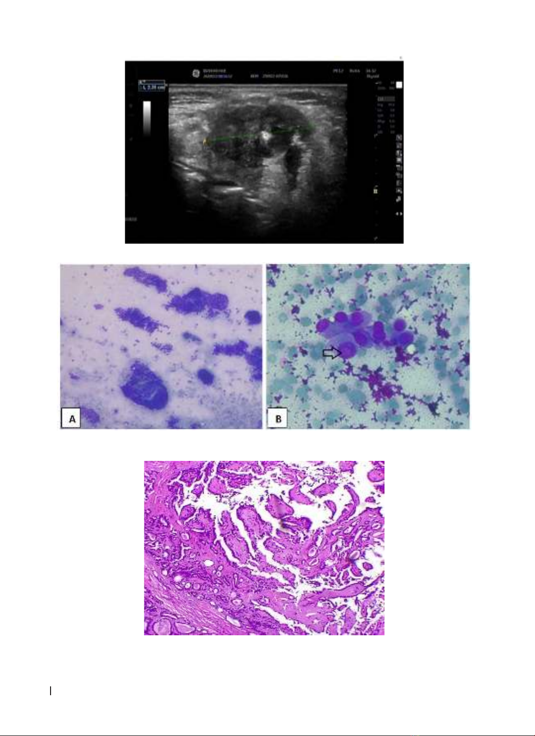
thyroid funtional tests. A neck and thyroid gland
ultrasound were performed which showed a well-
defined and heterogeneous lesion of 24x13 mm with
solid component occupying 50% in the midline of
the anterior neck area, between the hyoid bone and
thyroid cartilage (Figure 1). There were no clinical
and radiographic findings of thyroid gland disease
and associated cervical lymph nodes.
Fine-needle aspiration (FNA) demonstrated
classic features characteristic of papillary carcinoma
(including papillary formations, nuclear grooves and
nuclear pseudoinclusions) (Figure 2).
This patient underwent total thyroidectomy along
with SP. Intra-operatively, a 2.5x1.5 cm cystic tumor
with solid components was found. Intraoperative
biopsy indicated a papillary carcinoma that infiltrates
surrounding muscle tissue. Therefore, the tumor,
the thyroid gland, the hyoid bone and the bilateral
cervical lymph node were removed and sent for
further evaluation. The postoperative pathologic
report showed the papillary neoplasm appearing
in the TGDC with the presence of normal thyroid
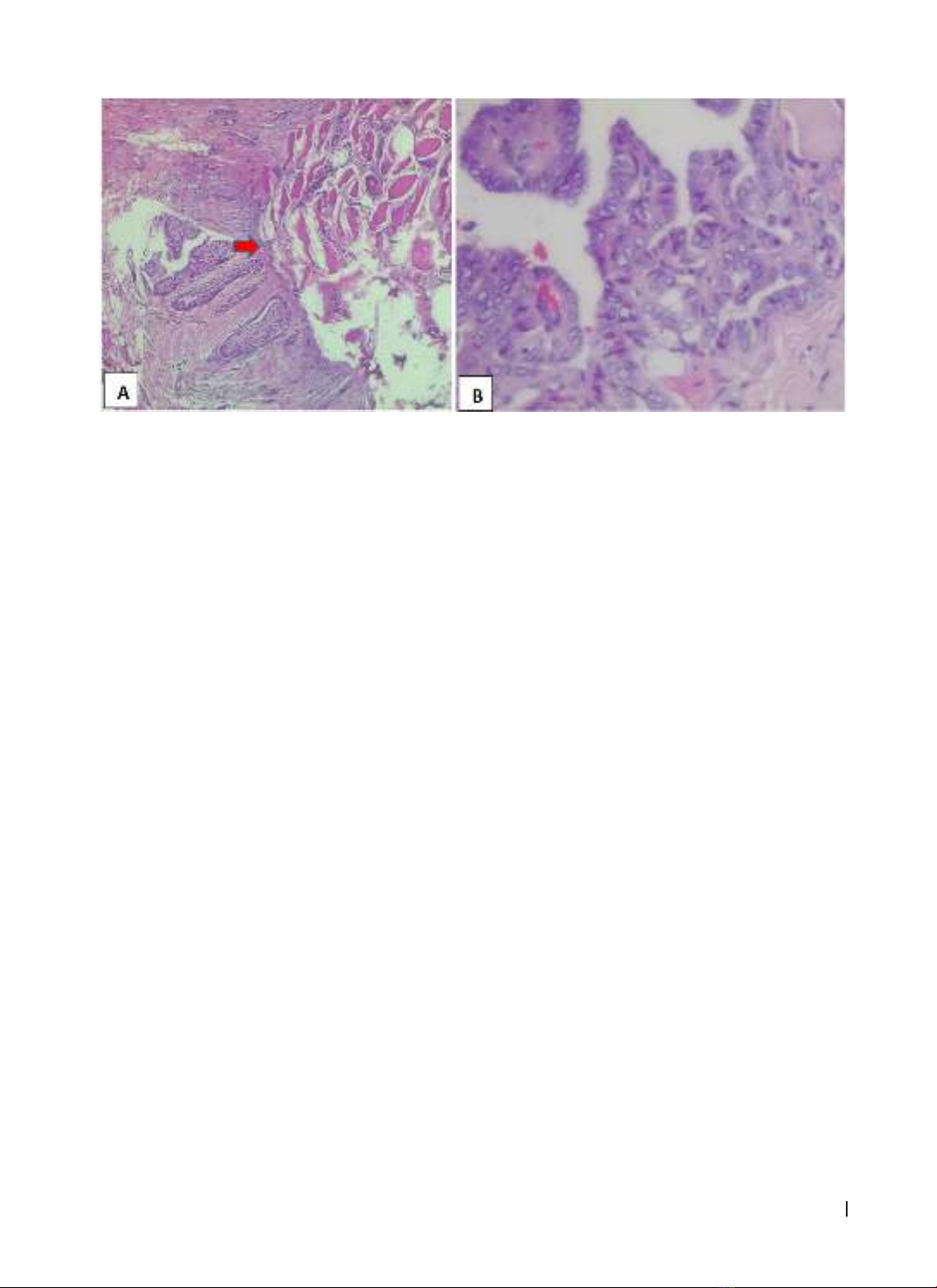
follicles within wall of the cyst (Figure 3). Microscopic
examination of the papillary neoplasm described the
presence of true papillae with fibrovascular cores
and the lining cells with nuclear features of papillary
carcinoma (Figure 4B). The tumor cells infiltrated into
the cystic wall and adjacent soft tissue (Figure 4A).
Additionally, entire remaining thyroid parenchyma
was confirmed that there were no abnormal findings.
Consequently, a diagnosis of papillary carcinoma
evolving from the TGDC was made.