HUE JOURNAL OF MEDICINE AND PHARMACY ISSN 1859-3836
20
Hue Journal of Medicine and Pharmacy, Volume 14, No.2-2024
Evaluating of the effect of low-level laser therapy on wound healing in
rabbits
Nguyen Thi Bich Phuong1*, Nguyen Ngoc Tuan1, Dinh Van Han1, Nguyen Nhu Lam1,
Nguyen Thi Huong1, Le Thi Hong Hanh2, Tran Quoc Tien3, Tong Quang Cong3*
(1) Le Huu Trac National Burn Hospital
(2) Vietnam Military Medical University
(3) Institute of Materials Science
Vietnamese Academy of Science and Technology, Hanoi, Vietnam
Abstract
Background: A large number of studies have demonstrated the wound-healing effects of LLLT in vitro, in
animal models, and in clinical practice. However, there are also differences in the study results, which are
dose and wavelength dependent of LLLT. Objective: Evaluation of the wound healing process in experimental
animals treated with low- level laser therapy in clinical and histopathology. Subjects and Methods: Prospective
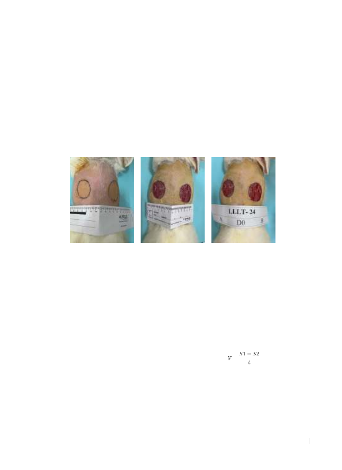
study on 30 rabbits, each rabbit created two full thickness of 2R = 4 cm wounds on both sides of the back:
wound A (treated with LLLT, 780 nm, 3 J/cm2 with 72 s irradiation time, 1 time per day), wound B (control: no
laser). Wounds are bandaged and laser irradiated once a day according to the procedure until the lesion is
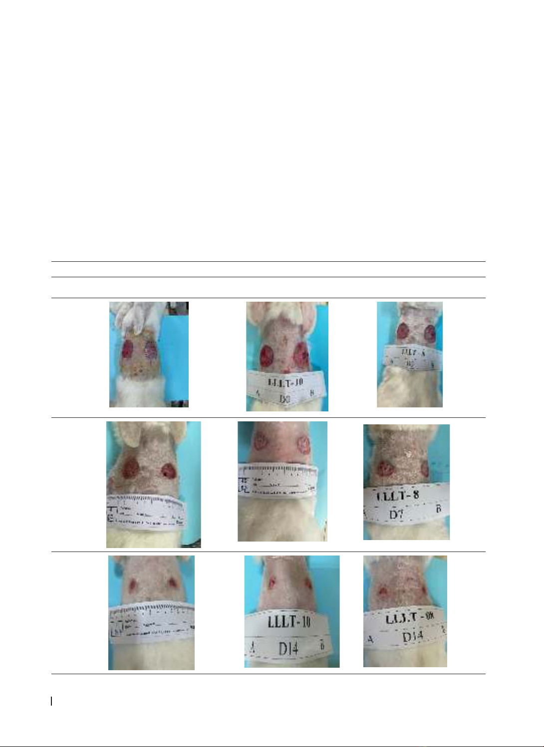
completely epithelialized. Wound biopsy was taken at: before treatment (D0), after 7 days (D7), after 14 days
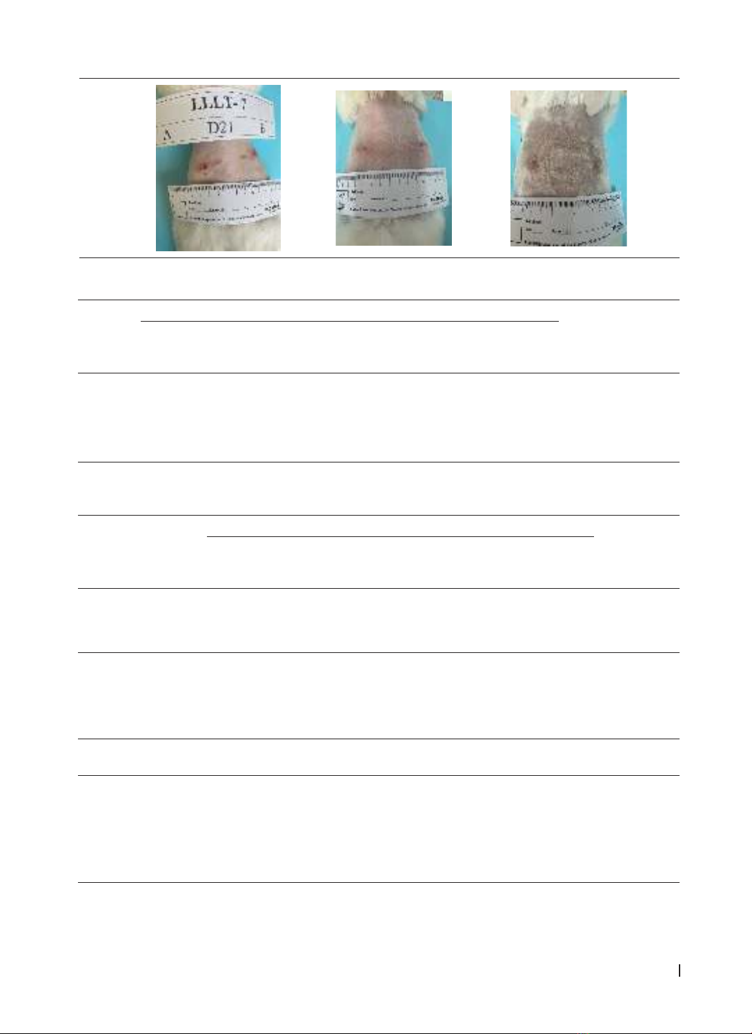
(D14) of treatment. Monitor and evaluate progress at the local wound. Results: The area and speed of wound
narrowing on the side of the laser site narrowed faster than the control side (p < 0.05). The results of rabbit
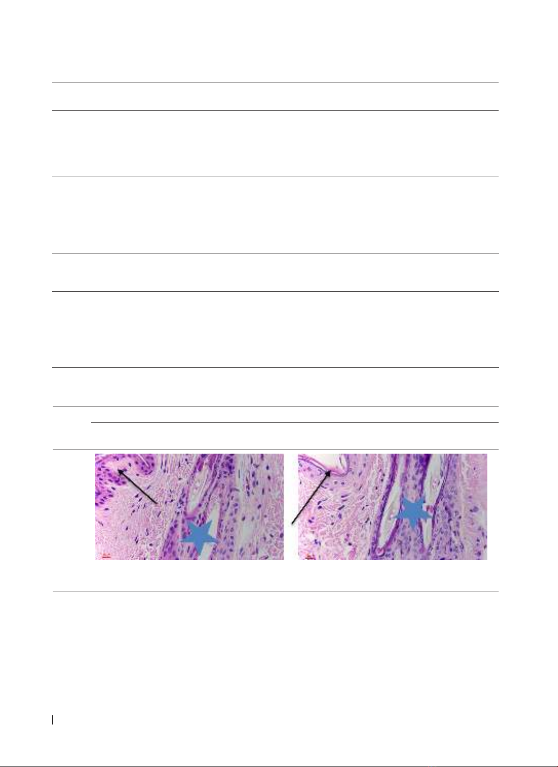
skin histopathology showed that the number of inflammatory cells on the laser side decreased significantly
compared with the non-laser side (p(D14) < 0.05), while the number of neovascular and fibroblasts increased
rapidly on the LLLT side when compared with the control side (p(D7) < 0.05). Conclusions: LLLT (780 nm, dose
3J/cm2) increased wound healing in experimental rabbit model. LLLT promotes wound narrowing, reduces
inflammation, stimulates angiogenesis, and increases collagen synthesis fibroblasts.
Keywords: low-level laser therapy, wound healing, experimental animals, histopathology.
Corresponding author: Tong Quang Cong; Email: congtq2004@gmail.com
Nguyen Thi Bich Phuong. Email: bsphuongvbqg@gmail.com
Recieved: 11/8/2023; Accepted: 19/2/2024; Published: 25/2/2024
DOI: 10.34071/jmp.2024.2.3
1. INTRODUCTION
Since the 1960s, the efficacy of low-level laser
therapy (LLLT) in wound treatment has been
reported. Adre Mester, a Hungarian physicist, was
the first to study the biological effectiveness of
LLLT. Numerous reports have been published over
the past few decades, demonstrating the positive
effects of low-level laser therapy (LLLT) in in vitro, in
vivo, and clinical studies [1,2,3]. The results of these
studies have varied. The wavelength mainly used
for phototherapy is from the red to near-infrared
region corresponding to the optical window of 600
nm - 1000 nm, with energy density ranging from 1
to several hundred mW/cm2 [4]. Contrary to the
thermal effects created by high-power laser beams
used in cosmetic and surgical procedures to destroy
tissue, the low-power semiconductor laser therapy
effect is a photobiomodulation effect. When the light
source comes into contact with the skin, it enables
photon energy to penetrate the tissue and interact
with different intracellular biomolecules, thereby
restoring cell function and improving the body’s
healing process [5,6,7]. The reason for the difference
in research results was pointed out to be due to
inconsistency, subjectivity in methods, and lack of
research standards. The parameters (most importantly
wavelength and dose) that are appropriate when
using LLLT will determine the biological effectiveness
on the wound healing process. In fact, to date there
are not many standard research protocols on LLLT
for wound treatment. The purpose of this study was
to evaluate the wound healing process in animals
experimentally treated with low-energy laser in
clinical and histopathological settings. The results are
systematically and methodically evaluated for future
clinical application research.
2. SUBJECTS AND METHODS
2.1. Subjects
30 New Zealand white rabbits - Vietnam, both
breeds, meeting experimental standards, healthy,
agile, smooth white fur, no skin and gastrointestinal
diseases, weight 2.2 - 2.7 kg. The animals were kept
separately.
HUE JOURNAL OF MEDICINE AND PHARMACY ISSN 1859-3836 21
Hue Journal of Medicine and Pharmacy, Volume 14, No.2-2024
2.2. Materials for research
- The LLLT device is made at the Institute of
Materials Science - Vietnam Academy of Science
and Technology (4-channel output, corresponding
to 4 wavelengths of 670 nm, 780 nm, 805 nm and
980 nm), Optical power is adjustable in the range:
0 - 300 mW, power supply: 100V - 240 V, 50/60 Hz.
- Sterile scalpel, Leica DM1000 optical
microscope, Optica Proview imaging software.
- Dressing rations (dressing equipment, cotton,
bandages, gauze), 1 x 1 cm reticle plastic sheet, to
determine the wound area, physiological saline NaCl
0.9%, Betadine solution 10%, vaseline of Le Huu Trac
National Burn Hospital.
2.3. Method: Self-controlled experimental study.
2.3.1. Experimental wounding method on animals
The rabbit was shaved clean and cleaned the
back area, then fixed onto a specialized laboratory
table. On the back of the same rabbit, we marked
the area creating a circular wound with a radius of
2R = 4 cm at two symmetrical positions on both sides
of the spine column. The rabbit was given general
anesthesia through the ear vein using Ketamine
solution and the surgical area was sterilized with
iodine alcohol. Afterward, the technician used a
scalpel to create an incision corresponding to the
designed area, removed the entire thickness of the
rabbit’s back skin, stopped bleeding, and covered
the wound tightly with vaseline gauze.
Figure 1. Image of the location where the wound was created on the rabbits back
2.3.2. The process of laser irradiation for
experimental wound treatment
Each rabbit had 2 experimental wounds,
wound A (on the left) was treated with LLLT, and
wound B (on the right) was not treated with laser
(control). Both wounds were bandaged once a day
according to the protocol until the tissue damage
was completely healed. Follow-up and evaluation
of the wound site’s progress were conducted, and
photographic evidence was taken.
The laser irradiation protocol for group A was as
follows: The research rabbit was fixed on the table.
The laser head was kept perpendicular to the wound
surface and positioned 1cm away from the surface
of the wound. The laser device was set to achieve
a photon energy density of 3 J/cm2 with a laser
wavelength of 780 nm. The corresponding settings
on the device were: Voltage: 10 V, continuous
radiation mode, irradiation time t = 72s, once a day
until the tissue damage was completely healed.
2.3.3. Evaluation of treatment effectiveness
At the site of the wound
- Daily monitoring of the following
developments: Inflammation, bleeding, exudate,
allergy, appearance of granulation tissue, wound
area, tissue healing process, and healing rate.
The wound area in cm2 is calculated by using the
grid method. A transparent glass grid (with each
square measuring 1cm2) is placed on the wound and
the wound area is traced onto the grid using a pen.
The rate of wound contraction is calculated using
the formula:
Where:
V: The rate of wound contraction is measured in
cm2/day
S1, S2: The wound area at the studied time
points (cm2).
t: Number of study days.
HUE JOURNAL OF MEDICINE AND PHARMACY ISSN 1859-3836
22
Hue Journal of Medicine and Pharmacy, Volume 14, No.2-2024
Histological examination of rabbit skin wounds
+ Biopsy the wound tissue using a biopsy tool
+ Location of sampling: at the edge of the wound
(including the remaining epidermal skin and the
wound area)
+ The biopsy specimen is immediately fixed in
10% formalin solution for about 4 hours. Next, the
specimen is processed using an automatic system.
The sample is then embedded in paraffin for
sectioning and stained with Hematoxylin – Eosin
+ Read histological lesions: observe under a
light microscope at 10X magnification, observe
and evaluate the overall image of the lesion, and
measure the thickness of the necrosis. At 40X
magnification, observe and count the inflammatory
cells, new blood vessels, and necrotic cells. Use the
Proview image capture software from Optica to
measure the thickness of the necrosis and count the
cells.
The sampling time: Before treatment (D0), after
7 days (D7), after 14 days (D14) of treatment.
2.3.4. Data analysis:
The research results were processed using SPSS
22 statistical software. The data were presented
as mean values (±) standard deviation. T-test, non-
parametric Mann-Whitney test, and Wilcoxon test
were used. The p-value was considered significant
when p < 0.05.
3. RESEARCH RESULTS
3.1. Impact of LLLT on wound healing in rabbits
Image of rabbit’s wound
Time Rabbit 07 Rabbit 10 Rabbit 08
D0
D7
D14
HUE JOURNAL OF MEDICINE AND PHARMACY ISSN 1859-3836 23
Hue Journal of Medicine and Pharmacy, Volume 14, No.2-2024
D21
Figure 2. Experimental wound at different study time points.
Table 1. Changes in wound area on rabbits (ꭓ ± SD) (*Mann-Whitney, **Wilcoxon test)
Time
Wound area (cm2)
p*
Position A
(laser-irradiated group: LLLT)
(n = 30)
Position B
(non-irradiated group)
(n = 30)
D0 11.2 ± 0.09 11.2 ± 0.08 pA-B = 0.548
D7 4.87 ± 1.24 5.87 ± 1.48 pA-B = 0.014
D14 0.66 ± 0.45 1.02 ± 0.42 pA-B = 0.002
P** p0-7, p7-14, p0-14 < 0.01 p0-7, p7-14, p0-14 < 0.01
Comment: The wound areas all decreased significantly after 14 days. At D7 and D14, the wound area at
position A was smaller than at position B, with a statistically significant difference (p < 0.05).
Table 2. Wound healing rate in rabbits ( ꭓ ± SD) (T test)
Time
The contraction rate of the wound in rabbits (cm2/day)
p
Position A
(laser-irradiated group: LLLT)
(n = 30)
Position B
(non-irradiated group)
(n = 30)
D0-D7 0.9 ± 0.17 0.76 ± 0.21 pA-B= 0.007
D7-D14 0.6 ± 0.16 0.69 ± 0.21 pA-B= 0.06
D0-D14 0.75 ± 0.33 0.73 ± 0.03 pA-B= 0.004
Comment: The contraction rate of the wound at position A was faster than position B in the first 7 days and
after 14 days, with statistically significant difference (p < 0.01)
3.2. The effect of LLLT on histological changes
Table 3. Changes in the number of capillaries at the wound site in rabbits. (ꭓ ± SD)
(*Mann-Whitney, **Wilcoxon test)
Time Position A
(laser-irradiated group: LLLT) (n = 17)
Position B
(non-irradiated group) (n = 17) p*
D0 2.41 ± 1 2.18 ± 0.73 pA-B = 0.558
D7 9.29 ± 3,93 6.82 ± 2.45 pA-B = 0.046
D14 7 ± 4.92 4.06 ± 2.045 pA-B = 0.068
p** p0-7, p0-14 < 0.01
p7-14 > 0.05 p0-7, p7-14, p0-14 < 0.01
Comment: The number of new vessels increased at both positions after 14 days. At time point D7: the
number of new vessels at position A was significantly higher than position B, with a significant difference
(p < 0.05). At time point D14: the number of new vessels at position A also increased more than position B,
however, the difference was not statistically significant with p > 0.05.
HUE JOURNAL OF MEDICINE AND PHARMACY ISSN 1859-3836
24
Hue Journal of Medicine and Pharmacy, Volume 14, No.2-2024
Table 4. Changes in the number of fibroblast cell at the wound site in rabbits. ( ꭓ ± SD)
(*Mann-Whitney, **Wilcoxon test)
Time Position A
(laser-irradiated group: LLLT) (n = 17)
Position B
(non-irradiated group) (n = 17) p*
D0 0 0
D7 15.06 ± 6.3 10 ± 5.73 pA-B= 0.006
D14 7.47 ± 6.53 8.29 ± 6.66 pA-B= 0.64
p** p0-7, p7-14, p0-14 < 0.01 p0-7, p7-14, p0-14 < 0.01
Comment: The number of fibroblast cell in both areas increased at D7, D14; with the highest increase at D7.
At D7: fibroblast cell at position A increased significantly more than position B, with a significant difference
with p = 0.006. At D14: There was no significant difference in the number of fibroblast cell between the two
areas with p > 0.05.
Table 5. Changes in the number of inflammatory cells at the wound site in rabbits. (ꭓ ± SD)
(*Mann-Whitney, **Wilcoxon test)
Time
Position A
(laser-irradiated group: LLLT)
(n = 17)
Position B
(non-irradiated group)
(n = 17)
p*
D0 5.53 ± 6.02 2.47 ± 2.26 pA-B = 0.312
D7 29.82 ± 19.16 52.88 ± 42.39 pA-B= 0.076
D14 11 ± 9.79 21.24 ± 14.99 pA-B= 0.03
p** p0-7, p7-14 < 0.01
p0-14 = 0.135 p0-7, p7-14, p0-14 < 0.01
Comment: The number of inflammatory cells in both areas was quite low at the time of wound creation,
then increased at D7 and decreased rapidly at D14. The number of inflammatory cells at position A was lower
than position B at both D7 and D14, with a significant difference at D14 (p < 0.05)
Time
Wound
Position A
(laser-irradiated group: LLLT) (n = 17))
Position B
(non-irradiated group) (n = 17)
D0
The skin epidermis (indicated by the arrow) shows distinct layers of cells. The hind leg has many
hair follicles (star shape). H&E staining, 40X magnification.