HUE JOURNAL OF MEDICINE AND PHARMACY ISSN 1859-3836
66
Hue Journal of Medicine and Pharmacy, Volume 14, No.2-2024
Research on the model of mandibular alveolar bone defect in rabbits
Nguyen Thi Thuy Duong1, Ngo Thi Quynh Trang1, Nguyen Mai Anh2,
Tran Tan Tai1, Nguyen Thanh Tung2*
(1) Odonto-stomatology Faculty, Hue University of Medicine and Pharmacy, Hue University
(2) Regenerative Medicine group, Faculty of Basic Science,
University of Medicine and Pharmacy, Hue University
Abstract
Objectives: The purpose of this study was to create an animal model of a mandibular alveolar bone defect
without compromising the animal’s well-being. Materials and methods: A total of 24 New Zealand white
rabbits underwent surgery to create mandibular alveolar bone defects. The animals were sacrificed at 2, 4, 6,
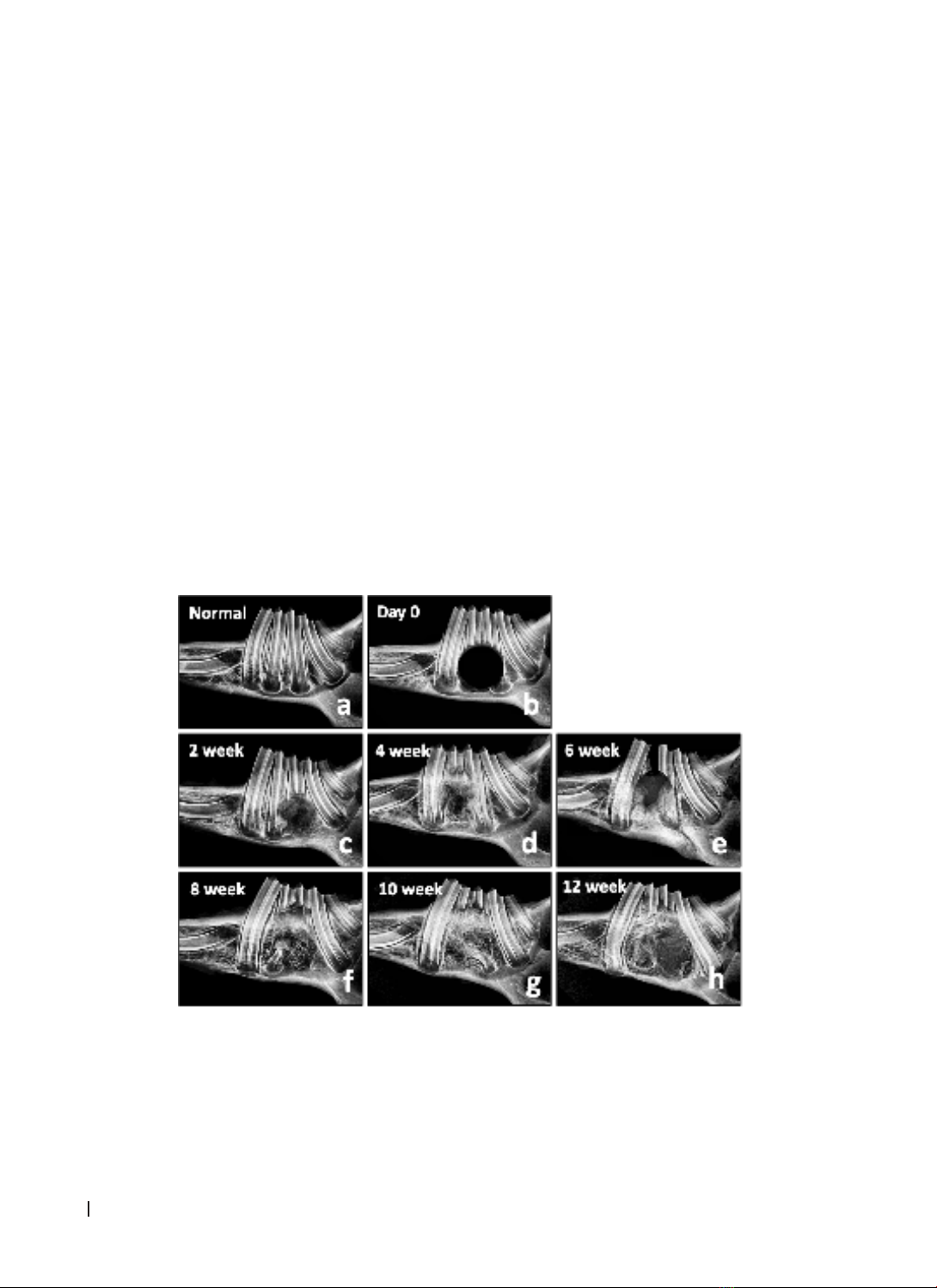
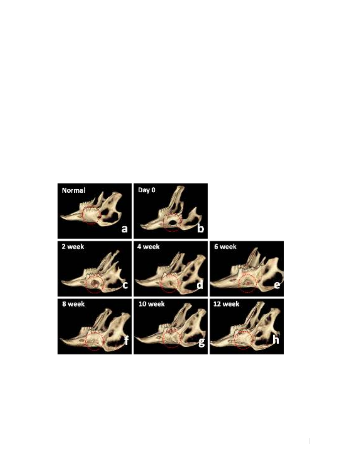
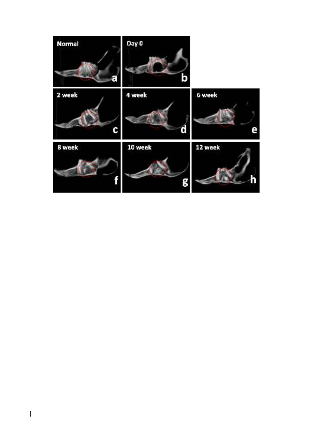
8, 10, and 12 weeks post-surgery. To assess bone regeneration at the surgical site, radiography, dental cone-
beam computed tomography (CT), and histological examination using Hematoxylin and Eosin staining were
performed on the skull. Results: A straightforward and easily executable method was devised to create the
rabbit mandibular alveolar defect model. After 10 weeks, complete soft tissue and bone regeneration were
observed. X-ray and cone-beam CT evaluations demonstrated a progressive increase in bone density from
weeks 2 to 12. Histological examination revealed that the alveolar bone structure was formed incrementally
at the surgical site. The bone and connective tissue had filled the defect after 8 weeks. Conclusion: The
creation of a model of mandibular alveolar bone defects in rabbits is a straightforward process that can
be used to assess the regeneration of alveolar bone at the defect site. This animal model can serve as the
foundation for tests to evaluate the capacity of biomaterials to regenerate the alveolar bone.
Keywords: mandibular alveolar bone, Alveolar bone defects, animal models, bone regenerative medicine,
tissue engineering.
Corresponding author: Nguyen Thanh Tung;
Email: nguyenthanhtung@hueuni.edu.vn; nttung@huemed-univ.edu.vn
Recieved: 1/12/2023; Accepted: 19/2/2024; Published: 25/2/2024
DOI: 10.34071/jmp.2024.2.9
1. INTRODUCTION
The alveolar bone, which is a component of the
upper and lower jawbones, encircles and supports
the teeth. In certain instances, the alveolar bone
may be damaged by trauma, jaw tumors and
cysts, infection, or tooth loss [1]. Furthermore,
periodontitis is another factor that contributes to
bone loss and alveolar bone defect development
[2]. Alterations in the shape and structure of the
alveolar bone not only affect the ability to chew, but
can also lead to aesthetic, comfort, and confidence
issues for patients, necessitating re-treatment.
Therefore, the restoration of alveolar bone defects
in patients is essential. In the context of replacing
missing teeth, reconstructing the bone morphology
in the jaw ridge is crucial for ensuring the stability
of the restoration and fulfilling the aesthetic and
functional requirements of the patient [3].
Alveolar bone defects are a prevalent issue in
Maxillofacial Surgery due to a variety of reasons [4].
These defects can heal slowly or not at all because
of factors such as large size, unstable physiological
characteristics, subpar surgical techniques, or
external influences such as metabolism, hormones,
nutrition, and stress [5]. Therefore, reconstructing
alveolar bone defects to restore both function and
aesthetics is a major challenge for maxillofacial
surgeons. Addressing alveolar bone defects typically
involves surgical intervention and the use of bone
grafting techniques and other healing aids [6]. Bone
grafting aims to stimulate or facilitate new bone
growth to fill defect [7].
Researchers have investigated various materials,
including autologous bone, tissue-engineered
materials, stem cells, and growth factors, to address
bone defects [8]. Autologous bone derived from the
patient’s own body is considered the optimal choice
because of its ease of use, low cost, and ability
to perform bone graft surgery simultaneously.
However, the removal of autologous bone can result
in significant consequences for the patient, such as
prolonged recovery time, infection, bleeding, and
nerve damage [9]. To overcome these limitations,
artificial bone powders with desirable biological
properties such as Hydroxyapatite and Beta-
Tricalcium Phosphate have been developed. Biphasic
Calcium Phosphate, a mixture of Hydroxyapatite
and Beta-Tricalcium Phosphate, have been