HUE JOURNAL OF MEDICINE AND PHARMACY ISSN 3030-4318; eISSN: 3030-4326
66
Hue Journal of Medicine and Pharmacy, Volume 14, No.4/2024
Microscopic characteristics and antioxidant activity of Uvaria boniana
Nguyen Khanh Thuy Linh1*, Nguyen Dinh Quynh Phu1,
Doan Quoc Tuan1, Nguyen Thi To Quyen1
(1) University of Medicine and Pharmacy, Hue University
Abstract
Background and objectives: Uvaria, a genus within the Annonaceae family, encompasses around 150
species of flowering plants. Uvaria boniana is extensively found throughout Vietnam and is utilized in
traditional medicine practices. The aim of this study was to determine the microscopic characteristics and
evaluate the antioxidant activity of Uvaria boniana. Materials and methods: The stems and leaves of Uvaria
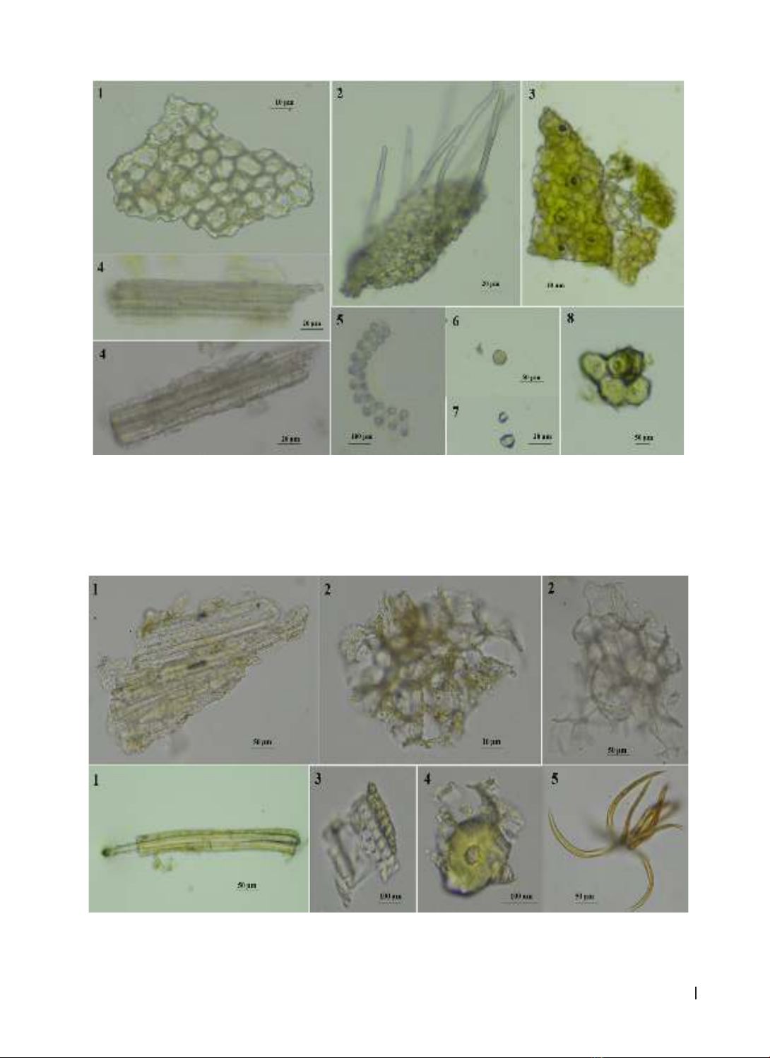
boniana were collected in Huong Tra district, Thua Thien Hue province in October 2023. Micro-morphology of
stems, leaves and powder properties were determined by the microscopic method. Antioxidant activity was
assessed using the DPPH assay. Results: The microscopic characteristics of this species have been described.
The methanol extract from the stem of U. boniana exhibited stronger antioxidant activity than the methanol
extract from the leaf, with IC50 values of 45.19 ± 0.68 µg/mL and 70.94 ± 0.19 µg/mL, respectively. Conclusion:
This is the first report on the microscopic characteristics and antioxidant activities of Uvaria boniana.
Keywords: Uvaria boniana, anatomic structures, powder properties, microscopic characteristics, antioxidant.
1. BACKGROUND
There are over 150 species of flowering plants in
the Uvaria genus, which is part of the Annonaceae
family. Predominantly, these species are either
climbing shrubs or diminutive trees. They thrive in
the moist, tropical climates found across Southeast
Asia, tropical Africa, Northern Australia, Madagascar,
and Indochina [1].
Uvaria is a large genus of the Annonaceae
family, with 17 species found in Vietnam [2].
Uvaria boniana, as described by Fin. & Gagnep,
is extensively found throughout Vietnam and is
utilized in ethnomedicine [2]. The crushed leaves
emit an aroma similar to that of cinnamon bark,
and a decoction made from them can be ingested
directly. Additionally, the fruits are employed in
the treatment of intestinal ulcers [3]. The root’s
water decoction is specifically used for managing
postpartum infections in women [4]. The number
of studies related to this species is very limited.
Thanh Tam Nguyen’s research has isolated
and determined the structures of five pure
compounds, including: uvaridacol G, 4-methyl-
4-[(2Z)-3’-phenylprop-2’-en1’-yl]cyclohex-2-
en-1-one, 3,7-dimethoxy quercetin 4’-O-[α-L-
rhamnopyranosyl-(1→2)-β-D-glucopyranoside,
β-sitosterol, and stigmasterol [5]. Son Ninh The
reported on the chemical composition, anti-
inflammatory and antibacterial activities of U.
boniana essential oil [6]. As far as we know,
there have been no studies on the microscopic
characteristics and the antioxidant activity of this
species. Therefore, the purpose of this study is
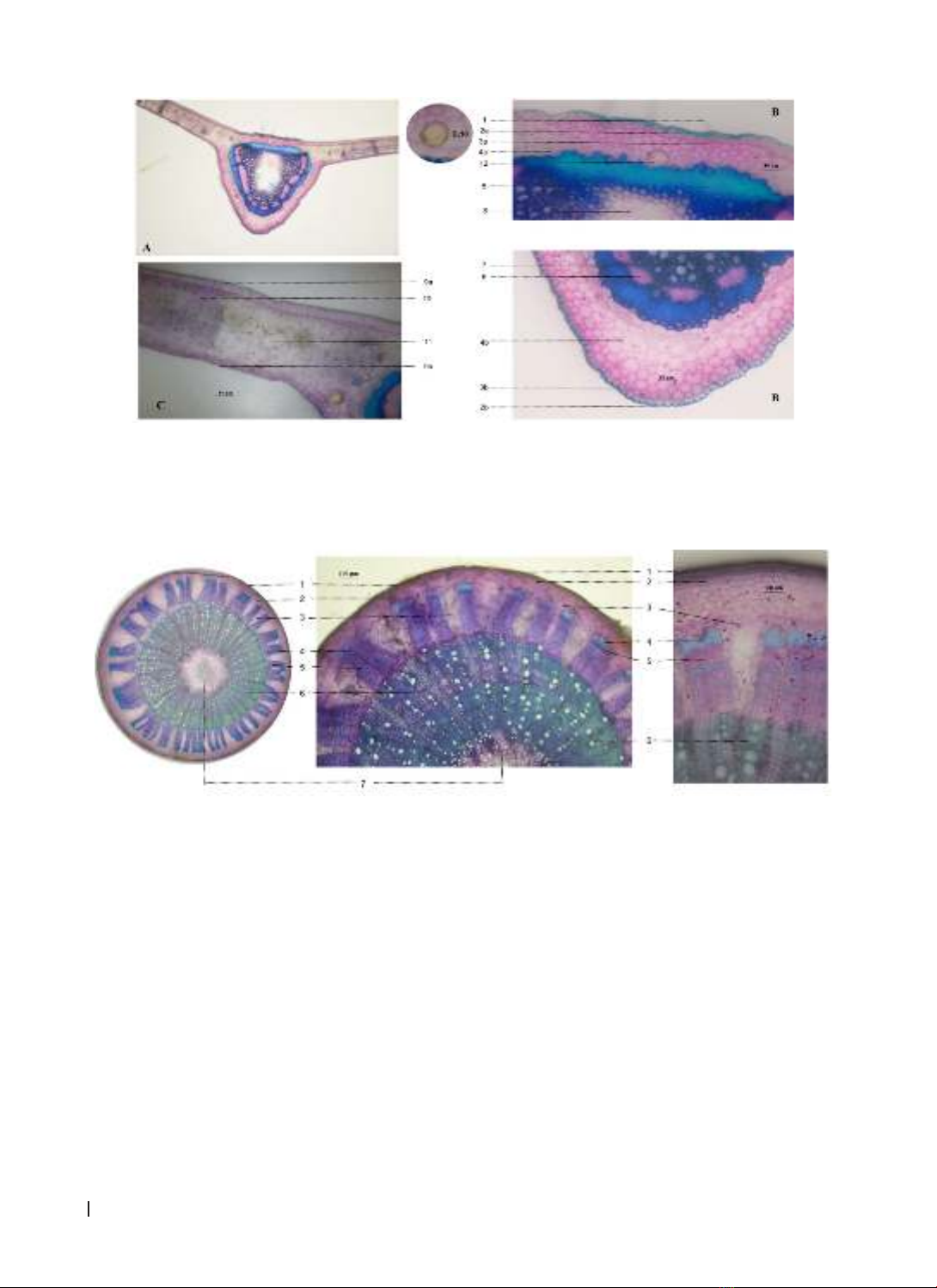
to provide data on microscopic characteristics of
the stem and leaf of this species and evaluate the
antioxidant activities of U. boniana.
2. MATERIALS AND METHODS
2.1. Materials
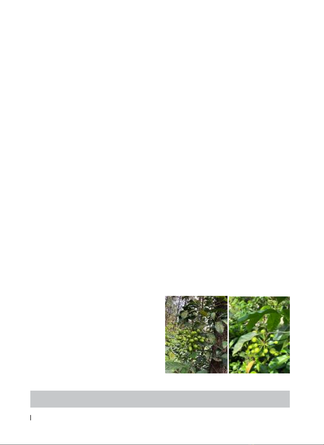
The stems and leaves of Uvaria boniana were
collected in Huong Tra district, Thua Thien Hue
province in October 2023. The plant material was
identified by Dr Anh Tuan Le (Mientrung Institute for
Scientific Research, Vietnam National Museum of
Nature, VAST, Vietnam). Voucher specimen has been
deposited at the Faculty of Pharmacy, University of
Medicine and Pharmacy, Hue University, Vietnam.
Some of pictures of the Uvaria boniana was
displayed in figure 1.
Figure 1. The image of the Uvaria boniana
Corresponding author: Nguyen Khanh Thuy Linh; Email: nktlinh@huemed-univ.edu.vn
Received: 21/2/2024; Accepted: 15/6/2024; Published: 25/6/2024
DOI: 10.34071/jmp.2024.4.9