HUE JOURNAL OF MEDICINE AND PHARMACY ISSN 1859-3836 79
Hue Journal of Medicine and Pharmacy, Volume 13, No.6-2023
Microscopic characteristics, chemical compositions and bioactivities of
Alpinia vietnamica
Nguyen Dinh Quynh Phu1*, Nguyen Hoai Bao Chau1, Doan Quoc Tuan1,
Nguyen Khanh Thuy Linh1, Tran Van Nguyen2, Le Thi Khanh Linh2
(1) University of Medicine and Pharmacy, Hue University
(2) Family Hospital, Danang
Abstract
Background: The genus Alpinia is one of the diverse genera in Thua Thien Hue province, in which many
species have been used as medicine. But until now, studies on A. vietnamica have rarely been reported.
Objectives: The present study was aimed at the determination of microscopic characteristics and chemical
compositions as well as evaluating the antioxidant and acetylcholinesterase inhibitory activities of A.
vietnamica. Materials and methods: A. vietnamica was collected in Phong Dien district, Thua Thien Hue
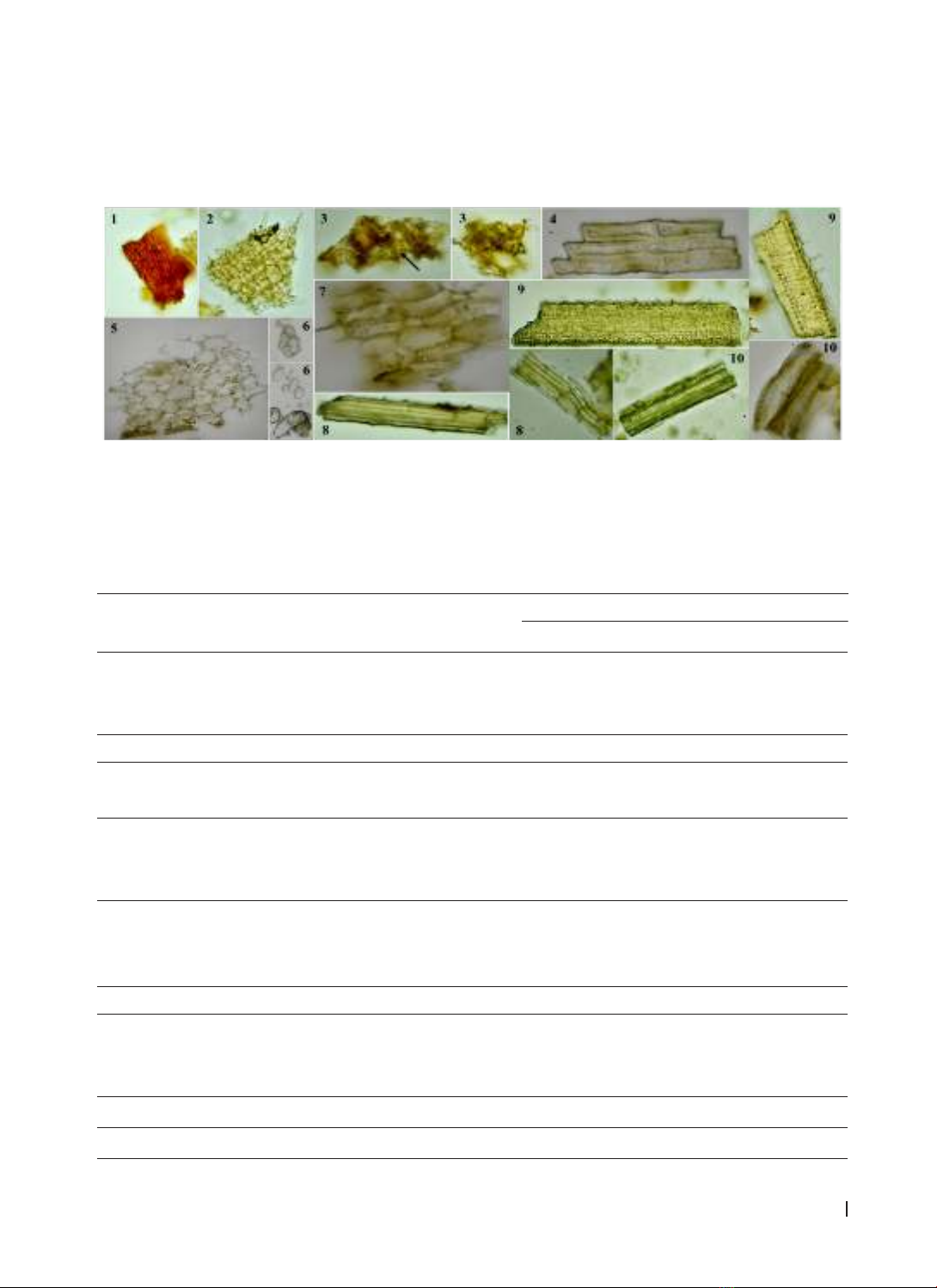
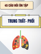
province. Anatomic structures and powder properties were determined by the microscopic method.
Phytochemical screening was conducted by specific chemical reactions. The Folin-Ciocalteau method and the
aluminum chloride-flavonoid assay, respectively, were used to quantify the total polyphenol (TPC) and total
flavonoid contents (TFC). Antioxidant activity was assessed using the DPPH assay, while acetylcholinesterase
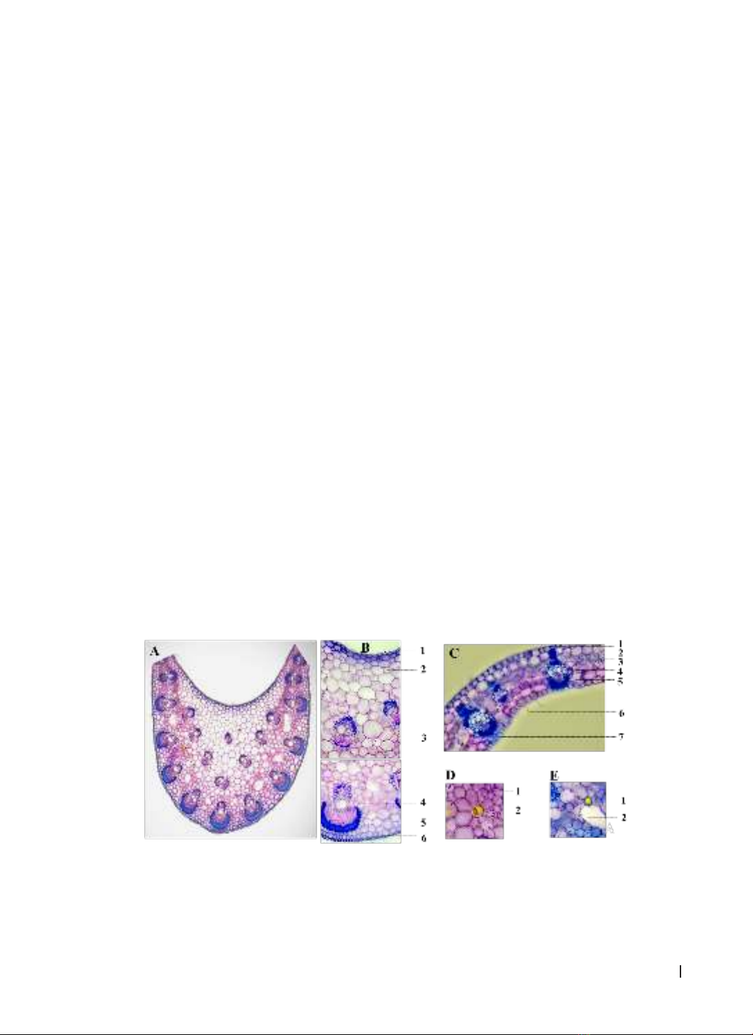
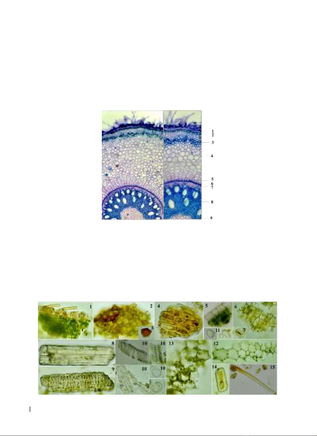
(AChE) inhibitory activity was evaluated using the Ellman method. Results: The microscopic characteristics
of this species have been described. Phytochemical analysis results revealed the presence of flavonoids,
coumarins, and tannins in A. vietnamica. The ethanol extract from the aerial part of A. vietnamica had higher
polyphenol and flavonoid contents than the underground part extract. Moreover, this extract also displayed
a stronger DPPH radical scavenging and exhibited AChE inhibitory activities. Conclusion: This is the first
report on the microscopic characteristics, chemical compositions, and biological activities of A. vietnamica.
Keywords: Alpinia vietnamica, microscopic characteristics, chemical constituents, antioxidant activity,
acetylcholinesterase inhibitory
Corresponding author: Nguyen Dinh Quynh Phu. Email: ndqphu@huemed-univ.edu.vn
Recieved: 15/9/2023; Accepted: 12/12/2023; Published: 31/12/2023
DOI: 10.34071/jmp.2023.6.10
1. BACKGROUND
Alpinia is a large genus of the Ginger family
(Zingiberaceae) with over 250 species that are
widely distributed in Asia. In many countries around
the world, species in this genus have been used
for traditional medicine, food, and spices. Fruits,
seeds, leaves, and rhizomes of these medicinal
plants are frequently used to treat digestive
system diseases such as indigestion, stomach
pain, and vomiting, or as anti-inflammatory drugs.
Terpenoids, diarylheptanoids, lignans, flavonoids,
alkaloids, essential oils, and other compounds are
found in Alpinia genus. Extracts and compounds
isolated from this genus have a variety of beneficial
biological properties, such as the ability to inhibit the
growth of cancer cells, antioxidant, antimicrobial,
cardioprotective activities, and reduce blood sugar
levels, etc [1].
There are approximately 31 species of Alpinia
grown or living under the canopy of forests,
streams, and wet places in Vietnam. Many species
have been used as medicine, spices, and considered
raw materials for essential oil extraction. Studies
on the genus Alpinia in Vietnam mainly focus on
analyzing the essential oil composition of some
species, such as A. oblongifolia, A. malaccaensis,
A. menghaiensis, A. pinnanensis, A. polyantha, A.
strobiliformis and A. tonkinensis [2]. Alpinia is well
known as a biodiversity genus in Thua Thien Hue
province, with many valuable medicinal species,
including A. vietnamica [3]. It was discovered in
Central Vietnam and is a newly identified species
in 2019 [4]. As far as we know, there has not been
any study about the microscopic characteristics,
chemical compositions, and bioactivities of A.
vietnamica. Therefore, the objective of this study
is to determine the microscopic characteristics,
preliminary phytochemical screening, and evaluate
the total polyphenol and total flavonoid contents as
well as the antioxidant and anti-acetylcholinesterase
activities of A. vietnamica.
2. MATERIALS AND METHODS
2.1. Materials
The whole of Alpinia vietnamica H.D. Tran,
Luu & Škorničk. (Zingiberaceae) was collected in
HUE JOURNAL OF MEDICINE AND PHARMACY ISSN 1859-3836
80
Hue Journal of Medicine and Pharmacy, Volume 13, No.6-2023
Phong Dien district, Thua Thien Hue province in
June 2022. The plant material was identified by Dr.
Thao Xuan Hoang (Faculty of Biology, University
of Education, Hue University). Voucher specimen
(AV01) has been deposited at the Faculty of
Pharmacy, University of Medicine and Pharmacy,
Hue University, Vietnam.
2.2. Methods
2.2.1. Identification of microscopic
characteristics
Microsurgery: Fresh leaves and roots were
cut into thin sections with a razor blade. The
sections were soaked in 5% sodium hypochlorite
for approximately 30 min and washed with water.
Immersed sections in 1% acetic acid for 3-5 min,
and rinsed with water. The sections were then dyed
with methylene blue and carmine red solution
at a suitable time and washed several times with
water. After that, they were placed on a glass slide,
mounted with a few drops of 10% glycerol, and a
cover glass was applied. The final sections were
observed using a microscope (Eclipse E100, Nikon,
Japan), and photographed with an attached camera
(Nikon, D5100) [5].
Powder characteristics: The aerial and
underground parts of the plant were powdered and
passed through hand sieve with a mesh size of 0.125
mm to obtain a fine powder. The powder was placed
on slides with several drops of 10% glycerol and
covered with a coverslip. Observations were made
with an optical microscope (Eclipse E100, Nikon,
Japan), and pictures were taken with a camera
(Nikon, D5100) [5].
2.2.2. Preliminary phytochemical screening
The extracts of the aerial and underground
parts of A. vietnamica were tested for the presence
of alkaloids, anthranoids, coumarins, cardiac
glycosides, flavonoids, saponins, tannins, organic
acids, and steroids by using specific chemical
reactions [6].
2.2.3. Preparation of the extract
The powder of dried aerial and underground
parts of A. vietnamica (10.0 g, each sample) was
macerated with ethanol (EtOH) (100 mL x 3 times)
at room temperature for three days, shaken, and
stirred occasionally. The EtOH extract was filtered
through cotton and filter paper, then recovered the
solvent until it was completely ethanol-free.
2.2.4. Determination of total polyphenol and
flavonoid contents
2.2.4.1. Determination of total polyphenol
content
The total polyphenol content was evaluated
by the Folin-Ciocalteu method with slight
modifications. Tested samples (0.2 mL) were mixed
with 0.8 mL distilled water and 1.0 mL of 10% Folin-
Ciocalteu reagent and then shaken for 5 min. Then,
a volume of 2.5 mL of 7.5% Na2CO3 was added. After
incubation at room temperature in a dark condition
for 30 min, the absorbance was measured at 760
nm. Gallic acid (GAE) was used as a standard for the
calibration curve [7]. All samples were analyzed in
3 replicates and the total polyphenol content was
expressed as milligrams of gallic acid equivalents per
gram of extract (mg GAE/g extract) according to the
formula (1):
TPC = (1)
where TPC was the total polyphenol content in
mg/g, in GAE (Gallic acid equivalent), C1 was the
concentration of Gallic acid established from the
curve in mg/mL, V was the initial volume of the
extract in mL, k was the dilution factor, and m was
the weight of the plant extract in g.
2.2.4.2. Determination of total flavonoid content
The total flavonoid content (TFC) was measured
by the aluminum chloride colorimetric method.
Tested samples (2.0 mL) were mixed with 2.0 mL of
2% AlCl3. After incubation at room temperature for
10 min, the absorbance was recorded at 430 nm.
Rutin (RE) was used as a standard for the calibration
curve [8]. TFC was expressed as mg of rutin
equivalents per gram of extract (mg RE/g) according
to the formula (2):
TFC = (2)
where TFC was total flavonoid content in mg/g,
in RE (Rutin equivalent), C1 was the concentration
of Rutin established from the curve in mg/mL, V
was the initial volume of the extract in mL, k was
the dilution factor, and m is the weight of the plant
extract in g.
2.2.5. Evaluation of antioxidant and anti-
acetylcholine esterase activities
2.2.5.1. Evaluation of antioxidant activity
The antioxidant activity was evaluated using the
DPPH method with minor modifications, and the
absorbance was measured at 517 nm [9]. Quercetin
was used as a positive control. The ability of the
sample to scavenge DPPH radicals (%) was calculated
using the following formula (3):
DPPH scavenging effect = [(Acontrol – Asample) /
Acontrol] x 100% (3)
where Acontrol was the absorbance of DPPH control
solution and Asample was the absorbance of DPPH
HUE JOURNAL OF MEDICINE AND PHARMACY ISSN 1859-3836 81
Hue Journal of Medicine and Pharmacy, Volume 13, No.6-2023
solution in the presence of tested samples. The tests
were carried out three times. The IC50 value is the
concentration of 50% free radical neutralizer DPPH
calculated in Microsoft Excel.
2.2.5.2. Evaluation of acetylcholinesterase
(AChE) inhibitory activity
AChE inhibitory activity was measured based
on the method of Ellman et al. with some minor
modifications [10, 11]. Briefly, for each well of a 96-
well microtiter plate, 140 µL of phosphate buffer
(pH = 8.0), 20 µL tested samples, and 20 µL of 0.25
IU/mL AChE were mixed and incubated for 15 min
at room temperature. Post-incubation, 10 µL of 2.4
mM acetylthiocholineiodide (ATCI) and 10 µL of
2.4 mM 5,5’-dithio-bis-2-nitrobenzoic acid (DTNB)
were added. The reaction mixture was mixed and
incubated for 24 min at room temperature. The
absorbance was determined at 405 nm (Elisa EMR-
500, Labomed Inc., USA). A control sample contained
all the aforementioned constituents without the
test extract. A blank sample was performed in the
absence of AChE. Berberine was used as the positive
control. The AChE inhibitory activity was determined
using the formula (4):
%I = [1 – (As – Ab/s) / (Ac – Ab/c)] x 100% (4)
where %I was the percent of AChE activity
inhibited; As and Ab/s were the absorbance of the test
sample and the blank of the test sample, respectively.
Ac and Ab/c were the absorbance of the control sample
and the blank of the control sample, respectively.
2.2.6. Statistical analysis
All the data in this research was analyzed using
the Microsoft Excel program in triplicate. Values were
expressed as mean ± standard deviation (SD) for three
replicates for each sample.
3. RESULTS
3.1. Microscopic characteristics
3.1.1. Anatomy structure
Leaf midrib [Fig. 1A, 1B, 1D]: The midrib was
concave on the upper side and convex on the lower
side in cross section. The upper and lower epidermis
(B1, B6) consisted of a layer of rectangular cells
arranged adjacently. The parenchyma (B2, B5) was
comprised of many layers of polygonal cells that
were different-sized, thin-walled, and arranged
randomly. In the parenchyma region (D1), there
were some cells with a spherical oil droplet inside
(D2). The phloem-xylem vasculars were divided
into two types, including ovoid-shaped and spoon-
shaped bundles. The ovoid-shaped bundles (B3)
were located in the center of the midrib, and the
spoon-shaped bundles (B4) were adjacent to the
lower epidermis.
Leaf blade [Fig. 1C, 1E]: The upper and lower
epidermis (C1, C7) had a similar structure to the
epidermis in the leaf midrib. The lower epidermis
had trichomes (C6). The parenchyma (C2) was
under the upper epidermis and comprised a layer
of oval-shaped cells, thin-walled and large-sized.
The palisade and spongy layers (C3, C5) were clearly
separated. The bundles of phloem-xylem (C4)
were irregular in size. In the spongy parenchyma
(E2), there were some cells containing yellow oil
droplets (E1).
Figure 1. Microscopic characteristics of leaf cross-section of Alpinia vietnamica
A, B: Leaf midrib (1. Upper epidermis, 2, 5. Parenchyma, 3. Ovoid-shaped bundle of phloem-xylem,
4. Spoon-shaped bundle of phloem-xylem, 6. Lower epidermis), C: Leaf blade (1. Upper epidermis, 2.
Parenchyma, 3. Palisade parenchyma, 4. Bundle of phloem-xylem, 5. Spongy parenchyma, 6. Trichome,
7. Lower epidermis), D: Parenchyma in leaf midrib (1. Parenchyma cell, 2. Essential oil cell), E: Spongy
parenchyma in leaf blade (1. Essential oil cell, 2. Spongy parenchyma cell)
HUE JOURNAL OF MEDICINE AND PHARMACY ISSN 1859-3836
82
Hue Journal of Medicine and Pharmacy, Volume 13, No.6-2023
Figure 2. Microscopic characteristics of root cross-section of Alpinia vietnamica
1. Epidermis, 2. Suberoid, 3. Sclereid, 4. Cortical parenchyma, 5. Endoderm, 6. Pericycle, 7. Phloem,
8. Xylem, 9. Pith parenchyma
3.1.2. Powder features
The aerial part [Fig. 3]: A green powder had the characteristic of pleasant and aromatic odour. Powder
features from the aerial part were observed under a light microscope at 10X and 40X magnifications. The
powder had several microscopic characteristics: fragment of epidermis contained trichomes and essential
oil (1), fragment of epidermis contained color fragment (2), color fragment (3), fragment of epidermis (4),
fragment of epidermis and palisade parenchyma (5), fragment of epidermis contained stomata (6), stomata
(7), bundle of fiber (8, 9), fragment of vessel (10), starch (11), fragment of parenchyma (12), fragment of
parenchyma contained starch (13), sclereid (14), and trichome (15).
Figure 3. Microscopic features of the aerial part of Alpinia vietnamica
Root [Fig. 2]: The root of A. vietnamica had a
circular cross-section; the cortical area occupied
more than a half of the microsurgery radius,
and the layers from outer to inner included: The
epidermis (B1) consisted of a layer of rectangular
cells arranged adjacently. The suberoid layer (B2)
was made up of a layer of closely spaced polygonal
cells. There were many sclereids (B3) close to the
outer layer of the cortex. The cortical parenchyma
(B4) comprised several layers of parenchyma that
were polygonal, unequal-sized and thin-walled.
The endoderm (B5) was a thick U-shaped layer
of cells forming a caspary belt. The tissues in the
innermost part of the endodermis that formed the
stele region called the pericycle (B6) comprised
a single layer of thin-walled cells. The vascular
tissues consisted of many patches of phloem (B7)
and xylem (B8) arranged radially. The pith region
(B9) was present at the centre of the internal
structure and comprised parenchymatous cells
along with intercellular spaces composed of many
polygonal cells and randomly arranged.
HUE JOURNAL OF MEDICINE AND PHARMACY ISSN 1859-3836 83
Hue Journal of Medicine and Pharmacy, Volume 13, No.6-2023
The underground part [Fig. 4]: A brown-yellow powder had the characteristic of pleasant and aromatic
odour. Some microscopic features of the underground part powder were observed under a light microscope
at 10X and 40X magnifications, including: fragment of phellem (1), fragment of epidemis (2), fragment of
epidemis contained essential oil cell (3), sclereid (4), fragment of parenchyma (5), starch (6), fragment of
parenchyma contained starch (7), bundle of fiber (8, 9), and fragment of vessel (10).
Figure 4. Microscopic features of the underground part of Alpinia vietnamica
3.2. Chemical compositions
3.2.1. Phytochemical screening
The phytochemical screening results of A. vietnamica revealed that coumarins, flavonoids, and tannins
were present in both the aerial and underground parts, while steroids were only found in the aerial part, as
shown in Table 1.
Table 1. Phytochemical screening results of Alpinia vietnamica
No. Class of
phytochemicals Test/Reagent Results
The underground part The aerial part
1Alkaloids
Mayer - -
Dragendorff - -
Bouchardat - -
2Anthranoids Bontraeger - -
3Coumarins Lactone ring-opening + +
Diazo + ++
4Cardiac glycosides
Liebermann -+
Legal - -
Keller-Killiani - -
5Flavonoids Cyanidin + +++
10% NaOH +++ +++
5% FeCl3++ +++
6Saponins Foam - -
7Tannins
10% FeCl3 + ++
10% (CH3COO)2Pb + ++
1% Gelatin + +
8Organic acids Na2CO3- -
9Steroids Liebermann -+
Notes: (+): mildly positive, (++): moderately positive, (+++): highly positive, (-): negative.