
RESEARC H Open Access
A pilot histomorphology and hemodynamic of
vasculogenic mimicry in gallbladder carcinomas
in vivo and in vitro
Wei Sun, Yue Z Fan
*
, Wen Z Zhang and Chun Y Ge
Abstract
Background: Vasculogenic mimicry (VM), as a new blood supply for tumor growth and hematogenous metastases,
has been recently described in highly aggressive human melanoma cells, etc. We previously reported VM in human
gallbladder carcinomas and its clinical significance. In this study, we further studied histomorphology and
hemodynamic of VM in gallbladder carcinomas in vivo and in vitro.
Methods: The invasive potential of human gallbladder carcinoma cell lines GBC-SD and SGC-996 were identified
by Transwell membrane. The vasculogenic-like network structures and the signal intensities i.e. hemodynamic in
gallbladder carcinomas stimulated via the three-dimensional matrix of GBC-SD or SGC-996 cells in vitro, the nude
mouse xenografts of GBC-SD or SGC-996 cells in vivo were observed by immunohistochemistry (H&E staining and
CD
31
-PAS double staining), electron microscopy and micro-MRA with HAS-Gd-DTPA, respectively.
Results: Highly aggressive GBC-SD or poorly aggressive SGC-996 cells preconditioned by highly aggressive GBC-SD
cells could form patterned networks containing hollow matrix channels. 85.7% (6/7) of GBC-SD nude mouse
xenografts existed the evidence of VM, 5.7% (17/300) channels contained red blood cells among these tumor cell-
lined vasculatures. GBC-SD xenografts showed multiple high-intensity spots similar with the intensity observed at
tumor marginal, a result consistent with pathological VM.
Conclusions: VM existed in gallbladder carcinomas by both three-dimensional matrix of highly aggressive GBC-SD
or poorly aggressive SGC-996 cells preconditioned by highly aggressive GBC-SD cells in vitro and GBC-SD nude
mouse xenografts in vivo.
Keywords: Gallbladder neoplasm vasculogenic mimicry, 3-dimensional matrix, Xenograft model, Histomorphology,
Hemodynamic
Background
The formation of a microcirculation (blood supply)
occurs via the traditionally recognized mechanisms of
vasculogenesis (the differentiation of precursor cells to
endothelial cells that develop de novo vascular net-
works) and angiogenesis (the sprouting of new vessels
from preexisting vasculature in response to external
chemical stimulation). Tumors require a blood supply
for growth and hematogenous metastasis, and much
attention has been focused on the role of angiogenesis
[1]. Recently, the concept of “vasculogenic mimicry
(VM)”was introduced to describe the unique ability of
highly aggressive tumor cells, but not to poorly aggressive
cells, to express endothelium and epithelium-associated
genes, mimic endothelial cells, and form vascular chan-
nel-like which could convey blood plasma and red blood
cells without the participation of endothelial cells (ECs)
[2]. VM consists of three formations: the plasticity of
malignant tumor cells, remodelling of the extracellular
matrix (ECM), and the connection of the VM channels
to the host microcirculation system [3-5]. Currently, two
distinctive types of VM have been described, including
tube (a PAS-positive pattern) and patterned matrix types
[6]. VM, a secondary circulation system, has increasingly
been recognized as an important form of vasculogenic
* Correspondence: fanyuezu_shtj@yahoo.com.cn
Department of Surgery, Tongji Hospital, Tongji University School of
Medicine, Shanghai, China
Sun et al.Journal of Experimental & Clinical Cancer Research 2011, 30:46
http://www.jeccr.com/content/30/1/46
© 2011 Sun et al; licensee BioMed Central Ltd. This is an Open Access article distributed under the terms of the Creative Commons
Attribution License (http://creativecommons.org/licenses/by/2.0), which permits unrestricted use, distribution, and reproduction in
any medium, provided the original work is properly cited.

structure in solid tumors [2]. A lot of approaches have
suggested that these VM channels are thought to pro-
vide a mechanism of perfusion and dissemination route
within the tumor that functions either independently of
or, simultaneously with angiogenesis [7-11]. VM chan-
nels and periodic acid-Schiff-positive (PAS) patterns are
also associated with a poor prognosis, worse survival
and the highest risk of cancer recurrence for the
patients with melanoma [2,12], cell renal cell carcinoma
[13], breast cancer [14], ovarian carcinoma [15], hepato-
cellular carcinoma [16-18], laryngeal squamous cell car-
cinoma [19], glioblastomas [20], gastric adenocarcinoma
[21] colorectal cancer [22] and gastrointestinal stromal
carcinoma [23].
Gallbladder carcinoma (GBC) is the most common
malignancy of the biliary tract and the fifth common
malignant neoplasm of the digestive tract in western
countries [24,25]. It is also the most common malig-
nant lesion of the biliary tract, the sixth common
malignant tumor of the digestive tract and the leading
cause of cancer-related deaths in China and in Shang-
hai [26]. 5-year survival for the patients lies between
0% and 10% in most reported series [26,27]. The poor
prognosis of GBC patients is related to diagnostic
delay, low surgical excision rate, high local recurrence
and distant metastasis, and biological behavior of the
tumor. Therefore, it is an urgent task to reveal the
precise special biological behavior of GBC develop-
ment, and provide a novel perspective for anticancer
therapeutics. We previously reported the existence of
VM in human primary GBC specimens and its correc-
tion with the patient’s poor prognosis [28]. In addition,
the human primary gallbladder carcinoma cell lines
SGC-996, isolated from the primary mastoid adenocar-
cinoma of the gallbladder obtained from a 61-year-old
female patient in Tongji Hospital were successfully
established by our groups in 2003, the doubling time
of cell proliferation was 48 h. Furthermore, we found
SGC-996 cells accorded with the general characteristic
of the cell line in vivo and in vitro. Based on these
results, we hypothesized that the two different tumor
cell lines, including GBC-SD and SGC-996, can exhibit
significant different invasive ability and possess discre-
pancy of VM channels formation.
In this study, we show evidence that VM exists in the
three-dimensional matrixes of human GBC cell lines
GBC-SD (highly aggressive) and SGC-996 (poorly
aggressive, but when placed on the aggressive cell-pre-
conditioned matrix) in vitro,andinthenudemouse
xenografts of GBC-SD cells in vivo. Taken together,
these results advance our present knowledge concerning
the biological characteristic of primary GBC and provide
the basis for new therapeutic intervention.
Methods
Cell culture
Two established human gallbladder carcinoma cell lines
used in this study were GBC-SD (Shanghai Cell Biology
Research Institute of Chinese Academy of Sciences,
CAS, China) and SGC-996 (a generous gift from
Dr. Yao-Qing Yang, Tumor Cell Biology Research Insti-
tute of Tongji University, China). These cells were
maintained and propagated in Dulbecco’smodified
Eagle’s media (DMEM, Gibco Company, USA) supple-
mented with 10% fetal bovine serum (FBS, Hangzhou
Sijiqing Bioproducts, China) and 0.1% gentamicin sulfate
(Gemini Bioproducts, Calabasas, Calif). Cells were main-
tained at log phase at 37°C with 5% carbon dioxide.
Invasion assay in vitro
The 35-mm, 6-well Transwell membranes (Coster
Company, USA) were used to measure the in vitro inva-
siveness of two tumor cells. Briefly, a polyester (PET)
membrane with 8-μm pores was uniformity coated with
a defined basement membrane matrix consisting of 50
μl Matrigel mixture which diluted with serum-free
DMEM (2 volumes versus 1 volume) over night at 4°C
and used as the intervening barrier to invasion. Upper
wells of chamber were respectively filled with 1 ml
serum-free DMEM containing 2 × 10
5
·ml
-1
tumor cells
(GBC-SD or SGC-996 cells, n = 3), lower wells of cham-
ber were filled with 3 ml serum-free DMEM containing
1 × MITO+ (Collaborative Biomedical, Bedford, MA).
After 24 hr in a humidified incubator at 37°C with 5%
carbon dioxide, cells that had invaded through the base-
ment membrane were stained with H&E, and counted
by light microscopy. Invasiveness was calculated as the
number of cells that had successfully invaded through
the matrix-coated membrane to the lower wells. Quanti-
fication was done by counting the number of cells in 5
independent microscopic fields at a 400-fold magnifica-
tion. Experiments were performed in duplicate and
repeated three times with consistent results.
Network formation assay in vitro
Thick gel of rat-tail collagen typeⅠwas made by mixing
together ice-cold gelation solution, seven volumes of
rat-tail collagen typeⅠ(2.0 mg·ml
-1
,SigmaCompany,
Germany) were mixed with two volumes of 10 × con-
centrated DMEM and one volume of NaHCO
3
(11.76
mg·ml
-1
). Then 50 μl cold thick gel of rat-tail collage-
nⅠand Matrigel (Becton Dickinson Company, USA) were
respectively dropped into a sterilized 35 mm culture
dish (one 18 × 18 mm
2
glass coverslips placed on the
bottom of dish) and allowed to polymerize for 30 min at
room temperature, then 30 min at 37°C in a humidified
5% carbon dioxide incubator. The 7.5 × 10
5
tumor cells
Sun et al.Journal of Experimental & Clinical Cancer Research 2011, 30:46
http://www.jeccr.com/content/30/1/46
Page 2 of 12

were then seeded onto the gels and incubated at 37°C
with 5% carbon dioxide and humidity. The cultures
were maintained in DMEM supplemented with 10% FBS
and 0.1% gentamicin sulfate. The culture medium was
changed every 2 days. In addition, on the premise of dif-
ferent invasion of two kinds of tumor cells, for experi-
ments designed to analyze the ability of poorly
aggressive tumor cells to engage in VM when placed on
a matrix preconditioned by the highly aggressive tumor
cells, which were removed after three days with 20 mM
NH
4
OH followed by three quick washes with distilled
water, phosphate buffered saline (PBS), and then com-
plete medium. Followed by this experimental protocol,
the highly aggressive tumor cells were cultured on a
matrix preconditioned by the poorly aggressive tumor
cells to explore the changes of remodeling capabilities.
For experiments designed to analyze the ability of the
cells to engage in VM using phase contrast microscopy
(Olympus IX70, Japan). The images were taken digitally
using a Zeiss Televal invertal microscopy (Carl Zeiss,
Inc., Thornwood, NY) and camera (Nickon, Japan) at
the time indicated.
Tumor Xenograft assay in vivo
All of procedures were performed on nude mice accord-
ing to the official recommendations of Chinese Commu-
nity Guidelines. BALB/C nu/nu mice, 4 weeks old and
about 20 grams, the ratio of male and female was 1:1 in
this study. All mice were provided by Shanghai Labora-
tory Animal Center, Chinese Academy of Sciences
(SLACCAS) and housed in specific pathogen free (SPF)
condition. A volume of 0.2 ml serum-free medium con-
taining single-cell suspensions of GBC-SD and SGC-996
(7.5 × 10
6
·ml
-1
) were respectively injected subcuta-
neously into the right axilback of nu/nu mice. In addi-
tion, the maximum diameter (a) and minimum diameter
(b) were measured with calipers two times each week.
The tumor volume was calculated by the following for-
mula: V (cm
3
)=∏ab
2
/6. The present study was carried
out with approval from Research Ethical Review Broad
in Tongji University (Shanghai, China).
Immunohistochemistry in vitro and in vivo
For H&E staining: 12 paraffin-embedded tissue speci-
mens of tumor xenografts were deparaffinized, hydrated,
and stained with H&E. Companion serial section were
stained with double staining of CD31 and PAS.
For CD
31
and PAS double staining: Briefly, 12 paraf-
fin-embedded tissue specimens (5 μm thickness) of the
tumor xenografts were mounted on slides and deparaffi-
nized in three successive xylene baths for 5 min, then
each section was hydrated in ethanol baths with differ-
ent concentrations. They were air-dried; endogenous
peroxide activity was blocked with 3% hydrogen
peroxide for 10 min at room temperature. The slides
were washed in PBS (pH7.4), then pretreated with
citratc buffer (0.01 M citric acid, pH6.0) for twice 5 min
each time at 100°C in a microwave oven, then the slides
were allowed to cool at room temperature and washed
in PBS again, the sections were incubated with mouse
monoclonal anti-CD
31
protein IgG (Neomarkers, USA,
dilution: 1:50) at 4°C overnight. After being rinsed with
PBS again, the sections were incubated with goat anti-
mouse Envision Kit (Genetech, USA) for 40 min at 37°C
followed by incubation with 3, 3-diaminobenzidine
(DAB) chromogen for 5 min at room temperature and
washing with distilled water, then the section were incu-
bated with 0.5% PAS for 10 min in a dark chamber and
washing with distilled water for 3 min, finally all of
these sections were counterstained with hematoxylin.
TheMicrovesselinmarginalareaoftumorxenografts
was determined by light microscopy examination of
CD
31
-stained sections at the site with the greatest num-
ber of capillaries and small venules. The average vessel
count of five fields (×400) with the greatest neovascular-
ization was regarded as the microvessel density (MVD).
After glass coverslips with samples of three-dimen-
sional culture were taken out, the samples were fixed in
4% formalin for 2 hr followed by rinsing with 0.01 M
PBS for 5 min. The cultures were respectively stained
with H&E and PAS (without hematoxylin counterstain).
The outcome of immunohistochemistry was observed
under light microscope with ×10 and ×40 objectives
(Olympus CH-2, Japan).
Electron microscopy in vitro and in vivo
For transmission electron microscopy (TEM), fresh
tumor xenograft tissues (0.5 mm
3
) were fixed in cold
2.5% glutaraldehyde in 0.1 mol·L
-1
of sodium cacodylate
buffer and postfixed in a solution of 1% osmium tetrox-
ide, dehydrated, and embedded in a standard fashion.
The specimens were then embedded, sectioned, and
stained by routine means for a JEOL-1230 TEM.
Dynamic MRA with intravascular contrast
agent for xenografts in vivo
On day 21, when all the tumors of xenografts had
reached at least 1.0 cm in diameter, they were examined
by dynamic micro-magnetic resonance angiography
(micro-MRA), MRI is a 1.5 T superconductive magnet
unit (Marconic Company, USA). Two kinds of tumor
xenograft nude mice (n = 2, for each, 7 weeks old, 35 ±
3 grams), anesthetized with 2% nembutal (45 mg·kg
-1
)
intraperitoneal injection and placed at the center of the
coils, were respectively injected I.V. in the tail vein with
human adult serum gadopentetic acid dimeglumine salt
injection (HAS-Gd-DTPA, 0.50 mmol (Gd)·l
-1
, Mr = 60-
100kD, 0.1 mmol (Gd)·kg
-1
,giftfromDepartmentof
Sun et al.Journal of Experimental & Clinical Cancer Research 2011, 30:46
http://www.jeccr.com/content/30/1/46
Page 3 of 12

Radiology, Tongji Hospital of Tongji University, China)
before sacrifice. Micro-MRA was performed to analyze
hemodynamic in the VM (central tumor) and angiogen-
esis (marginal tumor) regions. The images were acquired
before injection of the contrast agents and 2, 5, and 15
min after injection. Three regions of interest (ROI) in
the central area and the marginal area of the xeno-
grafted tumors and counted time-coursed pixel numbers
per mm
3
. Two experiments were performed on these
three gated ROI. All of the data (n = 6) were obtained
directly from the MRA analyzer and were expressed as
the mean ± SD.
Statistical analysis
All data were expressed as mean ± SD and performed
using SAS version 9.0 software (SAS Institute Inc., Cary,
NC, USA). Statistical analyses to determine significance
were tested with the c2 or Student-Newman-Keuls
ttests. P< 0.05 was considered statistically significant.
Results
Invasive potential of GBC-SD and SGC-996 cells in vitro
The Transwell plates were used to measure the in vitro
ability of cells to invade a basement membrane matrix–
an important step in the metastatic cascade. We found
the GBC-SD cells were mainly composed of spindle-
shaped and polygonal cells. However, the SGC-996 cells
could mainly form multi-layered colonies. The invasion
results are summarized in Figure 1A. Both GBC-SD and
SGC-996 cells could successfully invade through the
matrix-coated membrane to the lower wells. However,
the number of GBC-SD cells were much more than that
of SGC-996 cells (137.81 ± 16.40 vs. 97.81 ± 37.66, t=
3.660, P= 0.0013). Hence, GBC-SD cells were defined
as highly invasive cell lines, whereas SGC-996 cells were
defined as poorly invasive cell lines (Figure 1B).
Vessel-like structure formation in three-dimensional
culture of GBC-SD and SGC-996 cells in vitro
As shown in Figure 2, highly aggressive gallbladder
carcinoma GBC-SD cells wereabletoformnetworkof
hollow tubular structures when cultured on Matrigel
and rat-tail collagen typeⅠcomposed of the ECM gel in
the absence of endothelial cells and fibroblasts. The
tumor-formed networks initiated formation within 48 hr
after seeding the cells onto the matrix with optimal
structure formation achieved by two weeks. Microscopic
analysis demonstrated that the networks consisted of
tubular structures surrounding cluster of tumor cells.
During formation, the tubular networks became mature
channelized or hollowed vasculogenic-like structure
at two weeks after seeding the cells onto the gels. How-
ever, poorly aggressive SGC-996 cells were unable to
form the tubular-like structures with the same
conditions. After three days of incubation with the
aggressive GBC-SD cells, these cells were removed, and
poorly aggressive SGC-996 cells did assume a vasculo-
genic phenotype and initiated the formation of
patterned, vessel-like networks when seeded onto a
three-dimensional matrix preconditioned by aggressive
GBC-SD cells (Figure 2b5). GBC-SD cells could still form
hollowed vasculogenic-like structures when cultured on a
matrix preconditioned by SGC-996 cells (Figure 2a5).
The three-dimensional cultures of GBC-SD cells
stained with H&E showed the vasculogenic-like struc-
ture at two weeks (Figure 2a3). To address the role of
the PAS positive materials in tubular networks forma-
tion, the three-dimensional cultures of GBC-SD cells
were stained with PAS without hematoxylin counter-
stain. GBC-SD cells could secret PAS positive materials
and the PAS positive materials appeared around the sin-
gle cell or cell clusters. As an ingredient of the base-
membrane of VM, PAS positive materials were located
in granules and patches in the tumor cells cytoplasm
(Figure 2a4). In contrast, the similar phenomenon didn’t
occur in SGC-996 cells (Figure 2b3, 2b4).
VM’s histomorphology of GBC-SD and SGC-996
xenografts in vivo
The tumor appeared gradually in subcutaneous area of
right axilback of nude mice from the 6th day after inocu-
lation. After 3 weeks, the tumor formation rates of nude
mouse xenografts were 100% (7/7) for GBC-SD and
71.4% (5/7) for SGC-996 respectively. In addition, the
medium tumor volume of GBC-SD xenografs was 2.95 ±
1.40 cm
3
(mean ± SD, range 1.73 to 4.86 cm
3
), while was
3.41 ± 0.56 cm
3
(mean ± SD, range 2.85 to 4.05 cm
3
)in
SGC-996 xenografts, there was no significant difference
between the two groups (Figure 3a1b1, P > 0.05).
H&E staining, dual-staining with CD
31
-PAS and TEM
were used for xenografts to observe the morphology
characteristic. Microscopically, in GBC-SD xenografts
(n = 7, 4 μm-thick serial tissue specimens per nude
mice model), the red blood cells were surrounded by
tumor cell-lined channel and tumor cells presented var-
ious and obviously heteromorphism, necrosis was not
observed in the center of the tumor (Figure 3a3a4). The
channel consisted of tumor cells was negative of CD
31
and positive PAS. Abundant microvessels appeared
around the tumor, above all, in the marginal of the
tumor. VM positive rate was 85.7% (6/7). Among 24 tis-
sue sections, 10 high-power fields in each section were
counted to estimate the proportion of vessels that were
lined by tumor cells, 5.7% (17/300) channels were seen
to contain red blood cells among these tumor cell-lined
vasculatures. However, in SGC-996 xenografts (n = 5, 4
μm-thick serial tissue specimens per nude mice model),
the phenomenon of tumor cell-lined channel containing
Sun et al.Journal of Experimental & Clinical Cancer Research 2011, 30:46
http://www.jeccr.com/content/30/1/46
Page 4 of 12
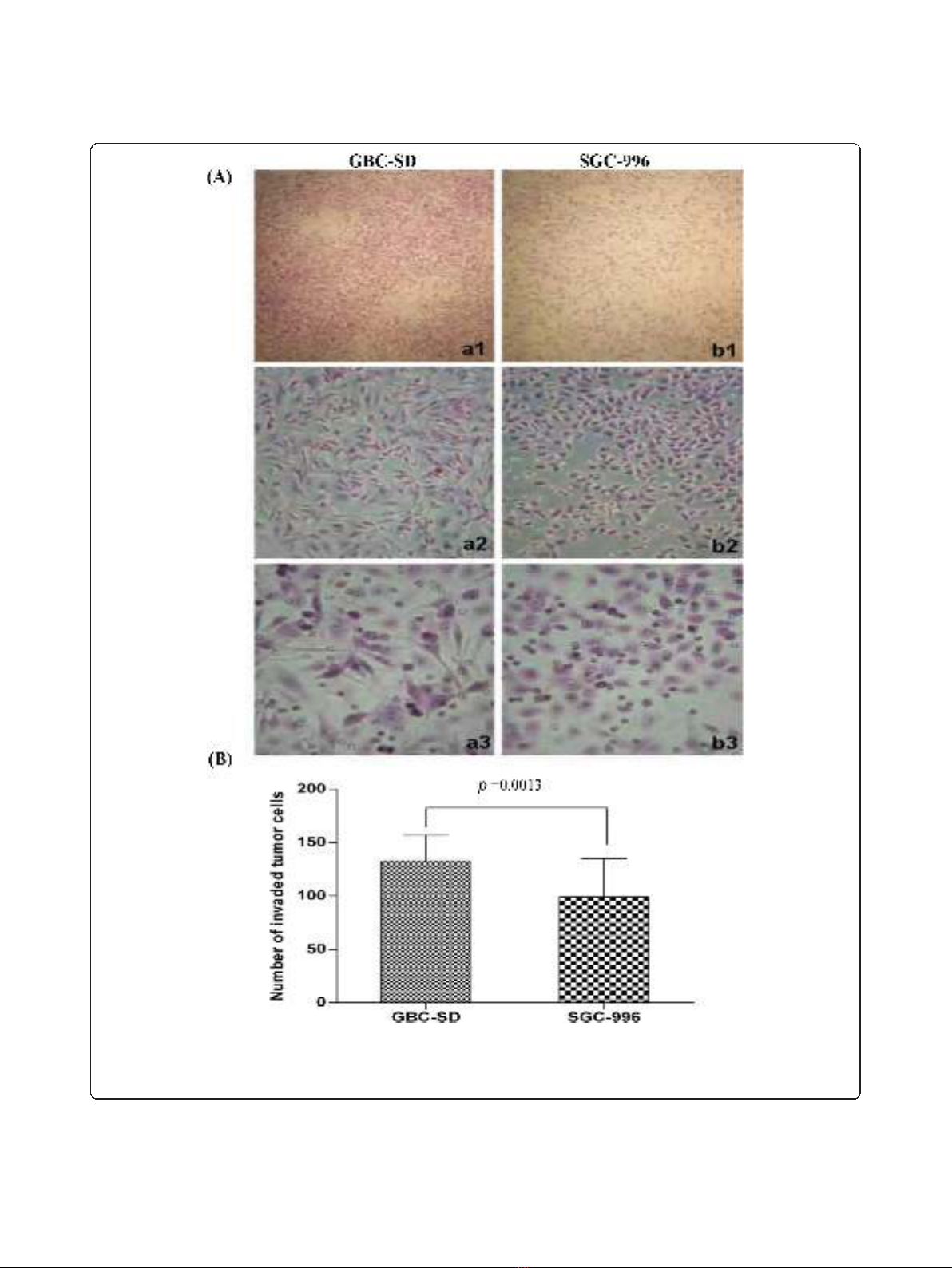
Figure 1 Invasive potential of human gallbladder carcinoma cell lines GBC-SD and SGC-996 in vitro.(A) Representative phase contrast
microscopy pictures of GBC-SD cells (a
1-3
; original magnification, a
1
× 100, a
2
× 200, a
3
× 400) and SGC-996 cells (b
1-3
; original magnification, b
1
× 100, b
2
× 200, b
3
× 400) with HE staining. Both GBC-SD and SGC-996 cells could invade through the matrix-coated membrane to the lower
wells of Transwell plates. (B) The invaded number of GBC-SD cells were much more than that of SGC-996 cells (P= 0.0013).
Sun et al.Journal of Experimental & Clinical Cancer Research 2011, 30:46
http://www.jeccr.com/content/30/1/46
Page 5 of 12


![PET/CT trong ung thư phổi: Báo cáo [Năm]](https://cdn.tailieu.vn/images/document/thumbnail/2024/20240705/sanhobien01/135x160/8121720150427.jpg)


