RESEARCH Open Access
Pyrosequencing, a method approved to detect
the two major EGFR mutations for anti EGFR
therapy in NSCLC
Sandrine Dufort
1,2
, Marie-Jeanne Richard
1,2
, Sylvie Lantuejoul
2,3
and Florence de Fraipont
1,2*
Abstract
Background: Epidermal Growth Factor Receptor (EGFR) mutations, especially in-frame deletions in exon 19 (ΔLRE)
and a point mutation in exon 21 (L858R) predict gefitinib sensitivity in patients with non-small cell lung cancer.
Several methods are currently described for their detection but the gold standard for tissue samples remains direct
DNA sequencing, which requires samples containing at least 50% of tumor cells.
Methods: We designed a pyrosequencing assay based on nested PCR for the characterization of theses mutations
on formalin-fixed and paraffin-embedded tumor tissue.
Results: This method is highly specific and permits precise characterization of all the exon 19 deletions. Its
sensitivity is higher than that of “BigDye terminator”sequencing and enabled detection of 3 additional mutations
in the 58 NSCLC tested. The concordance between the two methods was very good (97.4%). In the prospective
analysis of 213 samples, 7 (3.3%) samples were not analyzed and EGFR mutations were detected in 18 (8.7%)
patients. However, we observed a deficit of mutation detection when the samples were very poor in tumor cells.
Conclusions: pyrosequencing is then a highly accurate method for detecting ΔLRE and L858R EGFR mutations in
patients with NSCLC when the samples contain at least 20% of tumor cells.
Introduction
Detection of mutations of the epidermal growth factor
receptor (EGFR) gene is critical for predicting the
response to therapy with tyrosine kinase inhibitors
(TKIs, e.g.: gefitinib and erlotinib) in patients with non-
small-cell lung cancer (NSCLC) [1]. Practically all muta-
tions are on exons 18 through 21 where they affect the
ATP-binding cleft of EGFR [2]. In vitro studies have
shown that EGFR mutants have constitutive TK activity
and, therefore, a greater sensitivity to anti-EGFR inhibi-
tion. Two classes of mutation account for approximately
90% of EGFR mutations reported to date in lung adeno-
carcinoma [3]. The class I mutations are in-frame dele-
tions in exon 19, which almost always include amino-
acid residues leucine 747 to glutamic acid 749 (ΔLRE).
The second mutation is a single-point mutation in exon
21, which substitutes an arginine for a leucine at codon
858 (L858R).
Thus far, the direct DNA sequencing method is the
most common and conventional method used for the
detection and identification of mutations in tumor cells.
However, its sensitivity is suboptimal for clinical tumor
samples. Mutant DNA needs to comprise ≥25% of the
total DNA to be easily detected [4]. All new techniques
claim to be more sensitive with the ability to detect
mutations in samples containing ≤10% mutant alleles.
Pyrosequencing is a non-electrophoretic real time
sequencing technology with luminometric detection [5].
Not only can it detect mutations but it also permits a
mutation to be characterized and to quantify the per-
centage of mutated alleles in a sample. We have pre-
viously shown that it is a robust method to characterize
the KRAS codon 12 and 13 mutations in paraffin-
embedded samples in daily practice [6].
Here we also show that pyrosequencing is a simple
and sensitive method to detect the two most common
mutations of the EGFR TK domain, and demonstrate its
* Correspondence: fdefraipont@chu-grenoble.fr
1
UM Biochimie des Cancers et Biothérapies, CHU Grenoble, Institut de
Biologie et Pathologie, parvis Belledonne, 38 043 Grenoble, France
Full list of author information is available at the end of the article
Dufort et al.Journal of Experimental & Clinical Cancer Research 2011, 30:57
http://www.jeccr.com/content/30/1/57
© 2011 Dufort et al; licensee BioMed Central Ltd. This is an Open Access article distributed under the terms of the Creative Commons
Attribution License (http://creativecommons.org/licenses/by/2.0), which permits unrestricted use, distribution, and reproduction in
any medium, provided the original work is properly cited.

usefulness for detecting such mutations in clinical lung
tumor samples, in a large prospective series.
Materials and methods
Cell lines
The human lung cancer cell lines NCI-H1650 and NCI-
H1975 were obtained from the American Type Culture
Collection (ATCC). Both cell lines were cultured in
RPMI 1640 supplemented with 10% fetal bovine serum
at 37°C in air containing 5% CO
2
. Peripheral Blood
Lymphocytes (PBL) used as negative control were
obtained from healthy volunteers.
Clinical samples
Between 1
st
January and 30 June 2010, 213 tumor sam-
ples were collected from consecutive patients with an
advanced lung adenocarcinoma, DNA extracted and
their EGFR mutation status determined for selection for
anti EGFR treatments by clinicians. All analyses were
conducted with full respect of patients’rights to confi-
dentiality and according to procedures approved by the
local authorities responsible for ethics in research. All
samples were histologically analyzed by an experienced
thoracic pathologist and classified according to the
WHO classification of lung cancer. For each sample, the
percent of tumor cells was determined.
DNA extraction
The DNAeasy kit (Qiagen) was used according to the
manufacturer’sinstructionstoextractgenomicDNA
from cells and from tumor tissues. A prolonged (48H)
proteinase K digestion was used for paraffin-embedded
tissues [6].
PCR amplification of exons 19 and 21 of the EGFR gene
PCR and sequencing primers were designed using the
PSQ assay design (Biotage) and are described in table 1.
100 ng of tumor DNA was amplified using a nested
PCR to amplify almost all samples independent of the
type of tissue fixative or of the fixative conditions.
The first PCR product was amplified at 58°C for 20
(exon 19) or 10 (exon 21) cycles. The second PCR pro-
cedure was carried out in a total volume of 50 μlcon-
taining 2 μl of the first PCR, 20 pmol of each primer,
1.5 mmol/l MgCl
2
and 1.25 U of FastStart Taq DNA
polymerase (Roche). PCR conditions consisted of initial
denaturing at 95°C for 15 min, 45 cycles at 95°C for 20
s, 62°C (exon 19) or 61°C (exon 21) for 20 s, 72°C for
20 s and a final extension at 72°C for 10 min. The PCR
products (10 μl) were analyzed by electrophoresis in a
3% agarose gel to confirm the successful amplification
of the 180-bp or the 195-bp PCR product.
Pyrosequencing analysis
40 μl of PCR product were bound to streptavidin
Sepharose HP (GE Healthcare), purified, washed, dena-
turedusinga0.2mol/lNaOHsolution,andwashed
again. Then 0.3 μmol/l pyrosequencing primer was
annealed to the purified single-stranded PCR product
and the pyrosequencing was performed on a PyroMark
ID system (Qiagen) following the manufacturer’s
instructions. The nucleotide dispensation order was
GTATCAGACATGAC for analysis of exon 19 and
CTGCGTGTCA for analysis of exon 21.
Results
Pyrosequencing assay of exon 19 deletions
In order to test the pyrosequencing method for the ana-
lysis of exon 19 deletions, we used DNA from the NCI-
H1650 cell line as positive control and DNA extracted
from human peripheral blood lymphocytes (PBL) as
wild-type control. We choose a particular pyrosequen-
cing program with the oligonucleotide dispensation
order (GTATCAGACATGAC) because it permits to
distinguish wild type and mutated alleles (table 2) gener-
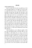
ating for each sample a specific pyrogram (Figure 1A
and 1B and Figure 2). These pyrograms correspond to a
mix of wild type and mutated alleles. We quantitatively
evaluated the exon 19 deletion (c.2235-2249del; p.
Glu746-Ala750del) by determining the ratio between the
peak areas of the two adenines dispensed in positions 6
(A
6
)and8(A
8
). We tested the reproducibility of the
Table 1 Sequences of primers used for pyrosequencing analysis
Exon 19 Exon 21
primer sequence T° of
hybridation
primer sequence T° of
hybridation
First 5’-GCAATATCAGCCTTAGGTGCGGCTC-3’58°C 5’-CTAACGTTCGCCAGCCATAAGTCC-3’58°C
PCR 5’-CATAGAAAGTGAACATTTAGGATGTG-3’5’-
GCTGCGAGCTCACCCAGAATGTCTGG-3’
second 5’-CATGTGGCACCATCTCACAAT-3’62°C 5’-GAATTCGGATGCAGAGCTTCTT-3’61°C
PCR 5’-Biotin-CCCACA CAGCAA
AGCAGAAACT-3’
5’-Biotin-CTTTCTCTTCCGCACCCA
primer for sequence
reaction
5’-TAAAATTCCCGTCGC-3’5’-CATGTCAAGACTACAGATT-3’
Dufort et al.Journal of Experimental & Clinical Cancer Research 2011, 30:57
http://www.jeccr.com/content/30/1/57
Page 2 of 7
technique by analyzing each DNA in 20 consecutive and
independent runs. We found an A
6
/A
8
ratio of 1.06 ±
0.04 for the wild type sample and 4.59 ± 0.33 for the
sample with the deletion. The relative standard deviation
(RSD) was respectively 3.9% and 7.2%. Thus, a sample
could be considered as mutated if A
6
/A
8
was superior to
1.2 (corresponding to [the mean + 3 standard devia-
tions] of the wild type sample). To demonstrate the
assay sensitivity, we also quantified the A
6
/A
8
ratio in
variousmixtures(10/0,9/1,8/2,7/3,6/4,5/5,4/6,3/7,
2/8, 1/9 and 0/10) of DNA from the NCI-H1650 cell
line with DNA from peripheral blood lymphocytes (Fig-
ure 1C). Each mixture was analyzed 5 times in the same
runandwefoundanA
6
/A
8
ratio varying from 5.27 ±
0.38 (mixture 10/0) to 1.11 ± 0.05 (mixture 0/10). We
determined that all the mixtures containing at least 20%
of NCI-H1650 DNA have an A
6
/A
8
ratio superior to 1.2
and could be considered as mutated.
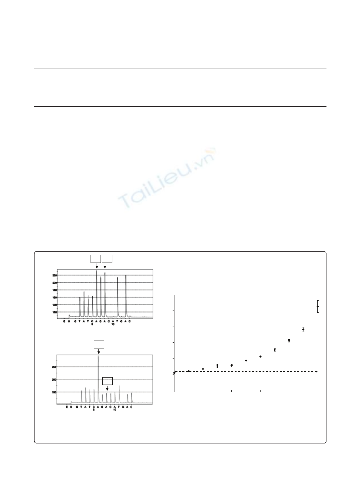
Moreover, the pyrosequencing program that analyzed
the deletions in exon 19 was designed to detect almost
all types of deletion (figure 2). In comparison with the
graph obtained with the wild type sample, the diminu-
tion of several peaks (marked *) and the emergence of
new ones (marked ◊) were considered as specific of a
deletion (table 2).
Pyrosequencing assay of L858R exon 21 point mutation
L858R-specific pyrosequencing was performed using the
NCI-H1975 cell line and a percentage of T > G muta-
tion was determined (Figure 3). The result obtained
with 20 consecutive runs, was 46.2 ± 3% with good
reproducibility (RSD = 6.4%). We also determined the
Table 2 Sequencing of wild type and mutated alleles with a particular program of pyrosquencing
nucleotide dispensation during pyrosequencing G T A T C A G A C A T G A C
WT T A T C AA GG AA TT AA
allelic c.2235-2249del TATCAAAA C A T C
sequence of c.2236-2250del TATC AA G ACAT C
c.2237-2251del TATC AA GG CA T C
c.2240-2257del T A T C AA GG AA T C
Bold letters correspond to the nucleotides identical in wild type and mutated alleles; italic letters correspond to the nucleotides specific of mutated alleles.
0
1
2
3
4
5
6
0 20406080100
proportion of H1650 DNA (%
)
A
6
/A
8
A
B
C
A6
A8
A6A8
A6/A8= 1.06 0.04; RSD=3.9%
A
6
/A
8
= 4.59 0.33; RSD=7.2%
Figure 1 Analysis of exon 19 deletions by pyrosequencing. The analysis was performed with PBL DNA (A) as wild-type control and with
NCI-H1650 DNA (B) as deletion control. The deletion was quantified by determining the ratio between the A
8
and A
6
peak areas. (C) The
sensitivity was characterized by measuring A8/A6 ratio in different mixtures of NCI-H1650 DNA and PBL DNA.
Dufort et al.Journal of Experimental & Clinical Cancer Research 2011, 30:57
http://www.jeccr.com/content/30/1/57
Page 3 of 7
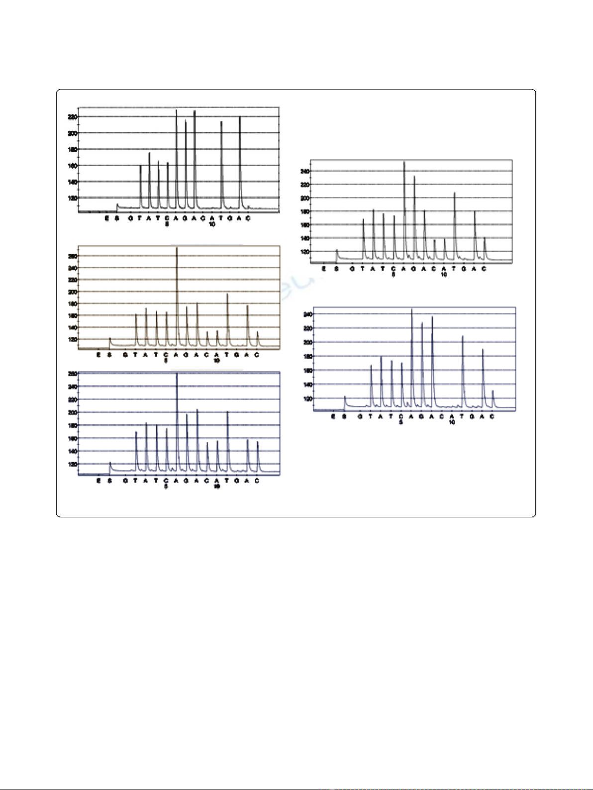
repeatability and the sensitivity of this method with var-
ious mixtures (10/0, 9/1, 8/2, 7/3, 6/4, 5/5, 4/6, 3/7, 2/8,
1/9 and 0/10) of DNA from the NCI-H1975 cell line
and DNA from peripheral blood lymphocytes (Figure
3C). We detected the percentage of T > G mutation
with a linear variation (R
2
= 0.99) from 39.6 ± 0.6%
(mixture 10/0) to 7.7 ± 1.7% (mixture 4/6) and a relative
standard deviation varying from 1.4 to 15.9%. We also
determined a% of mutation for the mixtures 3/7 and 2/8
with a CV largely higher then 20%.
EGFR mutation in tumor samples
We compared the results obtained previously by con-
ventional BigDye Terminator sequencing [7] using the
method described by Pao et al [8] and those obtained by
pyrosequencing 58 of these tumor samples (Table 3). All
mutated samples were confirmed twice, starting from
independent polymerase chain reactions. We observed a
very high concordance between the two methods (56/58
(96.6%) for exon 19 and 57/58 (98.3%) for exon 21 ana-
lysis). For 3 samples (3/58; 5%), results were discordant
and mutations were detected only by pyrosequencing
and not by Big Dye terminator sequencing, reflecting
the lower sensitivity of the classical sequencing method.
Indeed, the two samples with an exon 19 deletion have
an A
6
/A
8
ratio of 1.7 and 1.8 which correspond to less
of 25% of mutated alleles (figure 1C). For the sample
with a L858R mutation detected only by pyrosequen-
cing, we found that only 22.5% of the DNA was
mutated.
We then determined the EGFR status of 213 patients
with advanced or metastatic lung adenocarcinomas for
c.2235_2249del; p.Glu746_Ala750del
c.2236_2250del; p.Glu746_Ala750del
c.2237_2251del; p.Glu746_Thr751delinsAla
c.2240_2257del; p.Leu747_Pro753delinsSe
r
Wild type
** * *
** *
*
***
**
¸¸ ¸
¸¸ ¸
¸¸ ¸
¸
Figure 2 Comparison of different pyrograms observed for exon 19 analyses in different tumor tissues. The exon 19 status were
described as wild type or deleted (*: peak diminished in the deleted samples; ◊: peak increased in the deleted samples).
Dufort et al.Journal of Experimental & Clinical Cancer Research 2011, 30:57
http://www.jeccr.com/content/30/1/57
Page 4 of 7
selection of to anti EGFR therapies (table 4). Seven
(3.3%) samples were inconclusive due to poor DNA
quality with no DNA amplification. Of the 206 remain-
ing samples, 18 EGFR mutations were detected (8 of
exon 19 and 10 of exon 21) (18/206; 8.7%). Among
these 206 specimens, 36 had less than 20% of tumor
cells and only one with a mutation was detected (1/36;
2.8%). For the 170 specimens containing more than 20%
of tumor cells, 17 with mutations were found (17/170;
10%).
Discussion
Pyrosequencing is sensitive and enables accurate detec-
tion of mutations. A previous study has described the
capacity of this method to detect small insertions [9]
but this study is the first to demonstrate the application
of pyrosequencing to exon 19 deletions. Analysis of
exon 21 by pyrosequencing had been succinctly
described by Takano et al. [10,11], but without any data
about the specificity, the repeatability or the sensitivity.
We first investigated the characteristics of EGFR
mutations in the lung cancer cell lines NCI-H1650 and
NCI-H1975 and used them as positive controls for the
deletion in exon19 and the point mutation in exon 21
respectively. Moreover we used the DNA of these cells
mixed with DNA isolated from blood samples from
healthy volunteers to evaluate the basic properties of
our novel method. We didn’t observe strict linearity
because the two cell lines (NCI-H1650 and NCI-H1975)
have respectively 4 and 2.8 EGFR gene copies [12] but
we found good sensitivity.
In routine daily practice fixed paraffin-embedded spe-
cimens, most often of small size, are the only samples
available for both diagnosis and molecular analyses. The
DNA is frequently fragmented, which could hamper
PCR amplification. However, the PCR conditions
described in this study allowed analysis of 96.7% of the
paraffin-embedded tissues whatever the type of fixative
used or the duration of the fixation. When the samples
could be amplified and analyzed, results were concor-
dant (97.4%) with those obtained by conventional Big-
Dye terminator sequencing. The difference in sensitivity
between the two methods is illustrated by the 3 samples
characterized as mutated only by pyrosequencing. The
frequency of deletions in exon 19 and mutations in
exon 21 among the NSCLC patients was almost
R
2
= 0,99
0
5
10
15
20
25
30
35
40
45
0 10203040506070809010
0
proportion of H1975 DNA (%)
% of mutation
*
A
B
C
Figure 3 Analysis of c.2573T > G; p.Leu858Arg exon 21 mutation by pyrosequencing. Examples of pyrosequencing profiles obtained with
PBL (A) and NCI-H1975 (B) DNA.* represented the T > G mutation. (C) Sensitivity curve established with different mixtures of NCI-H1975 and PBL
DNA.
Table 3 Comparison of EGFR status (wild type (WT) or mutant (M)) of exon 19 and exon 21 determined by big dye
sequencing or by pyrosequencing on 58 NSCLC tissues
Exon 19 big dye sequencing Exon 21 big dye sequencing
WT M WT M
pyrosequencing WT 47 / pyrosequencing WT 53 /
M2 9 M1 4
Dufort et al.Journal of Experimental & Clinical Cancer Research 2011, 30:57
http://www.jeccr.com/content/30/1/57
Page 5 of 7