HUE JOURNAL OF MEDICINE AND PHARMACY ISSN 1859-3836
76
Hue Journal of Medicine and Pharmacy, Volume 14, No.2-2024
Secondary hyperparathyroidity in patients with progressive chronic
kidney disease
Dinh Thi Minh Hao1, Vo Tam1*
(1) Nephrology Department, Hue Central Hospital, Vietnam
Abstract
In patients with end-stage chronic kidney failure, there are a number of disorders that cause bone damage.
In particular, secondary hyperparathyroidism (SHPT) is related to chronic kidney failure, a calcium-phosphorus
metabolism disorder that causes bone disorder. Secondary HPT occurs when parathyroid hormone(PTH) is
continuously produced in response to chronically low serum calcium levels, commonly seen in patients with
progressive chronic kidney disease. In this article we present a case of secondary HPT causing facial and
thoracic bone changes.
Key words: end-stage chronic kidney failure, chronic kidney, secondary hyperparathyroidism (SHPT).
Corresponding author: Vo Tam. Email: vtam@huemed-univ.edu.vn
Recieved: 8/11/2023; Accepted: 19/2/2024; Published: 25/2/2024
DOI: 10.34071/jmp.2024.2.10
1. INTRODUCTION
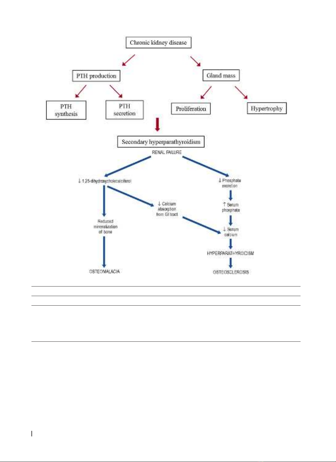
In patients with chronic kidney disease, there is
a spiral of calcium-phosphorus disorders involving
the kidney-gut axis: insufficient 1,325-(OH)2D3
produced in kidneys, causing vitamin D to not be
absorbed, leading to low serum calcium levels and
increased PTH response of the parathyroid glands,
eventually causes secondary hyperparathyroidism.
In chronic renal failure, secondary or tertiary
hyperparathyroidism may occur. Secondary
hyperparathyroidism can affect many different
bones and is most common in flattened bone plates
that change the pattern of bone trabeculae. In its
most severe forms, it can cause bone hypertrophy or
fibrocystic osteomyelitis, all of which are collectively
known as renal osteomalacia [1].
In this article, we introduce a case of secondary
HPT related to changes in the maxillofacial and
thoracic bones:
Case report:
A 29-year-old male patient with end-stage chronic
kidney disease has been receiving peritoneal dialysis
for about 5 years. He comes to our department to
do tests preparing for a kidney transplant. While
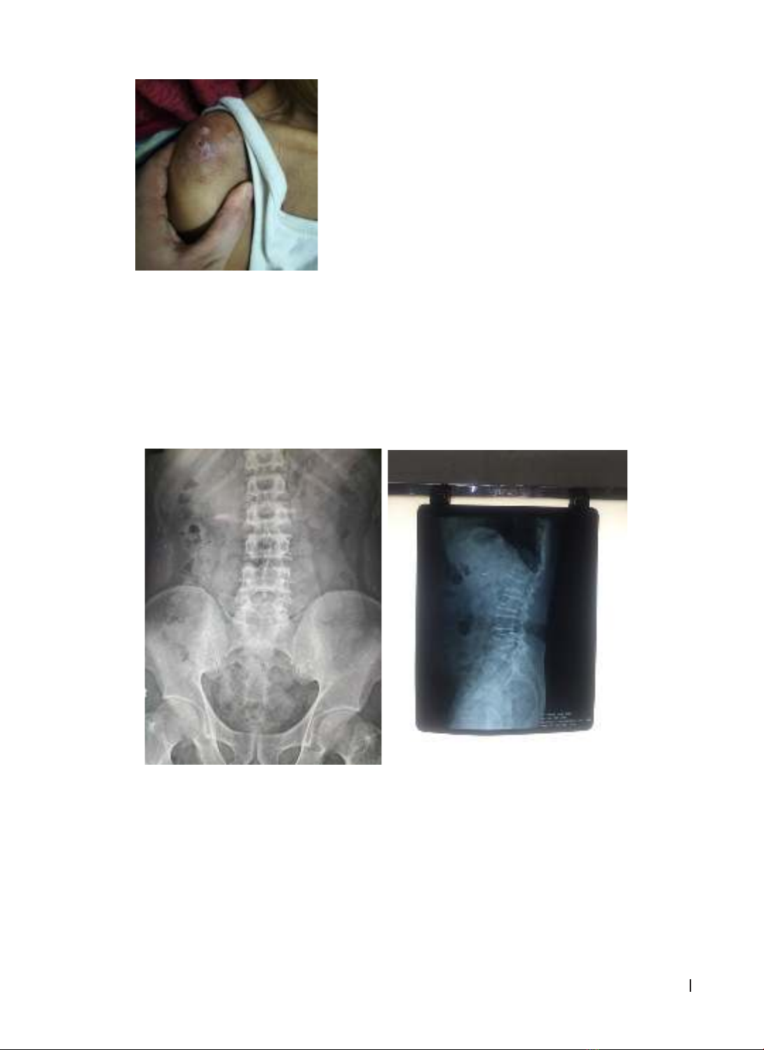
examined, the patient was found to have abnormally
deformed bone areas, mainly focusing on flat bone
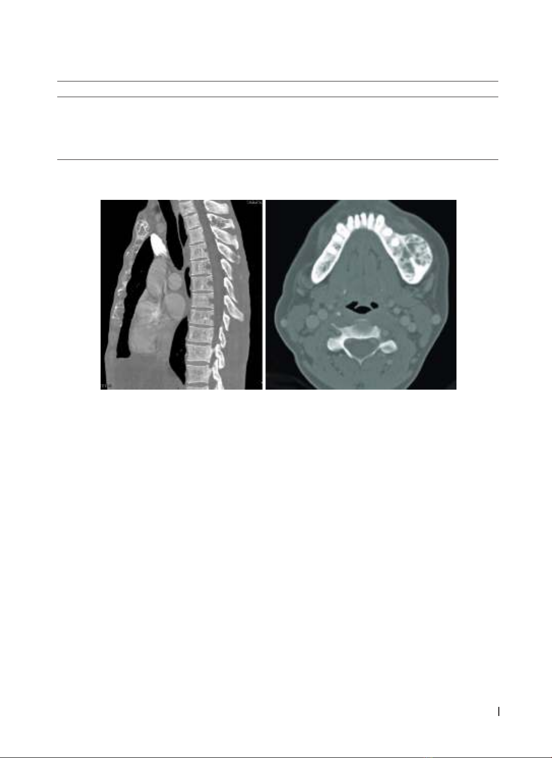
areas: jawbone, sternum, ribs, unrelated to trauma.
The patient has no other medical history. The
deformed bone areas have appeared for nearly 3
years but he was no pain, had no other symptoms and
had not received any treatment. The results of dental
and facial examination showed no abnormalities
other than cystic jaw bone changes.
Figure 1. patient with face deformity
HUE JOURNAL OF MEDICINE AND PHARMACY ISSN 1859-3836 77
Hue Journal of Medicine and Pharmacy, Volume 14, No.2-2024
He was then given tests related to bone metabolic disorders in people with CKD. The result is a severe
Ca-P disorder as figure below
Blood test Patient result Normal range
Calcium 1.86 2.15 - 2.50 mmol/l
Phosphorus 1.64 0.8 - 1.6 mmol/l
Alkaline Phosphatase 520 30 - 120 U/l
PTH 3204 15 - 65 pg/ml
Accompanied by images of bone dystrophy and severe bone cysts in the jaw and sternum areas. In
addition, measuring bone density also detects osteoporosis in the bones of the spine. Ultrasound and MRI
scan detected parathyroid adenoma.
Figure 2. images on thoracic MRI and CT of patient’s jaw
Ultimately, the patient was diagnosed with
secondary hyperparathyroidism - a condition
caused by a calcium-phosphorus disorder common
seen in patients with progressive CKD and ESKD,
he was received conservative medical treatment
with calcitriol 0.25 mcg/day, cinacalcet 30 mg/day
and sevelamer 800 mg/day. He was examined and
retested every 3 months. Results after 3 months are
as follow: Ca 1.65 mmol/l, Phosphorus 1.20 mmol/l,
alkaline phosphatase 210 U/l, PTH 1500 pg/ml.
Discussion: So what is secondary
hyperparathyroidism in patient with CKD?
We know that hyperparathyroidism can be
primary, secondary, and even have tertiary and
quaternary. Among them, the primary type is mostly
due to parathyroid adenoma (80–90% of cases),
parathyroid hyperplasia, and parathyroid carcinoma.
Secondary hyperparathyroidism is an acquired
disorder, occurring secondary and commonly seen
in patients with chronic kidney disease, especially
patients with advanced chronic renal failure. Even
though the patient has received renal replacement
therapy. This disorder affects about 38% patients
in Stage 3 and 68% patients in Stage 4 of chronic
kidney disease [2].
Characteristics of HPT due to chronic kidney
disease are increased parathyroid function in
response to calcium-phosphorus imbalance caused
by impaired kidney function.
In CKD, hyperphosphatemia and decreased 1,25
(OH)2 D3 production lead to decreased ionized
calcium, causing the parathyroid glands to become
stimulated (secondary hyperparathyroidism)
and over time can become secondary or tertiary
hyperparathyroidism [1].
One of the first changes of the Parathyroid
gland in CKD is cell hyperplasia. These disorders
continuously increase blood PTH levels, leading
to increased CaxP products, increasing the risk of
calcium deposition in tissues, causing excessive
excretion of calcium and phosphorus through
the kidneys. Consequences: urinary stones,
chronic bone loss due to diffuse bone mineral
loss, pathological fractures, cystic bone lesions
(fibrous osteomyelitis and cystic fibrosis), renal
osteodystrophy [1],[5].
HUE JOURNAL OF MEDICINE AND PHARMACY ISSN 1859-3836
78
Hue Journal of Medicine and Pharmacy, Volume 14, No.2-2024
A characteristic of SHPT is that PTH concentrations are always elevated above 5 times the normal value
Lab comparison between HPT types
Hyperparathyroidism Calcium PTH Vitamin D Phosphate
Primary Elevated Elevated/ normal Elevated Decreased
Secondary Elevated/ normal Elevated Decreased Elevated/
decreased
Tertiary Elevated Elevated Decreased Elevated
Clinically, this disorder often has a gradual,
insidious onset and no obvious symptoms. Its
appear when serum PTH levels increased very high,
causing bone metabolism disorders, typically renal
osteodystrophy. Common symptoms are:
- Bone pain: The level can range from very mild
pain to very severe, unable to move. The pain is
often vague, with deep pain in the lumbosacral
area, hip joints, knees and both sides of the legs.
Pathological fractures are possible, often occurring
in the femoral neck and vertebrae collapse.
Muscle weakness: Muscle weakness, especially
the proximal muscles, can reduce the patient’s
ability to move.
- Calcium-induced skin necrosis: Skin necrosis can
occur due to peripheral ischemia or calcification of
arterioles causing ulcers. Periarthritis: Patients have
severe pain with swelling, redness and heat around
one or more joints. Pain may occur in the ankle or
foot without local signs of inflammation.[1][2]
HUE JOURNAL OF MEDICINE AND PHARMACY ISSN 1859-3836 79
Hue Journal of Medicine and Pharmacy, Volume 14, No.2-2024
Figure 3. image of patient with calcium-induced
skin necrosis- at Nephrology Department
- Spontaneous tendon rupture: Usually occurs
in the quadriceps, triceps, and extensor digitorum
tendons
- Bone deformities: Bending of the tibia, femur,
deformity of the bone ends, often seeing bulging
ends of long bones in adolescence.
-Extraskeletal calcification: Calcification of
medium-sized arteries; juxtaarticular calcification
and calcific tumor; calcification of internal organs:
heart, lungs, kidneys...
- Developmental delay: Seen in children with
chronic kidney failure
On diagnostic imaging:
- X-ray: Hyperparathyroidism leads to bone
dystrophy. The earliest change in bone is subperiosteal
bone resorption. The late stage has systemic
osteoporosis, bone destruction, bone cavities and
bone deformities.
- Bone density measurement: Bone density
measurement results may show reduced bone
density or osteoporosis.
- Parathyroid scan (Scintigraphy): Parathyroid scan
using radioactive isotopes Iodine-123, Thallium-201
or 99mTechnetium - methoxyisobutylisonitrile
(99mTc-MIBI) helps evaluate location, shape, size,
and function of the Parathyroid Gland.
- Bone biopsy: Bone histopathology results
help accurately diagnose bone disease due to
hyperparathyroidism, mixed bone disease or
osteomalacia
Figure 4. images of extra-skeletal calcification in patients with SHPT
at the Department of Nephrology - Hue Central Hospital)
However, this disorder context also needs to be
distinguished from other cases:
- Pseudohypercalcemia: hypercalcemia results
may be due to laboratory errors or because the
tourniquet time for blood collection is too long and
the test always needs to be repeated. Hypercalcemia
may be due to hyperproteinemia (as in dehydration),
so serum calcium should be measured as albumin.
- Hypercalcemia due to malignant tumors: many
malignant tumors (breast, lung, pancreatic, uterine,
kidney cancer) can cause hypercalcemia. In some
cases (especially breast carcinoma) there are bone
metastases. Although other cancers have not seen
bone metastases, blood calcium increases because
these tumors secrete PTHrP (parathyroid hormone-
related protein), a protein with a similar structure
to tertiary PTH. PTHrP also causes osteolysis and
hypercalcemia such as PTH. The clinical symptoms
HUE JOURNAL OF MEDICINE AND PHARMACY ISSN 1859-3836
80
Hue Journal of Medicine and Pharmacy, Volume 14, No.2-2024
of cancer-induced hypercalcemia are identical to
those of hyperparathyroidism. Serum phosphorus is
low but PTH levels are also low.
Regarding treatment, calcium-phosphorus
disorders are currently treated according to KDIGO
2017 guidelines [3].
a. Hypophosphatemia: Is the foundation of
SHPT prevention and treatment. Dietary phosphate
restriction and phosphate binders are effective
means of controlling phosphate.
- Phosphate-restricted diet: A phosphorus-poor
diet of 800 - 1000 mg/day (adjusted to a low-protein
diet), limiting protein and avoiding dairy products is
very important.
- Phosphate binders: Aluminum, magnesium,
iron, calcium, lanthanum salts and non-absorbable
intestinal polymers are effective drugs.
- Patients with CKD stage 3 - 4 often use
calcium-containing phosphorus inhibitors (calcium
carbonate, calcium acetate). CKD stage 5 can use
a calcium-containing phosphorus scavenger and/or
a calcium-free phosphorus scavenger (Sevelamer
hydrochloride).
- The total dose of elemental calcium from
calcium-containing phosphate binders must not
exceed 1500 mg/day. Calcium-free phosphorus
scavenger should be prescribed for dialysis patients
with hypercalcemia or with PTH < 16.5 pmol/l or
with severe extraskeletal calcification. When using
calcium-containing phosphorus scavengers, there
is a risk of aggravating hypercalcemia, then it is
necessary to reduce the calcium concentration in
the filtrate below 2.5 mEq/l.
b. Calcium control: When eGFR < 50 ml/min,
calcium absorption in the digestive system is
reduced due to reduced calcitriol synthesis in the
kidneys. Providing adequate amounts of calcium to
absorb phosphorus can help reverse the negative
balance of calcium. If the patient has well adjusted
phosphorus levels but has low blood calcium levels,
calcium supplementation is still needed [4].
Treatment goals: According to recommendations
of NKF - K/DOQI.
CKD stage 3 - 4: Calcium control within normal
limits
CKD stage 5: Keep blood calcium concentration
2.1 - 2.37 mmol/l.
Product Ca x P < 55 mg2/dl2.
c. Control of PTH: diet, calcimimetics, phosphorus
binders
CKD stage 3: keeps PTH at 35 - 70 pg/ml or 3.85
- 7.7 pmol/l. Measure serum PTH every 12 months
CKD stage 4: keeps PTH at 70 - 110 pg/ml or 7.7
- 12.1 pmol/l. Measure serum PTH every 3 months
CKD stage5/5D: keep PTH at 150 - 300 pg/ml or
16.5 - 33 pmol/l. Measure serum PTH every 3 months
d. Calcimimetics: Effective in treating
hyperparathyroidism in ESKD. In dialysis patients,
cinacalcet significantly reduces PTH concentrations.
Cinacalcet is indicated in patients with serum iPTH
> 300 pg/ml
e. Vitamin D: Supplement vitamin D (calcitriol)
or vitamin D analogues (doxercalciferol, alfacalcidol,
paricalcitol). Use of calcitriol depends on the severity
of CKD and the degree of hyperparathyroidism.
In the early stages, low doses of calcitriol may be
sufficient to limit hyperparathyroidism. In patients
with ESKD, high doses of calcitriol can be used.
f. When does the issue of parathyroidectomy arise?
Patients with ESKD, have severe
hyperparathyroidism (usually PTH > 800 pg/mL and
elevated AP) and additional symptoms:
- Persistent hypercalcemia
- Persistent hyperphosphatemia
- PTH increases continuously despite adequate
treatment
- Progressive extraskeletal calcification, including
calcifilaxis
- Persistent itching
Or, post-kidney transplant patients have increased
PTH accompanied by hypercalcemia with unexplained
decreased graft kidney function. These cases are
called tertiary hyperparathyroidism and need to be
resolved with partial parathyroidectomy [6].
5. CONCLUSION
SHPT is a very common condition in patients with
advanced chronic kidney disease. Symptoms appear
when parathyroid hormone level is increased. Early
medical treatment is the optimal choice for patients.
REFERENCE
1. Elmukhtar Habas Sr., Mohsen Eledrisi, Fahmi Khan,
Abdel-Naser Y Elzouki. Secondary Hyperparathyroidism
in Chronic Kidney Disease: Pathophysiology and
Management, Cureus. 2021 Jul; 13(7): e16388.
2. Dennis L Andress 1, Daniel W Coyne, Kamyar
Kalantar-Zadeh, Mark E Molitch, Farhad Zangeneh,
Stuart M Sprague. Management of Secondary
Hyperparathyroidism in CKD Stages 3 and 4: Is it Time for