RESEARCH Open Access
Down-regulation of TM4SF is associated with the
metastatic potential of gastric carcinoma TM4SF
members in gastric carcinoma
Zhouxun Chen
1,2*
, Suchen Gu
2
, Bogusz Trojanowicz
2
, Naxin Liu
1
, Guanbao Zhu
1
, Henning Dralle
2
and
Cuong Hoang-Vu
2
Abstract
Background: The aim of this study was to clarify the clinical significance of TM4SF members CD9, CD63 and CD82
in human gastric carcinoma.
Methods: By employing RT-PCR and immunohistochemistry, we studied the expression of CD9, CD63 and CD82 in
49 paired tissue specimens of normal gastric mucosa and carcinoma. All tissues were obtained from patients who
underwent curative surgery.
Results: All normal gastric epithelium and gastric ulcer tissues strongly expressed transcripts and proteins of CD9,
CD63 and CD82 as compared with corresponding controls. We found a significant correlation between CD63
mRNA level and different pM statuses (P = 0.036). Carcinomas in M0 stage revealed a stronger expression of CD63
than carcinomas in M1 stage. Expression of CD9 protein was found significantly stronger in pN0, pM0 than in
advanced pN stages (P = 0.03), pM1 (P = 0.013), respectively. We found the relationship between CD63 expression,
gender (p = 0.09) and nodal status (p = 0.028), respectively. Additionally, advanced and metastasized tumor tissues
revealed significantly down-regulated CD82 protein expression (p = 0.033 and p = 0, respectively), which correlated
with the tumor pTNM stage (p = 0.001).
Conclusion: The reduction of CD9, CD63 and CD82 expression are indicators for the metastatic potential of gastric
carcinoma cells. Unlike their expression in other tumor types, the constitutive expression of CD63 may indicate that
this factor does play a direct role in human gastric carcinogenesis.
Introduction
The TM4 superfamily (TM4SF) includes more than 20
core members and a number of additional proteins with
sequence similarities. Nearly all mammalian cells con-
tain one or more TM4SF proteins. The correct biologi-
cal functions of the TM4 superfamily could not have
been fully elucidated, but it has been reported that sev-
eral TM4SF members, such as CD9, CD63, CD81, CD82
and CD151 might be involved in cell signaling. Further-
more, recent data suggest some TM4SF members might
play roles in signal transduction pathways and to regu-
late cell activation, development, proliferation, and
motility [1]. For instance, CD9, CD82 and CD63 have
been reported to modulate the tumor progression or
metastasis [2-4]. As type III integral membrane glyco-
proteins, CD9, CD82 and CD63 have two divergent
extracellular loop domains, the larger of which contains
several conserved amino acid motifs, highly conserved
hydrophobic tetra-transmembrane domains and two
short cytoplasmic domains at the NH2 and COOH ter-
mini [5,6].
CD9 gene is located on human chromosome 12p13.3
and encodes 227 amino acids. It was described originally
as a 24-kDa surface protein of non-T acute lymphoblas-
tic leukemia cells and developing B-lymphocytes [7].
CD9 is also expressed in plasma membrane of various
cell types, including hematopoietic cells, endothelial
cells, normal epithelial cells, and several tumor cell
* Correspondence: zhouxun.chen@googlemail.com
1
Department of General Surgery, The first affiliated Hospital of Wenzhou
medical School, Wenzhou 325000, Zhejiang, PR. China
Full list of author information is available at the end of the article
Chen et al.World Journal of Surgical Oncology 2011, 9:43
http://www.wjso.com/content/9/1/43 WORLD JOURNAL OF
SURGICAL ONCOLOGY
© 2011 Chen et al; licensee BioMed Central Ltd. This is an Open Access article distributed under the terms of the Creative Commons
Attribution License (http://creativecommons.org/licenses/by/2.0), which permits unrestricted use, distribution, and reproduction in
any medium, provided the original work is properly cited.

types [8-12]. Some clinical and experimental studies
demonstrated that CD9 functions as a tumor metastatic
suppressor in various cancers, including non-small-cell
lung cancers, breast cancers, and colon cancers [13-15].
The CD82 gene is located on human chromosome
11p11.2 and encodes a 2.4 kb transcript which is trans-
lated into a N-glycosylated, transmembrane protein of
267 amino acids [3,16]. It attracted considerable atten-
tion as a tumor metastasis suppressor gene in prostatic
cancer. Recent and retrospective studies have shown
that decreased wild type CD82 expression could be a
useful marker for metastatic and has invasive potential
in some human tumor types, including pancreatic,
breast, colorectal, bladder and oral cancers [17-23].
CD63 is isolated from human chromosome 12p12-q13
has been implicated in phagocytic and intracellular lyso-
some-phagosome fusion events. CD63 plays a role in
the regulation of cell motility in melanoma cells and is
involved in cell adhesion events [24], and strongly
expressed on the cell surface in the early stage of malig-
nant melanoma but weakly in the more advanced stages
[25]. The data of our previous study demonstrated the
expression of CD82 was correlated significantly with the
metastatic status of human thyroid carcinoma. However,
CD63 expression pattern did not correlate with any
tumor staging [26].
The biological functions of these factors in human
gastric carcinoma are still not clearly understood. In this
retrospective study on staged human gastric carcinoma
tissues, we investigated the expression of these three
TM4SF members to determine whether they correlate
with the invasiveness and metastatic ability of gastric
carcinoma cells.
Materials and methods
Tissue specimens
No patient was required the perioperative neo/adjuvant
chemotherapy in this study. From each patient, one
representative primary tumor block, including tumor
centre and invasion front as well as tumor-associated
non-neoplastic mucosa, were examined by immuno-
histochemistry.
Forty-nine patients were included in this study who
with up to stage IV gastric carcinoma at the Department
of General, Visceral and Vascular Surgery of Martin-
Luther-University Halle-Wittenberg between 1994 and
2002. This study was approved by the local committee
of medical ethics and all patients gave written consent.
Tumor tissues were staged according to the Tumor-
node-metastasis (TNM) staging classification (UICC-
AJCC 1997). The clinical characteristics of the patients
with gastric carcinoma are presented in Table 1.
For employing Semi quantitative RT-PCR and immuno-
histochemistry, resected gastric tissues were immediately
frozen in liquid nitrogen and maintained at -80°C. Frozen
sections at 6 μm were cut by using Microm cryostat sys-
tem (Microm International GmbH, Walldorf, Germany)
on a cryostat and control sections were hematoxylin-eosin
stained.
Semi quantitative RT-PCR
To prevent crosscontamination of samples and carry-
over contamination, laser-assisted microdissection was
performed for subsequent isolation of genomic RNA (P.
A.L.M.
®
system, Bernried, Germany). Total RNA from
fresh tissue samples, SW480 cell line (human colon car-
cinoma cell line) and FTC-133 (human follicular thyroid
carcinoma cell line) was extracted by using the TRIZOL
reagent (Invitrogen, Carlsbad, USA) according to the
manufacturer’s protocol. First-strand cDNA synthesis
was performed with 1 μgoftotalRNAusingacDNA
synthesis kit (Gibco, Munich, Germany) following the
manufacturer’s protocol at 42°C for 30 min followed by
enzyme inactivation at 95°C for 5 min.
For PCR amplification, a 2 μl aliquot of the reaction
mixture was used. The following PCR primer pairs were
used to amplify a 800 bp amplicon of CD9 (sense 5’-
TGCATCTGTATCCAGCGCCA-3’/antisense 5’-CTC
AGGGATGTAAGCTGACT-3’; a 598 bp encoding
CD82 (sense 5’- GCA GTC ACT ATG CTC ATG G-3’/
antisense 5’-TGC TGT AGT CTT CGG AAT G-3’) and
a 347 bp amplicon for CD63 (sense 5’- CCC GAA AAA
CAA CCA CAC TGC-3’/antisense 5’-GAT GAG GAG
GCT GAG GAG ACC-3’),anda467bpampliconsfor
the housekeeping genes GAPDH (sense 5’-TGG TGA
AGGTCGGTGTGAAC-3’/antisense 5’-TTC CCA
TTCTCAGCCTTGAC-3’). All PCR reactions were
performed with AmpliTaq (for CD9, CD82 and 18 S)
and AmpliTaq-Gold (for CD63) (Amersham, USA). The
PCR profile was as follows: 30 sec at 94°C, 45 sec at
(CD9: 60°C; CD82:58°C; CD63:56°C, GAPDH:60°C) and
30 sec at 72°C. CD9, CD82, CD63 and GAPDH con-
sisted of 30 sec at 94°C, 30 sec at 60°C, 45 sec at 72°C,
and a final elongation step for 7 min at 72°C. 20 μl PCR
products were run visualized in a 1.5% agarose gel (Peq-
Lab), photographed with Kodak Image System 440 cf
and electronically evaluated with “TL100”Total Lab
software (Nonlinear Dynamics, UK). The expression of
positive control was set as 100% (Figure 1), the expres-
sion levels of all investigated specimens were classified
in comparison to the positive controls (for CD9 and
CD63: SW480; and for CD82: FTC-133-CD82 overex-
pressing clone) grey scale. The densitometric values
obtained for CD9, CD82 and CD63 bands in a given
tumor tissue sample were divided by the corresponding
valueofGAPDH,andtheratiowasreferredtoasthe
gene expression ratio for each gene. The evaluated value
of a specimen between 0%- 20% was defined as
Chen et al.World Journal of Surgical Oncology 2011, 9:43
http://www.wjso.com/content/9/1/43
Page 2 of 8
Table 1 Relation between CD9, CD63 and CD82 expression and various prognostic factors
clinicopathological
characteristics
No. of patients CD9 CD63 CD82
transcript protein transcript protein transcript protein
Gender avarage p-value average p-value avarage p-value average p-value avarage p-value average p-value
Male 29 83.10 0.707 3.11 0.238 112.56 0.616 4.40 0.009 87.90 0.66 3.19 0.54
Female 20 82.31 4.06 110.12 3.00 80.47 2.64
Age
≤65 20 81.10 0.867 3.78 0.477 113.94 0.842 4.14 0.323 83.12 0.884 3.32 0.551
>65 29 83.97 3.27 109.83 3.54 86.00 2.74
Tumor stage
T1 and T2 13 85.14 3.75 114.23 3.73 91.38 4.00
T3 11 89.98 0.79 3.43 0.215 107.64 0.462 3.17 0.81 78.34 0.866 2.39 0.033
T4 15 81.97 2.17 101.82 3.54 87.85 1.81
Nodal status
N0 5 74.61 5.60 105.13 4.25 67.82 4.40
N1 13 79.73 0.556 2.91 0.03 106.47 0.774 4.23 0.028 77.85 0.23 2.88 0.094
N2 15 94.81 (N2 and N3)2,571 111.05 2.77 109.72 2.33
N3 3 86.71 114.77 6.00 64.94 1.67
metastatic status
M0 11 90.68 0.403 4.64 0.013 121.84 0.036 3.90 0.137 90.40 0.77 4.35 0
M1 18 85.46 2.17 100.24 3.19 95.17 1.23
Differentiation
G1 and G2 5 81.93 4.20 118.89 5.25 83.67 3.75
G3 24 85.77 0.82 3.05 0.624 108.33 0.432 3.31 0.105 87.57 0.691 2.23 0.304
G4 8 86.09 3.50 114.50 3.67 71.39 3.10
pTNM stage
I and II 12 79.10 4.08 109.53 3.91 73.67 4.06
III 7 105.38 0.379 3.88 0.209 112.67 0.897 2.67 0.482 106.32 0.418 3.88 0.001
IV 19 69.66 2.50 96.80 3.68 46.35 0.95
Lauren’s classification
intestinal type 12 69.30 0.105 3.42 0.538 109.93 0.719 4.50 0.06 80.17 0.773 3.80 0.535
diffuse type 22 91.26 3.03 112.63 3.41 84.80 3.41
Chen et al.World Journal of Surgical Oncology 2011, 9:43
http://www.wjso.com/content/9/1/43
Page 3 of 8
“Negative"; 20%-50% “Decreased"; 50-75% “Moderate";
75% and more “Positive”.
Immunohistochemistry
Immunohistochemistry wasperformedbyusingDako
Coverplates (Dako, Germany) on frozen tissue sections of
6μm thickness. After 20 min fixation in a 1:4 mixture of
3% H2O2 in ice cold 90% Methanol, the slides were
washed in phosphate-buffered saline (PBS) and pre-incu-
bated for 10 min at room temperature with PBS - 1%
bovine serum albumin (BSA), which was also used as a
diluent for the antibodies. Successive sections were incu-
bated overnight at 4°C with the CD9 (mouse monoclonal,
MEM-61, abcam] at the dilutions of 1:200, the antibody
against human CD82 (mouse monoclonal, clone 50F11,
BD Pharmingen) at the dilutions of 1:300 and the antibody
against human CD63 (mouse monoclonal, NKI/C3, Novo-
castra Laboratories Ltd) at the dilutions of 1:200, respec-
tively. Negative control sections were only exposed to the
secondary antibody and processed as described above.
After 3 × 10 min washes in PBS, sections were incubated
for 30 min with a 1:1000 dilution of biotinylated goat anti-
mouse secondary antibody (Dako-anti-IgG-Kit) followed
by incubation with an avidin-biotin-peroxidase complex.
Specific immunostaining was visualized with a 15% diami-
nobenzidine (DAB) chromogenic solution (Dako, Aarhus,
Denmark). Finally, sections were lightly counterstained
with Mayer’s hematoxylin. Tissue sections from a normal
human tonsil (from patient who underwent tonsilectomy)
were used as positive controls.
Interpretation of immunostaining scoring
We employed the planimetric measurement features by
using the “PALM RoboSoftware 3.2”(PALM MicroLaser
Systems) software to determine the immunostaining
intensity. This software allows the user to encircle areas
for calculation (μm
2
). The sum of all immunopositive
cell squares (μm
2
) was calculated and compared with
the total section area. Subjective interpretation of immu-
nohistochemistry was minimized by using a modification
of the German immunoreactive score (IRS) method
(Table 2). The immunohistochemical scoring was
performed by two independent reviewers. A consensus
opinion was used to score the rare cases for divergent
opinions. We assigned an intensity score (0 to 3+) and a
distribution score (estimated percentage of reactive
cells) to describe staining of study cases. The criteria for
scoring staining intensity were listed in table 2: To cal-
culate the IRS, we assigned the following points for
staining distribution: 1, 1-25% of cells; 2, 26-50%; 3, 51-
75%; and 4, 76-100%. These points were then multiplied
bythestainingintensityscoretogivearangeofpoten-
tial IRSs from 0-12. Weak staining was defined as an
IRS that ranged from 1 to 3, and moderate/strong stain-
ing was 4-12.
Statistical analysis
Sigmaplot 8.0 was applyed for all graphs calculations.
Comparisons of the distributions of three TM4SF mem-
bers expression for different groups were performed
using the Wilcoxon-Mann-Whitney test (for two
groups) or the Kruskal-Wallis test (for more than two
groups). P-values of < 0.05 were considered to indicate
statistical significance.
Results
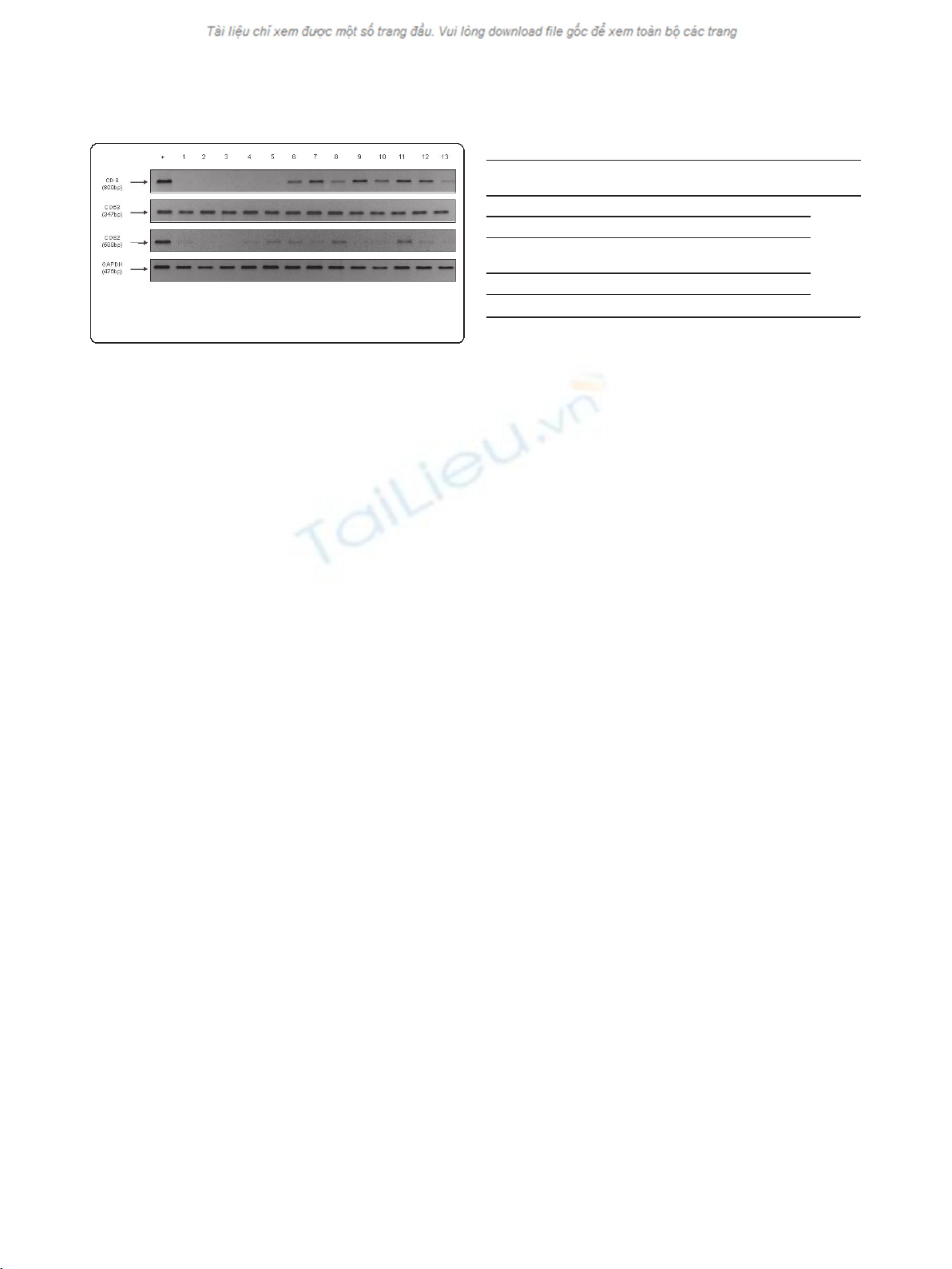
CD9, CD82 and CD63 gene expression in gastric cancer
tissues analyzed by RT-PCR
All normal gastric epithelium and gastric ulcer tissues
strongly expressed transcripts of CD9, CD63 and CD82.
Out of 49 gastric cancers tissues investigated, 17 carcino-
mas (34.7%) were evaluated as CD9 positive and 32 carci-
nomas (65.3%) as CD9 negative. Furthermore, 17
carcinoma tissues (34.7%) were evaluated as CD82 posi-
tive and 32 carcinomas (34.7%) as CD82 negative. Only 6
carcinomas (12.2%) were evaluated as CD63 negative, but
43 carcinomas (87.8%) were CD63 positive (Figure 1).
CD9, CD82 and CD63 protein expression analyzed by
immunohistochemistry
All normal gastric epithelium and gastric ulcer tissues
were strongly expressed immunostaning of CD9, CD63
and CD82. Out of 49 gastric cancer tissues were stu-
died by employing immunohistochemistry, 18 cases
(36.7%) were classified as CD9 positive. In these cases,
Table 2 Immunohistochemical scoring
A: Staining
intensity
B: Precentage of positive Tumor
cells
C: score
0 = no staining 0 = 0% positive cells
1 = weak staining 1 =< 10% positive cells
2 = moderate
staining
2 = 10 - 50% positive cells A × B =
C
3 = strong staining 3 = 51 - 80% positive cells
4 => 80% positive cells
Figure 1 1.5% Agarose gel electrophoresis of RT-PCR-amplified
CD9, CD63, CD82 and GAPDH. +: positive control; No.1-13: gastric
carcinoma tissue samples
Chen et al.World Journal of Surgical Oncology 2011, 9:43
http://www.wjso.com/content/9/1/43
Page 4 of 8
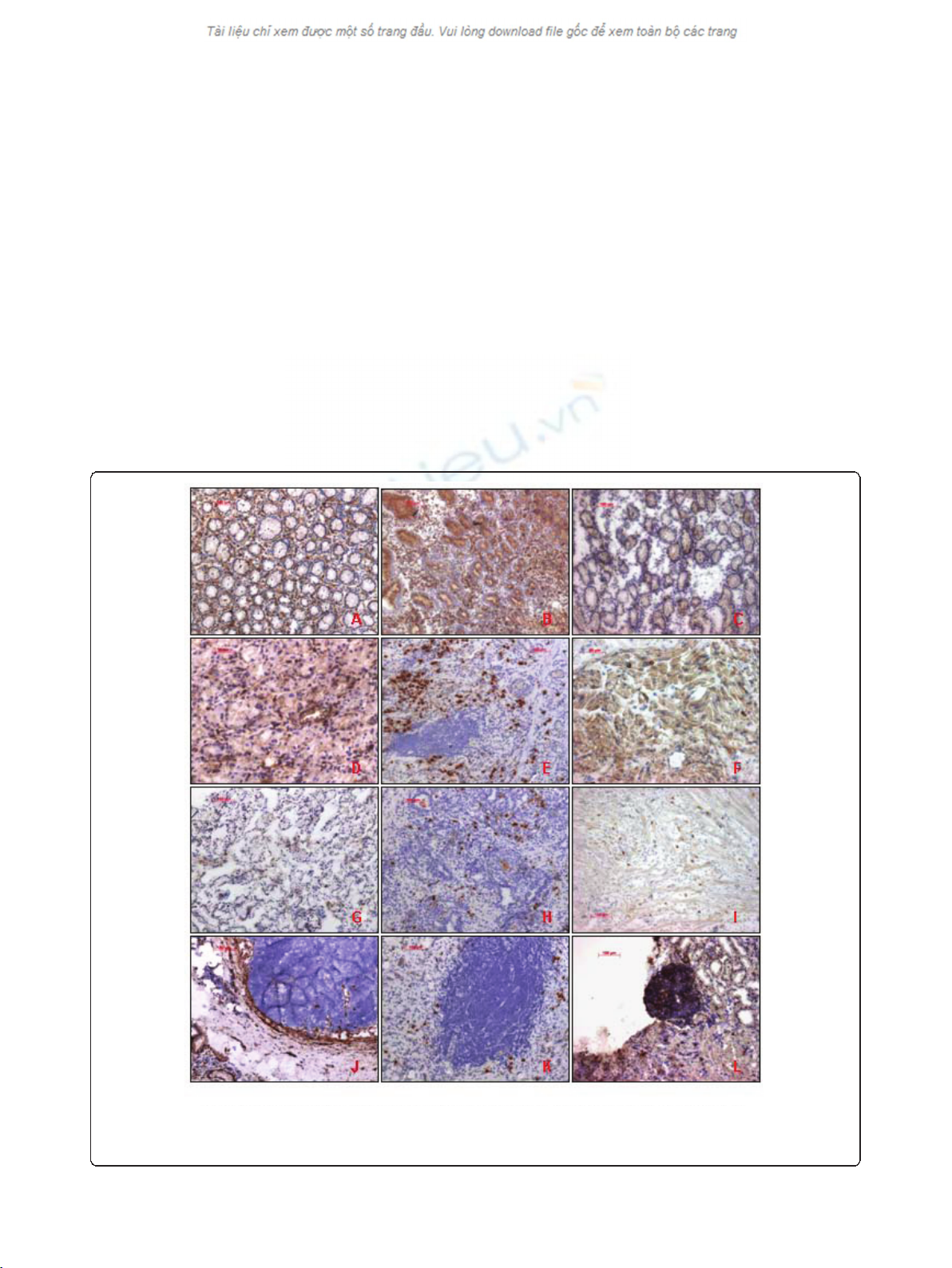
immunostaining of CD9 was intense and uniform on
the cell-surface membrane (Figure 2). 31 cases (63.3%)
revealed decreased CD9 expression, and the immunos-
taining in most of these tumors was heterogeneous.
The immunohistochemical results were agreed with
those of RT-PCR and 98.0% of the specimens coin-
cided directly.
Further investigations demonstrated 21 CD82 positive
cases (42.9%) and 27 CD82 negative cases (57.1%) (Fig-
ure 2). These results correlated with those of RT-PCR
and 91.8% of the specimens coincided directly.
We identified 30 cases (61.2%) positive for CD63 and
19 CD63 negative cases (38.8%) (Figure 2). These results
correlated with those of RT-PCR. However, only 73.5%
of the specimens coincided directly.
Relationship between CD9, CD82 and CD63 gene
expression and various prognostic factors
The relationship between CD9, CD63 and CD82 gene
expression and various prognostic factors are shown in
table 1. Analysis of CD9, revealed no statistically signifi-
cant correlations between gene expression and age, gen-
der, tumor status, differentiation, pTNM stage and
Lauren classification. Contrary, CD9 protein level was
associated with lymph node status (p = 0.03) as well as
with metastatic status (p = 0.013); Compared with 7
(63.6%) of N1 stage patients and 11(68.8%) of N2-3
stage patients, no N0 stage patients showed negative
gene expression. Furthermore, only 4(36.3%) of M0
stage patients had negative gene expression compared
with 13(72.2%) of M1 stage patients.
Figure 2 CD9, CD63 and CD82 immunohistochemical staining patterns. A,B,C: CD9, CD63 and CD82 expression in normal gastric mucosa; D,
E, F: CD9, CD63 and CD82 expression in Gastric tumor tissue (non-metastasized); G,H,I: CD9, CD63 and CD82 expression in Gastric tumour tissue
(metastasized); J,K,L:CD9, CD63 and CD82 expression in Lymph tissue (submucosa layer).
Chen et al.World Journal of Surgical Oncology 2011, 9:43
http://www.wjso.com/content/9/1/43
Page 5 of 8