HUE JOURNAL OF MEDICINE AND PHARMACY ISSN 3030-4318; eISSN: 3030-4326
76
Hue Journal of Medicine and Pharmacy, Volume 14, No.6/2024
Microscopic characteristics, total phenolic content and antioxidant
activity of Zingiber nudicarpum
Nguyen Dinh Quynh Phu1*, Doan Quoc Tuan1, Huynh Van Quynh1
(1) Hue University of Medicine and Pharmacy, Hue University
Abstract
Background: Zingiber Mill. is one of the most diverse genera in Vietnam with 36 recorded species,
many of which have been used as traditional medicine to treat several ailments. Reports of studies on Z.
nudicarpum D. Fang are relatively scarce. Objectives: This study aimed to determine the microscopic
characteristics, total phenolic content and antioxidant activity of Z. nudicarpum. Materials and methods: Z.
nudicarpum was collected in Phong Dien district, Thua Thien Hue province. Anatomic features and powder
characteristics were determined by the microscopic methods. The Folin-Ciocalteau method and 2,2-Diphenyl-
1-picrylhydrazyl (DPPH) assay were used to analysis the total phenolic content (TPC) and antioxidant
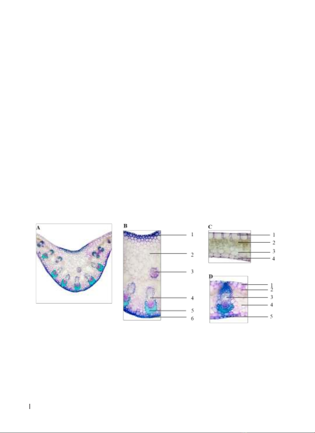
potential, respectively. Results: The microscopic characteristics of the leaves and roots of Z. nudicarpum
have been reported. The ethanol extract from the aerial part of Z. nudicarpum exhibited a notable amount
of phenolic content and antioxidant activity than the underground part extract. The total phenolic content in
the aerial and underground parts extract were 373.41 ± 1.50 mg GAE/g extract and 61.27 ± 1.65 mg GAE/g
extract, respectively. The highest DPPH radical scavenging effect was observed in the aerial part with IC50
value of 4.86 ± 0.08 µg/mL, while it was not found in the extract from the underground part (IC50 > 500 µg/
mL). Conclusion: This is the first report on the microscopic features, total phenolic content and antioxidant
capacity of Z. nudicarpum.
Keywords: Zingiber nudicarpum, microscopic characteristics, phenolic, DPPH.
Corresponding Author: Nguyen Dinh Quynh Phu. Email: ndqphu@huemed-univ.edu.vn
Received: 20/3/2024; Accepted: 10/10/2024; Published: 25/12/2024
DOI: 10.34071/jmp.2024.6.11
1. BACKGROUND
Zingiber Mill. is the third largest genus of the
Zingiberaceae family with over 140 species that are
extensively distributed in Asia, South America and
Africa. In particular, Southern China and the Indochina
peninsula are considered representative biodiversity
centers for this genus. Species in the Zingiber genus
have been used for ethnomedicine, food, and spices
in many countries. Recent research has identified a
wide variety of chemical components from Zingiber
plants, including volatile oils, organic acids, flavonoids,
terpenoids, etc… Modern pharmacological studies
have demonstrated that they possessed enormous
pharmacological applications such as antimicrobial,
antioxidant, anti-obesity, anti-inflammatory,
hypoglycemic, neuroprotective, cardiovascular
protective and anti-tumor effects [1].
There are currently at least 36 species of Zingiber
known to exist in Vietnam. Numerous species in this
genus have been widely used as medicinal plants
in folk and traditional medicine, as spices, and as a
source of raw material for the extraction of essential
oil [2]. Z. nudicarpum D. Fang has been considered
an endemic species in southern China. Recently, this
species was discovered in central Vietnam and has
been added to Vietnam’s flora. Z. nudicarpum has
been found in Nghe An, Quang Binh, Thua Thien Hue,
Quang Nam and Quang Ngai provinces [3]. Literature
review showed that studies on this plant mostly
focused on the chemical composition of essential
oil [4]. In this study, the microscopic characteristics,
total phenolic content and DPPH radical scavenging
effect Z. nudicarpum were investigated.
2. MATERIALS AND METHODS
2.1. Materials
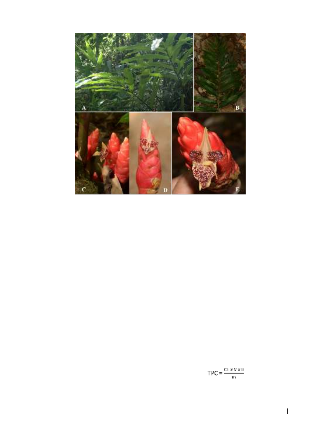
The plant of Zingiber nudicarpum D. Fang
(Zingiberaceae) (Figure 1) was collected in Phong
Dien district, Thua Thien Hue province in July 2023
and identified by Dr. Anh Tuan Le (Mientrung Institute
for Scientific Research, Vietnam National Museum of
Nature, VAST, Vietnam). Voucher specimen (PD-02)
has been deposited at the Faculty of Pharmacy, Hue
University of Medicine and Pharmacy, Vietnam.
HUE JOURNAL OF MEDICINE AND PHARMACY ISSN 3030-4318; eISSN: 3030-4326 77
Hue Journal of Medicine and Pharmacy, Volume 14, No.6/2024
Figure 1. Image of Zingiber nudicarpum
A: Plant, B: Leaves, C: Inflorescences, D, E: Flower
2.2. Methods
2.2.1. Identification of microscopic characteristics
Anatomical character: Fresh leaves and root
were cut into thin slices and soaked in 5% sodium
hypochlorite for approximately 30 minutes and washed
with water. Sections were submerged in 1% acetic acid
about 5 minutes before being washed with water. After
that, the pieces were colored with methylene blue and
carmine red solution at the appropriate time, and they
were repeatedly cleaned with water. The last sections
were put on a slide with a few drops of 10% glycerol
and observed under the microscope (Eclipse E100,
Nikon, Japan) and photographed with an attached
camera (Nikon, D5100) [5].
Powder character: The dried aerial and
underground parts of the plant were ground into
a powder and put through a 0.125-mesh hand
sieve. Slides are prepared and observed under the
microscope (Eclipse E100, Nikon, Japan) and the
images were taken (Nikon, D5100) [5].
2.2.2. Extraction
The aerial and underground parts of Z.
nudicarpum (10.0 g, each sample) were dried
and ground to afford a fine powder. From each
sample, the material was macerated with 100 mL
ethanol (EtOH) at room temperature for 24 hours
and shaken intermittently. The mixture was then
filtered through cotton. The process was repeated
twice and all filtrates were combined together then
evaporated under reduced pressure using a rotary
evaporator to obtain the EtOH extracts. These
dried extracts were stored at a refrigerator for the
following experiments.
2.2.3. Determination of total phenolic content
The total phenolic content (TPC) of EtOH extracts
were estimated by the Folin-Ciocalteu method
with slight modifications [6]. A 0.2 mL aliquot from
each extract was mixed to 0.8 mL distilled water
and 1.0 mL of 10% Folin-Ciocalteu reagent and
shaken. After 5 minutes, 2.5 mL of 7.5% Na2CO3
was added and allowed to react in dark condition at
room temperature in 30 minutes. The absorbance
of solution was measured at 760 nm by a UV-Vis
spectrophotometer. Quantification was done based
on a calibration curve of gallic acid. The results was
expressed as mg of gallic acid equivalents (mg GAE)
per gram of extract using following formula:
where TPC = total phenolic content in mg
GAE/g extract, C1 = the concentration of gallic acid
established from the calibration in mg/mL, V = the
HUE JOURNAL OF MEDICINE AND PHARMACY ISSN 3030-4318; eISSN: 3030-4326
78
Hue Journal of Medicine and Pharmacy, Volume 14, No.6/2024
initial volume of the sample test solution in mL, k is
the dilution factor and m is the weight of the plant
extract in g.
2.2.4. Determination of antioxidant activity
The antioxidant activity was evaluated using
the DPPH assay with a few minor modifications [7].
In brief, 2 mL of test sample solution at different
concentrations were mixed with 2 mL of 0.135 mM
DPPH. The reaction mixture was vortexed thoroughly
and then incubated in the dark at room temperature
in 30 minutes. Thereafter, the absorbance was read
at 517 nm. Quercetin was used as positive control
following same procedure as described above.
The percentage DPPH radical scavenging ability of
extracts were calculated and expressed in IC50 value,
which is the concentration at which the sample
removes 50% of DPPH free radicals:
DPPH free radical scavenging activity (%)
= [(Ac – As)/Ac] x 100
where Ac is the absorbance of the control solution
containing all reagents except test samples and As
is the absorbance of the DPPH solution containing
plant extract or reference standard.
2.2.5. Statistical analysis
All measurements were performed in triplicates
and analyzed by using Microsoft Excel program.
Experimental data were expressed as mean ± standard
deviation (SD) of three replicates.
3. RESULTS
3.1. Microscopic characteristics
3.1.1. Anatomy structure
Leaf midrib [Fig. 2A, 2B]: In transverse section,
the midrib was concave on adaxial surface and
convex on the abaxial surface. The upper and lower
epidermis (B1, B6) consisted of a layer of rectangular
cells and arranged adjacently with approximately
the same size. The parenchyma (B2) comprised of
many layers of polygonal cells, which were different
sizes and thin-walled. These cells were arranged
randomly with many intracellular spaces. There were
many large and small vascular bundles. The large
bundle (B4) was next to the lower epidermis, while
the small bundle (B3) was located in the middle
of the midrib. Large bundles were surrounded by
sclerenchyma cells (B5).
Leaf blade [Fig. 2C, 2D]: The structure of the
upper and lower epidermis (C1, D1 and C4, D5) was
comparable to that of the leaf midrib epidermis. The
size of the upper epidermis cells was bigger than
those of the lower epidermis. The palisade cells (C2,
D2) contained an abundance of chloroplasts. Vascular
bundles (D3) were surrounded by a parenchymal
sheath composed of large cells (C3, D4).
Figure 2. Microscopic characteristics of leaf cross-section of Z. nudicarpum
A, B: Leaf midrib (1. Upper epidermis, 2. Parenchyma, 3. Small bundle of phloem-xylem, 4. Large bundle
of phloem-xylem, 5. Sclerenchyma, 6. Lower epidermis); C: Leaf blade (1. Upper epidermis, 2. Palisade
parenchyma, 3. Parenchyma, 4. Lower epidermis); D: Leaf blade (1. Upper epidermis, 2. Palisade
parenchyma, 3. Bundle of phloem-xylem, 4. Parenchyma, 5. Lower epidermis)
Root [Fig. 3]: In transverse section of the root
of Z. nudicarpum, it was oval or broadly elliptical in
outline. The cortical area occupied more than half
of the microsurgery radius. The layers from outer to
inner showed the following features: The piliferous
layer (B1) was single layered, with polygonal cells
and the presence of unicellular root hairs. The
suberoid layer (B2) was composed of 2-3 layers
of polygonal, oval or irregularly shaped cells. The
cortical parenchyma (B3) comprised several layers
HUE JOURNAL OF MEDICINE AND PHARMACY ISSN 3030-4318; eISSN: 3030-4326 79
Hue Journal of Medicine and Pharmacy, Volume 14, No.6/2024
of parenchyma, with polygonal, unequal-sized, and
thin-walled cells. The oil cells were subrotund and
contained yellow or translucent oil droplets (C1).
Some of the cortical cells contained many starch
grains (B4, D1). The endodermis (B5) formed a
casparian strip with a thick U-shaped layer of cells.
The pericycle (B6) was a single layer of thin-walled
cells located underneath the endodermis. The
vascular tissues consisted of many patches of phloem
(B7) and xylem (B8) arranged radially. At the centre
of the internal structure was the pith parenchyma
(B9) made from thin-walled and polygonal cells.
Figure 3. Microscopic characteristics of root cross-section of Z. nudicarpum
B1. Piliferous layer, B2. Suberoid, B3. Cortical parenchyma, B4. Starch grain, B5. Endodermis, B6.
Pericycle, B7. Phloem, B8. Xylem, B9. Pith parenchyma, C1. Oil cell, D1. Starch grains
3.1.2. Powder character
The aerial part [Fig. 4]: A green powder with a pleasant and aromatic odour was covered in 10% glycerol
and observed under a light microscopic at 10X and 40X magnifications. The powder had several microscopic
features: fragment of epidermis containing stomata (1), fragment of epidermis (2), fragment of cork (3),
bundle of fiber (4), bundle of fiber containing vessel (5) and fragment of vessel (6).
Figure 4. Microscopic features of the aerial part of Z. nudicarpum
The underground part [Fig. 5]: A brown-yellow powder had pleasant and aromatic odour. Under the
light microscope at 10X and 40X magnifications, some microscopic characteristics were observed: fragment
of cork (1), fragment of vessel (2), bundle of fiber (3), fragment of parenchyma containing starch (4), starch
grains (5) and fragment of parenchyma (6).
HUE JOURNAL OF MEDICINE AND PHARMACY ISSN 3030-4318; eISSN: 3030-4326
80
Hue Journal of Medicine and Pharmacy, Volume 14, No.6/2024
3.2. Determination of total phenolic content
The total phenolic content (TPC) of the samples
was calculated from the regression equation of
the calibration curve (y = 3.8958x - 0.0319, R2 =
0.9898) and expressed in GAE as miligrams per
gram of the extract. The TPC of ethanol extracts
from Z. nudicarpum was shown in Table 1. The
results indicated that the TPC of ethanol extract
from the aerial and the underground parts of Z.
nudicarpum were 373.41 ± 1.50 mg GAE/g extract
and 61.27 ± 1.65 mg GAE/g extract, respectively.
According to the data, the TPC in the aerial part
was found significantly higher as compared to the
underground part.
Figure 5. Microscopic features of the underground part of Z. nudicarpum
Table 1. Total phenolic content and antioxidant activity of Z. nudicarpum
No. Sample Total phenolic content
(TPC ± SD (mg GAE/g extract))
Antioxidant activity
(IC50 ± SD (µg/mL))
1Aerial part 373.41 ± 1.50 4.86 ± 0.08
2Underground part 61.27 ± 1.65 > 500
Quercetin 3.38 ± 0.09
3.3. Evaluation of antioxidant activity
The antioxidant potential of Z. nudicarpum on
DPPH assay was also summarized in Table 1. The
findings revealed that the ethanol extract of the
aerial part demonstrated remarkable in vitro DPPH
radical scavenging activity with IC50 value of 4.86
± 0.08 µg/mL in comparison with quercetin used
as positive control (IC50 = 3.38 ± 0.09 µg/mL). In
contrast, the extract of the underground part was
not able to remove 50% of DPPH free radicals at a
concentration of 500 µg/mL.
4. DISCUSSION
Studies on the microscopic characteristics of
Zingiber species are relatively few and focus mainly
on morphological descriptions. Microscopic results
showed that the anatomy structure of leaf and
root of Z. nudicarpum have typical characteristics
such as many phloem-xylem vasculars in the midrib
and the casparian strip, as well as pericycle in the
root, besides the usual features such as epidermis,
parenchyma, sclerenchyma (in leaf) and piliferous
layer, suberoid, parenchyma, starch grains (in root).
The anatomical structure of root of Z. nudicarpum
is found to be quite similar to Z. officinale. The
powder characters were also comparable, except
for the starch grains in the underground part. Starch
granules in Z. nudicarpum were oblong or rounded,
while they were circular or oval in Z. officinale [8].
The present study provides a thorough detail of the
individual microcharacteristics of Z. nudicarpum for
the first time. Moreover, it can be used for determine
the identification and standardization of medicine
and contribute to the efficiency of the microscopic