Conserved structural determinants in three-fingered
protein domains
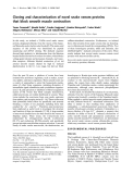
Andrzej Galat
1
, Gregory Gross
2
, Pascal Drevet
2
, Atsushi Sato
3
and Andre
´Me
´nez
4,
*
1 Institut de Biologie et de Technologies de Saclay, SIMOPRO ⁄DSV ⁄CEA, Gif-sur-Yvette, France
2 Institut de Biologie et de Technologies de Saclay, SBIGeM ⁄DSV ⁄CEA, Gif-sur-Yvette, France
3 Department of Information Science, Faculty of Liberal Arts, Tohoku-Gakuin University, Sendai, Japan
4 Muse
´um National d’Histoire Naturelle, Paris, France
To date, more than 45 000 protein three-dimensional
structures have been deposited in the Protein Data
Bank (PDB) [1], many of which have a high sequence
similarity to each other. Analyses of these structures
have revealed approximately 1000 diverse polypeptide
chain folds [2], as predicted about 10 years ago [3].
This number, however, may be subject to debate
because of the various possible ways of defining pro-
tein folds [4,5]. Nevertheless, it is accepted that the
space of protein folds is considerably smaller than that
of protein sequences [6,7]. However, how a given pro-
tein fold may evolve towards a novel function remains
obscure [6,7]. One way to approach such a complex
question is to analyse a set of functionally different
proteins recognized to adapt the same fold, and to
search for structural determinants that may reflect
both divergence and convergence criteria that are criti-
cal to the fold [5–9].
This study aims to identify the determinants associ-
ated with the three-dimensional structure of a fold that
characterizes a group of homologous proteins rich in
disulfides. According to the SCOP server (http://
scop.mrc-lmb.cam.ac.uk/scop) [2], approximately 75
folds are considered to be relatively small in size, and
about 50 are rich in disulfide bonds. In this study, we
focused our work on a group of proteins adapting the
fold originally discovered for snake neurotoxins, which
possesses three adjacent fingers rich in b-pleated sheets
Keywords
atomic interactions; cystine networks; three-
finger proteins; three-fingered protein; three-
fingered protein domain
Correspondence
A. Galat, Bat. 152, CE-Saclay, F-91191
Gif-sur-Yvette Cedex, France
Fax: +33 1 69 08 90 71
Tel: +33 1 69 08 84 67
E-mail: galat@dsvidf.cea.fr
*Deceased. The former President of the
Museum of Natural History, Paris, France
(Received 6 March 2008, revised 17 April
2008, accepted 18 April 2008)
doi:10.1111/j.1742-4658.2008.06473.x
The three-dimensional structures of some components of snake venoms
forming so-called ‘three-fingered protein’ domains (TFPDs) are similar to
those of the ectodomains of activin, bone morphogenetic protein and trans-
forming growth factor-breceptors, and to a variety of proteins encoded by
the Ly6 and Plaur genes. The analysis of sequences of diverse snake toxins,
various ectodomains of the receptors that bind activin and other cytokines,
and numerous gene products encoded by the Ly6 and Plaur families of
genes has revealed that they differ considerably from each other. The
sequences of TFPDs may consist of up to six disulfide bonds, three of
which have the same highly conserved topology. These three disulfide
bridges and an asparagine residue in the C-terminal part of TFPDs are
essential for the TFPD-like fold. Analyses of the three-dimensional struc-
tures of diverse TFPDs have revealed that the three highly conserved disul-
fides impose a major stabilizing contribution to the TFPD-like fold, in
both TFPDs contained in some snake venoms and ectodomains of several
cellular receptors, whereas the three remaining disulfide bonds impose
specific geometrical constraints in the three fingers of some TFPDs.
Abbreviations
Act-R, activin receptor; BMP-R, bone morphogenetic protein receptor; ECD, ectodomain; GPCR, G-protein-coupled receptor; ID, sequence
similarity score; MSA, multiple sequence alignment; TFP, three-fingered protein; TFPD, three-fingered protein domain; TGFb-R, transforming
growth factor-breceptor; TM, transmembrane segment; uPAR, urokinase ⁄plasminogen activator receptor; WGA, wheatgerm agglutinin.
FEBS Journal 275 (2008) 3207–3225 ª2008 The Authors Journal compilation ª2008 FEBS 3207

[10–12]. In order to provide proteins of this group with
a historically accepted name and a relevant topograph-
ical designation, we have called them three-fingered
proteins (TFPs), which all share one or more three-
fingered protein domains (TFPDs). In this article, we
describe the analyses of fifty three-dimensional struc-
tures of diverse TFPDs [1] and several hundreds of
sequences containing the TFPD-like motif.
A TFPD possesses the following features. Firstly, it
is made up of a single polypeptide chain of 60–100
amino acid residues, folded into three adjacent loops
emerging from a hydrophobic palm, which includes at
least three and, in the majority of cases, four disulfide
bonds. Secondly, it possesses five b-strands encompass-
ing the three loops or fingers. Thirdly, the TFPDs act
as monomers or multimers, and display substantial
variations in terms of loop size and shape, number of
extra disulfide bonds and additional secondary struc-
tures. Fourthly, the TFPDs display a wide distribution
in the eukaryotic kingdom. Fifthly, the TFPDs are
devoid of known enzymatic activities, but exert a wide
range of binding activities, varying from ligands
(including toxins that block or modulate the functions
of different receptors, ion channels and enzymes [13])
to receptors that are anchored to the cell surface mem-
brane [such as CD59 or urokinase ⁄plasminogen activa-
tor receptor (uPAR), also known as CD87]. Activin
(Act-R), bone morphogenetic protein (BMP-R) and
transforming growth factor-b(TGFb-R) receptors [14]
transmit signals through a transmembrane (TM)
segment to their cytoplasmic kinase domains.
Cheek et al. [15] have recently classified small
proteins rich in disulfide bonds into 41 different fold
groups. Three of these are called ‘knottin-like I, II and
III’, which are characterized by a structural core con-
sisting of four cysteine residues forming a disulfide
crossover. According to these authors, the TFPDs
belong to ‘knottin-like group II’. Interestingly, despite
the fact that some plant lectins, such as wheatgerm
agglutinin (WGA), are considered to share some topo-
graphical similarity with TFPDs [16], they have been
classified to a different fold, namely ‘knottin-like
group I’. According to Cheek et al. [15], the four cys-
tines are located on four elements that adapt different
spatial connections in groups I and II. In this work,
we have analysed in detail the conserved structural
elements of the TFPDs and examined whether or not
they are also present in some plant lectins.
We have found that all analysed TFPDs share a
conserved structural core that includes two small
b-sheets encompassing the three loops (fingers), a net-
work of three cystines and several clusters of inter-
atomic interactions, including one cluster that involves
a strictly conserved asparagine residue, which estab-
lishes several hydrogen bonds with the amino acids in
the three fingers. We have accumulated evidence sug-
gesting that the cystine that locks the third finger is
differently organized in the TFPDs that act as ligands
or receptors. Finally, our definition of the TFPD fold
has allowed for its clear distinction from the fold
typical of several plant lectins, such as WGA.
Results and Discussion
On the diversity of TFPDs
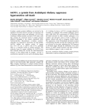
In Fig. 1, the three-dimensional structure (1IQ9) of a
typical TFP, i.e. a short-chain neurotoxin from snake
venom, is shown. The four disulfide bonds form a tight
network at the base of a palm, from which emerge
three long loops, called fingers F1, F2 and F3. A disul-
fide bridge tightly closes each finger. F1 is linked to F2
and F2 to F3 by b-turns called Lk1 and Lk2, respec-
tively. The Lk3 turn includes four amino acid residues
forming a b-turn closed by the last disulfide bridge of
the molecule. The b–sheet in F1 includes two b-strands
(b1–b2) linked by a b-turn at the tip of F1, whereas
the second small b-sheet involves three b-strands
(b3–b4–b5) located on F2 and F3. The three fingers
point approximately in the same direction.
In Table 1, data are summarized on the TFPDs
whose three-dimensional structures have been used in
this work. The 34 selected toxins from snake venoms
act as blockers or modulators of ligand-gated ion
channels (snake neurotoxins), integrin receptors (den-
droaspin), enzymes (fasciculins) or G-protein-coupled
receptors (GPCRs) interacting with muscarinic toxins.
Table 1 also includes 16 structures of cell surface
membrane-bound proteins, such as uPAR, Act-R and
TGFb-R. NIR represents the number of intramolecu-
lar atomic interactions calculated in the range
2.7–4.5 A
˚(2.7–4.0 A
˚). NIR is the sum of the intramo-
lecular interactions whose nature varies with the over-
all hydrophobicity of a given TFPD. There are about
28–31% interactions between diverse C and S atoms
(hydrophobic interactions) and 15–18% interactions
between diverse O and N atoms (hydrophilic interac-
tions); the remainder is caused by interactions
between the atoms from these two groups. Although,
the spatial organizations of some secondary structures
in the diverse TFPDs are similar, the distributions of
the atomic interactions vary. Thus, about 32–34%
interactions occur between atoms in the main chain,
22–31% between atoms of diverse side chains and the
remainder between main chain atoms and side chain
atoms.
Three-fingered protein domain A. Galat et al.
3208 FEBS Journal 275 (2008) 3207–3225 ª2008 The Authors Journal compilation ª2008 FEBS

The length of the polypeptide chain of a TFPD may
vary from 59 to 106 amino acids, except for uPAR
which contains three consecutive TFPDs. The number
of interatomic interactions shorter than 4.5 A
˚varies
from about 1100 pairs for an average sized short
neurotoxin structure to almost twice as many in the
larger ectodomain (ECD) of TGFb-RII. Obviously,
this number depends on several factors, including the
structural resolution. In this respect, NMR-based
structures must be considered with caution.
F1F1
F2F2
F3
F3
Lk1
Lk1
Lk3
Lk2
Lk3
Lk2
α-Bungarotoxin (1HC9)
Front Rear
B2a
Bucandin (1F94)
B1a
Front Rear
Front Rear
B1a
B1b
B1b
B1a
Front Rear
B3a
Activin receptor II (1S4Y)TGF-β - receptor II (1M9Z)
A
B
Fig. 1. (A) Stereoview of the tertiary structure of a TFP: the a-neurotoxin of Naja nigricollis (1IQ9). The structure was annotated as follows:
F1, F2 and F3 indicate the three successive fingers and Lk1, Lk2 and Lk3 denote the linkers that join F1 to F2, F2 to F3 and F3 to the C-ter-
minal, respectively. (B) Front and rear views of spatial positioning of the disulfides B1a, B2a, B2b and B3a.
A. Galat et al. Three-fingered protein domain
FEBS Journal 275 (2008) 3207–3225 ª2008 The Authors Journal compilation ª2008 FEBS 3209

Table 1. Crystallographic structures of diverse TFPDs. Ab, antibody; NIR, number of intramolecular atomic interactions below 4.5 A
˚(4 A
˚);
Norm-B factors show the most flexible parts of the molecule (calculated for the Caatoms); NR, number of amino acids used in the analysis.
No. PDB Protein (complex) Organism R(A
˚)NR
NIR ⁄4.5 A
˚
(4 A
˚) Norm-B Reference
Toxins from diverse snake venoms
T1 1IQ9 Toxin a Naja nigricollis 1.80 61 1128 (521) 18P, 19G, 48G [17]
T2 1VBO Atratoxin-B N. atra 0.92 61 1150 (575) 19G, 33G [18]
T3 1JE9 Neurotoxin II N. kaouthia NMR 61 964 (472) [19]
T4 2ERA Erabutoxin A, S8G Laticauda
semifasciata
1.80 62 1116 (536) 45TVK47 [20]
T5 1QKE Erabutoxin A L. semifasciata 1.50 62 1103 (532) 10E, 45TVK47 [21]
T6 6EBX Erabutoxin B L. semifasciata 1.70 62 1142 (552) 20G, 47KPG49 [22]
T7 1FAS Fasciculin-I Dendroaspis
angusticeps
1.80 61 1074 (498) 7TTTSRAI13 [23]
T8 1FSC Fasciculin-II D. angusticeps 2.00 61 1083 (503) 19G, 32K, 33M,
55S
[24]
T9 1FSS Fasciculin-II ⁄(AChE) D. angusticeps 1.90 61 1097 (513) 18GE19, 43P,
44G, 54T
[25]
T10 1F8U Fasciculin-II ⁄(AChM) D. angusticeps 2.90 61 1082 (543) 18GEN20, S55 [26]
T11 1FF4 Muscarinic toxin 2 D. angusticeps 1.50 65 1248 (562) 7KSIGG11 [27]
T12 1F94 Bucandin Bungarus candidus 0.97 63 1267 (610) 19AE20, 22T,
42T, 44TE45
[28]
T13 2H8U Bucain B. candidus 2.20 65 1022 (468) 32NPSGK [29]
T14 1JGK Candoxin B. candidus NMR 66 1027 (478) [30]
T15 2H5F Denmotoxin B. dendrophila 1.90 75 1225 (581) 41DENGE45 [31]
T16 2H7Z Iriditoxin B. dendrophila 1.50 75 1302 (578) 17TSSDCS [31]
T17 1TGX Cardiotoxin N. nigricollis 1.55 60 878 (373) 16K, 28A, 32V,
33P
[32]
T18 1CXO Cardiotoxin N. nigricollis NMR 60 1285 (643) [33]
T19 1H0J Cardiotoxin-3 N. atra 1.90 60 1083 (492) 12K, 16A, 17G,
23K, 24M, 49V
[34]
T20 2BHI Cardiotoxin A3 ⁄
sulfogalactoceramide
N. atra 2.31 60 1047 (486) 8PLF, 22Y, 31KV [35]
T21 1UG4 Cardiotoxin-IV N. atra 1.60 60 1033 (502) 28AAPLVP33 [36]
T22 1CDT Cardiotoxin N. mossambica 2.50 60 1059 (503) 29K [37]
T23 1KXI Cardiotoxin-V N. n. atra 2.19 62 971 (438) 17E, 29K, 30F [38]
T24 1CHV Cardiotoxin-(analogue) N. n. atra NMR 60 874 (415) [39]
T25 1CB9 Cardiotoxin N. oxiana NMR 60 823 (380) [40]
T26 2CTX a-Cobratoxin N. n. siamensis 2.40 71 1121 (510) 67-TRKRP-71 [41]
T27 1LXG a-Cobratoxin ⁄
(YRGWKHWVYYTCCPDTPYLhS)
N. n. kaouthia NMR 71 998 (515) [42]
T28 1YI5 a-Cobratoxin ⁄acetylcholine
binding protein (AChB)
N. n. siamensis 4.20 68 907 (396) [43]
T29 1HC9 a-Bungarotoxin ⁄
(WRYYESSLLPYPD)
B. multicinctus 1.80 74 1296 (551) 50SKKPY54,
C-term
[44]
T30 1NTN Neurotoxin-I N. n. oxiana 1.90 72 1110 (524) C-term [45]
T31 1KBA j-Bungarotoxin B. multicinctus 2.30 66 1222 (583) 15P, 16N, 17G,
35G
[46]
T32 1KFH a-Bungarotoxin B. multicinctus NMR 74 1612 (836) [47]
T33 1LSI Long neurotoxin L. semifasciata NMR 66 1162 (569) [48]
T34 1DRS Dendroaspin D. j. kaimose NMR 59 923 (443) [49]
Ectodomains of some receptors
R1 1CDR CD59 ⁄(disaccharide) Homo sapiens NMR 77 1256 (569) [50]
R2 2OFS CD59 H. sapiens 2.12 75 1512 (684) 32GLQ [51]
R3 1YWH Urokinase receptor ⁄
(KSDChaFskYLWSSK)
H. sapiens 2.70 268 4527 (1914) 79GNSGG,
C-term
[52]
R4 2FD6 uPAR ⁄plasminogen ⁄Ab H. sapiens 1.90 248 4642 (2091) 92L, 116SPEE,
229EPKNQSY
[53]
Three-fingered protein domain A. Galat et al.
3210 FEBS Journal 275 (2008) 3207–3225 ª2008 The Authors Journal compilation ª2008 FEBS

Conserved and variable sequence features
of TFPDs
In Fig. 2, an alignment of the non-redundant primary
structures of the three-fingered ligands and ECDs
listed in Table 1 is shown. Using the sequence of the
short neurotoxin from Naja nigricollis (1IQ9) as an
arbitrary reference, we calculated the pairwise sequence
similarity scores (IDs) with the remaining sequences of
the other TFPDs (Fig. 2), and found that they varied
between 86% and 30% for diverse snake toxins and
below 25% for the ECD sequences of some cell surface
receptors. This difference is caused, at least in part, by
the longer loops of the ECDs and extensive amino acid
substitutions in the fingers. In Fig. 2, a number of
strictly conserved sequence features are emphasized.
These include six half-cystines that form three disul-
fides, named B1, B2 and B4, five b-strands (coloured
yellow) located on fingers 1, 2 and 3, and an aspara-
gine residue adjacent to the last half-cystine of B4.
These are the minimal strictly conserved sequence and
structural features that define the TFPD based on the
alignment of sequences from the three-dimensional
structures.
Other sequence features are highly but not strictly
conserved. These include the cystine called B3, which
is only lacking in the first domain of uPAR (1YWH1),
a hydrophobic residue (often an aromatic residue)
adjacent downstream to the second half-cystine of B1,
and a glycine residue adjacent upstream to the second
half-cystine of B2. This glycine residue is strictly con-
served in all the toxins only. In addition, linker 1 usu-
ally comprises four to six amino acids, except for
several ECDs where it can be as long as nine amino
acids (ActRIIb). Similarly, linker 3 comprises four
amino acids, except in two cases where it can be five
amino acids (fasciculin). Other sequence elements of
TFPD tend to vary substantially from one protein to
another. These include the length and composition of
the fingers, small helical stretches and additional disul-
fides, which are labelled by a letter related to the disul-
fide that surrounds them (Fig. 2). With the exception
of B1a, the disulfide bridges seem to be specific to cer-
tain classes of TFPD (Fig. 2), such as B2a which
occurs in long neurotoxins and B3a which is found in
Act-RII. B1a is a more common feature and can be
seen in both ligands, such as bucandin, and in the
ECDs of receptors (e.g. TGFb-R); in contrast, B1b
only occurs in the ECDs of TGFb-RII (Fig. 1B).
On the conserved and variable three-dimensional
features of TFPDs
Conserved interaction clusters
To compare qualitatively and quantitatively the three-
dimensional structures of diverse TFPDs, distance
maps were constructed from the three-dimensional
structures (Table 1). Figure 3 illustrates such maps
calculated for two three-fingered ligands and two
three-fingered ECDs. Figure 3A shows a comparison
Table 1. Continued.
No. PDB Protein (complex) Organism R(A
˚)NR
NIR ⁄4.5 A
˚
(4 A
˚) Norm-B Reference
R5 2I9B uPAR ⁄plasminogen H. sapiens 2.60 265 4414 (1957) [54]
R6 1BTE Act-RIIA Musculus
musculus
1.50 97 1944 (787) 33G, 38R,
61LDDIN65
[56]
R7 1LX5 Act-RIIA ⁄(BMP7) H. sapiens 3.30 94 1304 (913) [56]
R8 1S4Y Act-RIIB ⁄(Inhibinba) M. musculus 2.30 91 1723 (790) 29GEQD32 [57]
R9 1NYU Act-RIIB ⁄(Inhibinba) Rattus norvegicus 3.10 92 1699 (760) 26T, 50EGE52,
67SG68
[58]
R10 2HLR BMP-RII Ovis aries 1.20 67 626 (434) 39PY, 78N [59]
R11 1REW BMP-RIA ⁄(BMP2) H. sapiens 1.86 89 1457 (677) 47DAIN50, 67DQ68,
109QYLQ112
[60]
R12 1ES7 (BMP-RAI)
2
⁄(BMP2) H. sapiens 2.90 83 1304 (585) 265ED266, 270270 [61]
R13 2H64 Act-RIIB ⁄BMPIRA ⁄BMP2 H. sapiens ⁄
M. musculus ⁄
H. sapiens
1.92 92 1476 (700) 67DQ [62]
R14 2GOO Act-RIIA ⁄BMPIRA ⁄BMP2 H. sapiens ⁄
M. musculus ⁄
H. sapiens
2.20 92 1860 (662) 60WL [63]
R15 1M9Z TGFb-RII H. sapiens 1.05 105 2030 (951) 104KKPG107, C-term [64]
R16 1KTZ TGFb-RII ⁄(TGFb3) H. sapiens 2.15 106 2064 (949) 25P, 91E [65]
A. Galat et al. Three-fingered protein domain
FEBS Journal 275 (2008) 3207–3225 ª2008 The Authors Journal compilation ª2008 FEBS 3211