
BioMed Central
Page 1 of 9
(page number not for citation purposes)
Theoretical Biology and Medical
Modelling
Open Access
Research
A mathematical model of venous neointimal hyperplasia formation
Paula Budu-Grajdeanu1, Richard C Schugart1, Avner Friedman*1,
Christopher Valentine2, Anil K Agarwal2 and Brad H Rovin2
Address: 1Mathematical Biosciences Institute, The Ohio State University, Columbus, OH, USA and 2Division of Nephrology, Department of
Internal Medicine at The Ohio State University College of Medicine, Columbus, OH, USA
Email: Paula Budu-Grajdeanu - pgrajdeanu@mbi.osu.edu; Richard C Schugart - rschugart@mbi.osu.edu;
Avner Friedman* - afriedman@mbi.osu.edu; Christopher Valentine - Christopher.Valentine@osumc.edu;
Anil K Agarwal - Anil.Agarwal@osumc.edu; Brad H Rovin - Brad.Rovin@osumc.ed
* Corresponding author
Abstract
Background: In hemodialysis patients, the most common cause of vascular access failure is
neointimal hyperplasia of vascular smooth muscle cells at the venous anastomosis of arteriovenous
fistulas and grafts. The release of growth factors due to surgical injury, oxidative stress and
turbulent flow has been suggested as a possible mechanism for neointimal hyperplasia.
Results: In this work, we construct a mathematical model which analyzes the role that growth
factors might play in the stenosis at the venous anastomosis. The model consists of a system of
partial differential equations describing the influence of oxidative stress and turbulent flow on
growth factors, the interaction among growth factors, smooth muscle cells, and extracellular
matrix, and the subsequent effect on the stenosis at the venous anastomosis, which, in turn, affects
the level of oxidative stress and degree of turbulent flow. Computer simulations suggest that our
model can be used to predict access stenosis as a function of the initial concentration of the growth
factors inside the intimal-luminal space.
Conclusion: The proposed model describes the formation of venous neointimal hyperplasia,
based on pathogenic mechanisms. The results suggest that interventions aimed at specific growth
factors may be successful in prolonging the life of the vascular access, while reducing the costs of
vascular access maintenance. The model may also provide indication of when invasive access
surveillance to repair stenosis should be undertaken.
Background
Vascular access dysfunction in chronic hemodialysis
patients
Healthy kidneys filter wastes from blood and regulate
electrolyte, acid-base, and volume homeostasis. When the
kidneys fail, one needs treatment to replace the work the
kidneys normally perform. One available treatment is
hemodialysis, which utilizes an artificial kidney. The
patients' blood is pumped into the artificial kidney where
metabolic waste products diffuse out of the blood, and the
cleansed blood is then returned back to the body. In order
to perform hemodialysis, the patient must have suitable
vascular access to allow adequate flow of blood to the
hemodialysis circuit.
Published: 23 January 2008
Theoretical Biology and Medical Modelling 2008, 5:2 doi:10.1186/1742-4682-5-2
Received: 18 September 2007
Accepted: 23 January 2008
This article is available from: http://www.tbiomed.com/content/5/1/2
© 2008 Budu-Grajdeanu et al; licensee BioMed Central Ltd.
This is an Open Access article distributed under the terms of the Creative Commons Attribution License (http://creativecommons.org/licenses/by/2.0),
which permits unrestricted use, distribution, and reproduction in any medium, provided the original work is properly cited.

Theoretical Biology and Medical Modelling 2008, 5:2 http://www.tbiomed.com/content/5/1/2
Page 2 of 9
(page number not for citation purposes)
The most common types of vascular access used for hemo-
dialysis are the arteriovenous (AV) fistula and the
expanded polytetrafluoroethylene (ePTFE) graft. A sur-
geon creates an AV fistula by directly connecting an artery
to a vein, usually in the forearm. The increased blood flow
causes the vein to hypertrophy so that it can be used for
repeated needle insertions. A graft connects an artery to a
vein by using a synthetic tube of ePTFE, usually in the
shape of a loop. It does not require as much time to
mature as a fistula, so it can be used soon after placement.
The direct purpose of the graft is to provide a vessel that is
close to the skin (unlike the arteries) and has a high
enough pressure to provide a sustained flow rate over 300
ml/min without collapsing (unlike the veins).
Both types of vascular access can have complications that
require further treatment or surgery [1,2]. The data analy-
sis of the Dialysis Outcomes Quality Initiative panel [2,3]
suggests a primary patency of 85% for AV fistulas at one
year and 75% at two years, whereas the ePTFE graft pat-
ency can be as low as 50% after one year and 20% at two
years. These data exclude fistulae that did not mature ade-
quately to support hemodialysis.
Over the last thirty years, hemodialysis vascular access
dysfunction has been a major cause of morbidity and hos-
pitalization among hemodialysis patients worldwide [4].
In the US alone, it is responsible for the hospitalization of
more than 20% of patients with end-stage renal disease, at
an annual cost of 1 billion dollars [2]. Novel monitoring
and intervention programs, such as balloon angioplasty
and surgery to open or bypass the stenosed segment, have
improved the patency of native fistulae as well as ePTFE
grafts, but at a significant financial cost. The expense of
creating and maintaining vascular access for patients on
dialysis accounts for a significant portion of any health
care system. The intervention rates for ePTFE grafts are cur-
rently running six times higher than for fistulae [5]. While
infections account for 10–15% of the failure of the ePTFE
grafts, the leading cause of access failure is from loss of
patency due to venous stenosis. Venous stenosis is the
result of neointimal hyperplasia and luminal narrowing
or occlusion [6-8], either at the site of venous anastomosis
or in the downstream (proximal) vein. We assume that
both AV fistulae and ePTFE grafts have similar mecha-
nisms of venous neointimal hyperplasia. However, these
accesses are inherently different with different flow char-
acteristics. The model described here is more likely to be
applicable to ePTFE grafts, rather than AV fistulae, due to
exuberant inflammation produced by synthetic ePTFE
graft.
Pathogenesis of venous neointimal hyperplasia (VNH)
The most important events initiating the pathogenesis of
VNH are: (a) surgical injury at the time of creation of the
vascular access, as the vein is often stretched and manipu-
lated; (b) hemodynamic stress at the graft-vein or artery-
vein anastomosis, as a result of a combination of high
shear stress and turbulence [2,9,10]; (c) the presence of
the ePTFE graft itself, as a foreign body, which can attract
macrophages that release cytokines and growth factors
[2,11]; and (d) vascular access injury from dialysis nee-
dles. Other possible causes for VNH formation are: (e) dif-
ferences in diameters between arteries and veins and less
defined intimal layer may cause harmful fluid ebbs and
backflow [2]; and (f) genetic predisposition of veins to
vasoconstriction and neointimal hyperplasia after injury
to endothelial and smooth muscle cells [12,13]. Treat-
ment of an initial stenosis is often accomplished by bal-
loon angioplasty. However, this treatment may inflict
endothelial and smooth muscle cell injury, predisposing
the vein to exaggerated VNH and repeated stenosis [2].
All the above stenosis-initiating events result in the activa-
tion of the smooth muscle cells and fibroblasts of the vas-
cular media and adventitia, with migration into the
intima and proliferation. In addition, there is a significant
adventitial angiogenesis and excessive intimal synthesis of
collagen [7,11]. This excess extracellular matrix (ECM)
creates a neointimal expansion that contributes to access
stenosis [14]. Access stenosis predisposes to access throm-
bosis and subsequently to access failure [15]. Thus, the so-
called neo-intima is composed of vascular smooth muscle
cells that are derived from all three layers of the vein.
Various groups [11,15-17] have demonstrated the expres-
sion of a number of chemical mediators during the patho-
genesis of VNH, some of which could be potential
therapeutic targets [2]. It has been demonstrated that (i)
transforming growth factor-beta (TGF-
β
) stimulates
smooth muscle cell growth and matrix production, and
inhibits the degradation of matrix proteins [15,18,19]; (ii)
platelet-derived growth factor (PDGF) has strong
mitogenic and chemotactic effects on smooth muscle cells
[7,20]; and (iii) endothelin-1 (ET-1) is a potent mitogenic
peptide, and causes constriction of smooth muscle cells
[16,21]. Each of these growth factors has been implicated
in the occurrence of neointimal hyperplasia [16]. Several
mechanisms have been suggested for enhanced produc-
tion of these growth factors in neointimal hyperplasia
including, in particular, oxidative stress [16] and turbu-
lent flow [7,22].
Oxidative stress is characterized by circulating tissue pro-
teins by oxidative activity [16]. Several studies have shown
that increased levels of oxidative stress induce the produc-
tion of TGF-
β
[16,23,24]. Other studies have implied that
increased oxidative stress levels contribute to the platelet-
activated release of PDGF and the production of ET-1 by
endothelial cells [16,25,26].
Theoretical Biology and Medical Modelling 2008, 5:2 http://www.tbiomed.com/content/5/1/2
Page 3 of 9
(page number not for citation purposes)
It has also been suggested that turbulent flow of blood
stimulates the mechanoreceptors on smooth muscle cells
and shear-stress receptors on endothelial cells [27,28].
Turbulent flow might also stimulate the production of
TGF-
β
since it is thought to be produced locally by
smooth muscle cells as well as by macrophages and lym-
phocytes within the lesion created by the intimal hyper-
plasia [29]. Blood flow rate and the corresponding wall-
shear stress can influence platelet aggregation, which, in
turn, effects the production of PDGF [7,22,27]. Also, ET-1
levels increase in response to increased blood flow in the
AV fistula [16,30].
Present work
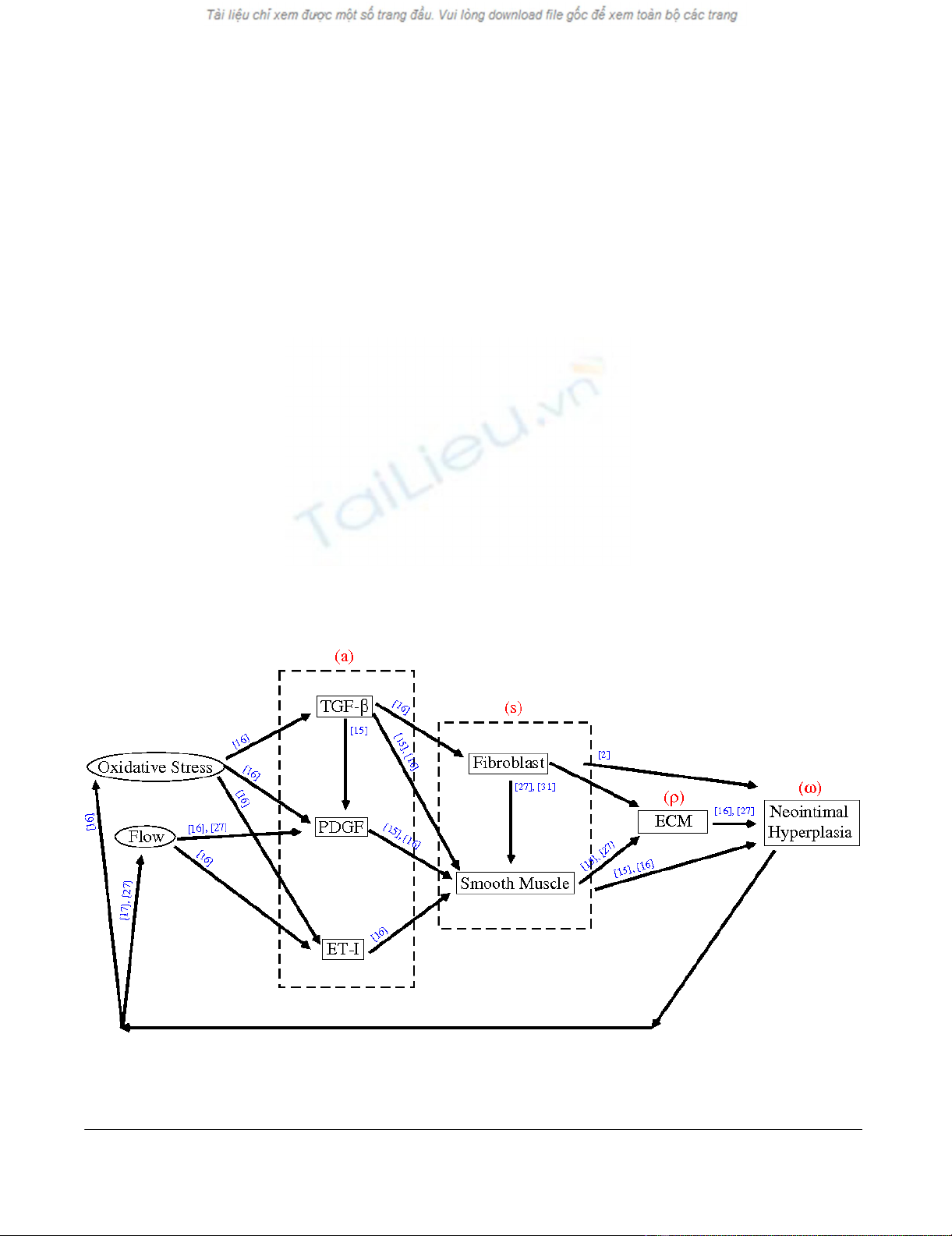
Based on the above cited work, a schematic diagram illus-
trating some causes and effects of VNH formation is rep-
resented in Figure 1. For simplicity, some of the
intermediate factors are not included in the diagram. For
example, we assume that fibroblasts produce basic fibrob-
last growth factors (bFGF) [31]; in turn, bFGFs stimulate
the production of smooth muscle cells [27]. These two
facts account for the arrow going from the fibroblast to
smooth muscle cells (i.e., the intermediate factor bFGF is
dropped out). Also, the fibroblasts contribute to the inti-
mal hyperplasia [2]. The fibroblasts in the neointima may
acquire a smooth muscle cell-like phenotype by express-
ing smooth muscle actin, and thus be called myofibrob-
lasts.
While the occurrence of VNH is well recognized, the
pathogenesis of it is complex and still not well under-
stood. Few studies have attempted to analyze the path-
ways that lead to dialysis access stenosis and direct
attention to potential therapies [2,11]. Computational
and mathematical tools have been applied to many areas
of biology resulting in descriptive models with predictive
capabilities. However, to our knowledge, there is no
mathematical model to account for cellular and molecu-
lar interactions relevant to hemodialysis vascular access
dysfunction. In the present work, we propose such a
model for venous neointimal hyperplasia development
describing:
• the interaction among growth factors, smooth muscle
cells, and fibroblasts;
• the effect of these interactions on the venous stenosis;
• the effect of the stenosis on the level of oxidative stress
and degree of turbulent flow;
• the influence of oxidative stress and turbulent flow on
growth factors.
A schematic diagram illustrating some causes and effects of intimal hyperplasiaFigure 1
A schematic diagram illustrating some causes and effects of intimal hyperplasia. The red letters represent the variables in our
model, while the blue numbers indicate the sources cited.

Theoretical Biology and Medical Modelling 2008, 5:2 http://www.tbiomed.com/content/5/1/2
Page 4 of 9
(page number not for citation purposes)
In the next section we introduce the mathematical model
and illustrate how the model can potentially be used to
predict vascular access failure based on the concentration
of growth factors. The goal of any surveillance method is
to detect access stenosis in a timely manner so that appro-
priate corrective steps can be undertaken prior to throm-
bosis. This is of critical importance, since the access
survival after an episode of thrombosis is markedly
reduced. With this in mind, we discuss possible applica-
tions of our results, not only to identify vascular access at
the risk of thrombosis, but also for using the model to
develop innovative strategies to prevent or delay vascular
access failure. We conclude the work with comments on
the mathematical model and future directions.
Methods and Results
Model description
The mathematical model that describes the VNH develop-
ment is based on a simplification of the network diagram
of Figure 1. However, we hope that the features retained
for discussion are those of greatest importance in the
present state of knowledge. The process of developing the
model will identify important parameters and relation-
ships that have not yet been investigated and can thus pro-
mote refinement in future studies.
To begin with, we identify the model variables and con-
sider their movement, production and death in a radially
symmetric control domain, Ω, that represents the intima
and the lumen of the blood vessel at a cross-section where
a stenosis develops. The geometry of the domain is speci-
fied by the radius L = R0 + dINT, where R0 is the average
radius of the lumen before the neointimal layer starts to
form, and dINT stands for the average thickness of the
venous intimal layer. In this setting, the boundary of the
domain, Γ, corresponds to the interface between the
media and the intima.
We now motivate our choice of the variables. For simplic-
ity, we lump together several chemical species elemental
to the process of neointima formation, as well as several
cells and extracellular matrix components:
• a(x, t), general chemical species (TGF-
β
, PDGF, ET-1);
• s(x, t), general cellular species (smooth muscle cells,
fibroblasts);
•
ρ
(x, t), extracellular matrix (collagen, fibronectin, elas-
tin).
The quantity a(x, t) represents the concentration (in g/
cm3) of growth factors at x ∈ Ω in time t. In the absence of
more detailed information on each factor, a accounts for
all growth factors that potentially have a chemotactic
effect on the cells. However, it is possible to separately
describe the mechanism of action of particular growth fac-
tors as the model expands.
The quantity s(x, t) represents the density (in g/cm3) of
cells at x ∈ Ω in time t. We do not distinguish between var-
ious cells that are known to be involved in the formation
of the neointimal hyperplasia, assuming instead that they
all follow the same process of diffusion, chemotaxis and
growth.
The quantity
ρ
(x, t) represents the density (in g/cm3) of
extracellular matrix at x ∈ Ω in time t. Although the matrix
ρ
and the cellular species s have different geometric fea-
tures, for the purpose of this paper we assume that they
both act as a source of material filling in the intimal-lumi-
nal space, and consequently we treat them in the same
way.
To study the impact of the chemicals, cells and ECM on
stenosis, we chose to monitor the reduction of the lumi-
nal volume
ω
(t), which is initially
ω
0 (according to clini-
cians, vascular access needs clinical intervention when the
neointimal hyperplasia obstructs more than 50% of the
initial luminal space, that is, when
ω
(t) =
ω
0/2). As the
luminal space gets partially filled with cells s and extracel-
lular matrix
ρ
, the boundary of the luminal space is not
clearly defined. We take the point of view that the more
material there is in the intimal-luminal domain, the
smaller the luminal space will be, and simply define
where k is a dimensional constant.
Applying the laws of mass conservation to each of our var-
iables we obtain the equations governing the evolution of
a, s and
ρ
.
Chemical species
At the time t > 0 and the position x ∈ Ω, the concentration
of chemicals changes according to
We assume that the chemical species undergo random
motion (i.e., diffusion). Although the diffusion coefficient
Da may in general depend on position, we take it here to
be constant. Due to chemical signaling, the chemical spe-
cies decrease through uptake by the cellular species. The
value of the parameter
λ
is determined by the receptivity
of cells to the growth factors. In the absence of more
detailed information, we simply assume that the produc-
ww r
() ( , ) ( , ) .tksxtxtdV=− +
()
∫
0Ω(1)
∂
∂=∇ ∇
()
−+−
a
txt D a as c t
a
diffusion removal p
(,) ( ())
N
lww
10
rroduction
.(2)

Theoretical Biology and Medical Modelling 2008, 5:2 http://www.tbiomed.com/content/5/1/2
Page 5 of 9
(page number not for citation purposes)
tion rate of all growth factors is proportional to
ω
0 -
ω
(t).
This term represents the observation that the production
of chemical species depends on the oxidative stress and
turbulent flow caused by the narrowing of the luminal
space. We assume that the smaller the luminal space, the
larger the oxidative pressure and shear flow, and also the
larger the concentration of growth factors. Thus, the pro-
duction of chemicals within the lesion is triggered by a
large number of factors, which includes inflammation,
hemodynamic and mechanical stresses.
Cellular species
The density of cells is assumed to follow the equation
The cellular species undergo random motion, are chemo-
tactically attracted to the chemicals in the presence of
extracellular matrix, and grow up to a maximal value S.
The chemotactic force is proportional to s∇a. We assume
that the movement of cells due to chemotaxis cannot
occur without extracellular matrix, which has maximum
density P. For simplicity, the diffusion coefficient Ds and
the chemotactic coefficient
χ
a are considered constants.
The parameter c2 of the logistic growth term depends on
the whole family of growth factors, but for simplicity we
have taken it to be constant. We note that in the expres-
sion for the chemotaxis we have lumped together all the
cells (by s) and all the growth factors (by a). In an
extended model one would quantify the effect of each spe-
cific growth factor on the proliferation of each cell type
when the growth factors are separately modeled.
Extracellular matrix
We assume that extracellular matrix is being produced by
cellular species, up to a maximum value P,
We assume that the overproduction of extracellular matrix
during the formation of VNH exceeds the degradation of
the extracellular matrix, so that there is a total gain of the
ECM density at rate c3, as long as the density is not satu-
rated; for simplicity, we assume that c3 is constant.
Boundary and initial conditions
To complete the description of our model, it remains to
specify the boundary and initial conditions for each of the
variables. To begin with, we denote by a(x, 0) = a0 > 0 the
initial concentration of growth factors in the proximal
vein, at a cross-section characterized by the radius R(0) =
R0. We further assume that no cellular species or extracel-
lular matrix are present in the intimal-luminal space at
time t = 0, hence s(x, 0) = 0 and
ρ
(x, 0) = 0.
If there is an influx of growth factors from the media-
adventitia into the intima, we assume it is negligible com-
pared to the production of the growth factors due to oxi-
dative stresses and turbulent flow.
Consequently, we do not model the contribution of any
factors from the medial-adventitial layers or nonvascular
wall tissues, and therefore take
At low concentrations of chemicals inside the domain,
there is no tendency for cells to cross the boundary into
the intima. As the concentration of growth factors
increase, a threshold concentration (a = A) is reached
inside the domain, triggering the migration of cellular
species from the medial-adventitial layers into the intima
through the media-intima boundary. We assume a con-
stant influx rate,
β
s, and write
although in a more general case, the rate of this influx of
cells could depend on the concentration of chemicals. The
term H(.) is the Heaviside step function, defined as H(v)
= 0 when v < 0 and H(v) = 1 for v ≥ 0, and it is used to rep-
resent the chemical signal that switches on as soon as the
density arises above a threshold A.
Finally, to account for the inability of extracellular matrix
to pass through the boundary, we impose a no-flux condi-
tion for
ρ
, namely
Parameter values
Table 1 gives a summary of the parameters and their
numerical values used in the computer simulations to
solve the PDE system (2)–(4) with the boundary condi-
tions (5)–(7). The model parameters were obtained from
a wide variety of experiments on many different human or
animal models. Whenever such data were not available,
we estimated the order of magnitude of the parameters
and made choices that gave biologically reasonable
results.
∂
∂=∇ ∇
()
−∇ − ∇
s
txt D s P
s
Ssa
s
diffusion
a
chemot
(,) ( )
cr
1
aaxis growth
cs s
S
+−
21().
(3)
∂
∂=−
rr
txt cs P
growth
(,) ( ).
31
(4)
∇=
∈
ax
xxt
x
(,) .
Γ
0(5)
∇=−−
∈
sx
xxt Ha A
x
s
(,) ( ),
Γ
b
(6)
∇=
∈
r
x
xxt
x
(,) .
Γ
0(7)


![Hình ảnh học bệnh não mạch máu nhỏ: Báo cáo [Năm]](https://cdn.tailieu.vn/images/document/thumbnail/2024/20240705/sanhobien01/135x160/1985290001.jpg)


