
RESEARC H Open Access
Inhibition of HIV-1 replication by small interfering
RNAs directed against Glioma Pathogenesis
Related Protein (GliPR) expression
Gianni Capalbo
1
, Thea Müller-Kuller
1
, Ursula Dietrich
2
, Dieter Hoelzer
1
, Oliver G Ottmann
1
, Urban J Scheuring
1*
Abstract
Background: Previously, we showed that glioma pathogenesis related protein (GliPR) is induced in CEM T cells
upon HIV-1 infection in vitro. To examine whether GliPR plays a role as HIV dependency factor (HDF), we tested the
effect of GliPR suppression by siRNA on HIV-1 replication.
Results: Induction of GliPR expression by HIV-1 was confirmed in P4-CCR5 cells. When GliPR was suppressed by
siRNA, HIV-1 replication was significantly reduced as measured by HIV-1 transcript levels, HIV-1 p24 protein levels,
and HIV-1 LTR-driven reporter gene expression, suggesting that GliPR is a cellular co-factor of HIV-1. Microarray
analysis of uninfected HeLa cells following knockdown of GliPR revealed, among a multitude of gene expression
alterations, a down-regulation of syndecan-1, syndecan-2, protein kinase C alpha (PRKCA), the catalytic subunit bof
cAMP-dependent protein kinase (PRKACB), nuclear receptor co-activator 3 (NCOA3), and cell surface protein CD59
(protectin), all genes having relevance for HIV-1 pathology.
Conclusions: The up-regulation of GliPR by HIV-1 and the early significant inhibition of HIV-1 replication mediated
by knockdown of GliPR reveal GliPR as an important HIV-1 dependency factor (HDF), which may be exploited for
HIV-1 inhibition.
Background
The replication of HIV-1 depends on specific host fac-
tors [1-4]. A recent report identified 273 cellular HIV-1
dependency factors (HDF), that are important for HIV-1
replication [5]. Furthermore, HIV-1 modifies the mRNA
expression of a relatively large number of host cell
genes, as shown by several reports [6-10]. Differential
display experiments suggested that the expression of
~700 host genes (approximately 3% of all cellular genes)
is modified by HIV-1 infection in vitro [9]. A microarray
analysis using a limited subset of 1500 cDNAs identified
20 differentially expressed mRNAs from several cellular
pathways [7]. Specific HIV-1 proteins including Tat,
Nef, gp120 and Vpr were examined to dissect their role
in modifying the transcription of cellular genes [11-14].
While some of the differentially expressed cellular genes
may play a role in host defense mechanisms, others may
facilitate HIV-1 replication, infectivity, species propaga-
tion and survival. A subgroup of differential cellular
gene expressions may even support both host defense
and viral replication, since HIV-1 replication is linked to
immune activation of CD4+ T cells. Due to evolutionary
selection, HIV-1 is expected to induce specific host fac-
tors, favorable for viral replication or propagation, and
to suppress unfavorable cellular gene products [15-17].
Therefore, the examination of host cell genes, that are
up-regulated upon HIV-1 infection, is expected to iden-
tify potential targets for inhibition of HIV-1 replication.
Previously, we found an early up-regulation of GliPR
expression by more than 5-fold in CEM T cells infected
with HIV-1 by a differential display screen [9]. There-
fore, we were interested in delineating the role of GliPR
for HIV-1 replication.
GliPR was identified originally in human glioblastomas
[18] and was also described as related to testes-specific,
vespid, and pathogenesis protein 1 (RTVP-1) [19].
Increased expression of GliPR was associated with mye-
lomonocytic differentiation in macrophages [20].
* Correspondence: u.scheuring@gmx.de
1
Department of Hematology/Oncology and Infectious Diseases, J. W.
Goethe-University Hospital, Theodor Stern Kai 7, 60590 Frankfurt/Main,
Germany
Capalbo et al.Retrovirology 2010, 7:26
http://www.retrovirology.com/content/7/1/26
© 2010 Capalbo et al; licensee BioMed Central Ltd. This is an Open Access article distributed under the terms of the Creative Commons
Attribution License (http://creativecommons.org/licenses/by/2.0), which permits unrestricted use, distribution, and reproduction in
any medium, provided the original work is properly cited.
Whereas GliPR has been reported to act as a tumor
suppressor gene inducing apoptosis in prostate cancer
[21-24], it appears to be an oncogene in glioblastomas
[25] and Wilms tumors [26]. RTVP-1 protein was
reported to contain a N-terminal signal peptide
sequence and a transmembrane domain [27]. Further-
more, homology studies revealed a putative active enzy-
matic center in GliPR [27]. GliPR is homologous to
group 1 plant pathogenesis-related proteins (PR-1) that
are implicated in plant defense responses to viral, bac-
terial, and fungal infection [28,29]. Since GliPR shows
structural similarities with its homologous plant PR-1
proteins, mammalian testis proteins (TPX1) and the
insect venom Ag-5 protein, which are secretory proteins
[29,30], it has been suspected that GliPR is also secreted.
GliPR’s homology with plant PR-1 proteins that have
been attributed with a defense function may raise the
question whether GliPR has an evolutionarily conserved
role in innate immune response and human host
defense of viral infection including HIV-1. Alternatively
or additionally, HIV-1 may induce and exploit GliPR for
viral replication.
The effect of GliPR knockdown on HIV-1 replication
was studied, in order to test the hypotheses of GliPR being
a host defense protein against or a co-factor of HIV-1.
Furthermore, in order to identify downstream targets of
GliPR, the effect of GliPR suppression on cellular gene
expression was also investigated using cDNA microarrays.
Results
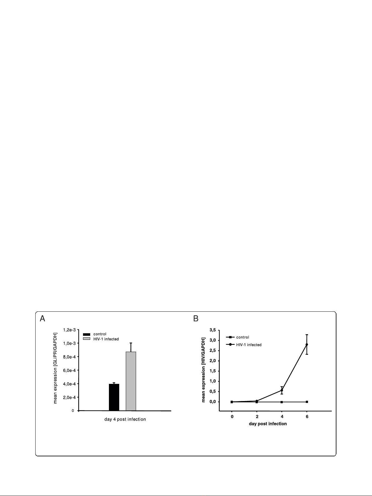
GliPR is induced upon HIV-1 infection in P4-CCR5 cells
Since HIV-1 infection induced GliPR expression in HIV-
1 infected human T cell line cells, as described
previously [9], we tested whether this modification
could be reproduced in P4-CCR5 HeLa cells infected
with HIV-1
LAI
. P4-CCR5 HeLa cells were employed for
thepresentstudybecausethey are more amenable to
efficient transfection of synthetic siRNA compared to
lymphocytic cell lines. Quantitative PCR demonstrated
an up-regulation of GliPR transcripts by approximately
2-fold at day 4 after infection compared to uninfected
cells (Fig. 1a). In order to display HIV-1 infection
kinetics, real-time quantitative PCR was also utilized to
determine levels of intracellular HIV-1 viral mRNA nor-
malized by cell number (house keeping gene GAPDH)
at different time points following infection (Fig. 1b). The
data show that HIV-1 replication is still in the early
logarithmic phase at day 4 in this cell culture system
and that GliPR expression is induced in this early phase.
Suppression of GliPR mediated by short interfering RNA
P4-CCR5 cells were transfected with siRNAs specific for
GliPR or a non-silencing siRNA, which was 5-prime
labeled with rhodamine. Flow cytometry analysis of cells
transfected with non-silencing siRNA 24 h post trans-
fection revealed transfection efficiencies on average of
90% in all samples. Forty-eight hours after transfection,
the relative levels of GliPR mRNA transcripts were
decreased by at least 90%, as shown by quantitative real-
time PCR (Fig. 2a). Even four and six days after trans-
fection a markedly reduced GliPR expression by at least
80% compared with non-transfected cells (mock) or
cells transfected with non-silencing siRNA was observed
(Fig. 2a).
Viability and proliferation rate of P4-CCR5 cells
transfected with siRNAs against GliPR or with the
Figure 1 Up-regulation of GliPR expression by HIV-1 infection.(A) HIV-1
LAI
-infected P4-CCR5 cells and controls were subjected to
quantitative PCR of GliPR expression at day 4 after HIV-1 infection. (B) In order to display HIV-1 infection kinetics, real-time quantitative PCR was
also utilized to determine levels of intracellular HIV-1 viral mRNA normalized by cell number (house keeping gene GAPDH) in triplicate at day 0,
2, 4 and 6 post infection. Bars represent the standard deviation of the mean of determinations.
Capalbo et al.Retrovirology 2010, 7:26
http://www.retrovirology.com/content/7/1/26
Page 2 of 10
non-silencing siRNA remained unchanged as deter-
mined by WST-1 cell proliferation assay (Fig. 2b).
In order to establish a test system in a T cell line as
well, a predominant type of host cell for HIV-1, Jurkat
cells were transfected with 2 different siRNAs targeting
GliPR, control non-silencing siRNA, or mock transfec-
tion without any siRNA. GliPR mRNA expression was
reduced by around 64% to 69% at 48 hours after trans-
fection with specific siRNAs compared to controls
(Fig. 2c). The less pronounced reduction of GliPR
expression compared to P4-CCR5 HeLa cells may be
attributed to the lower transfection efficiency generally
observed in T cell lines. In this experiment, approxi-
mately 70% of Jurkat cells were transfected, while 90%
of P4-CCR5 HeLa cells were transfected.
In general, GliPR-directed siRNAs reduced the expres-
sion of GliPR effectively in P4-CCR5 and Jurkat cells
without affecting cell viability.
Down-regulation of GliPR by siRNA inhibits HIV-1
replication in P4-CCR5 and Jurkat cells
In order to examine the effect of GliPR knockdown on
HIV-1 replication, P4-CCR5 cells were transfected with
GliPR-specific siRNAs and subsequently infected with
HIV-1
LAI
. As a negative control, the non-silencing
siRNA (si-nons-Rho) was utilized while a siRNA target-
ing HIV-1 p24 was used as a positive control, since it
was able to inhibit viral replication very effectively, as
previously demonstrated [31]. HIV-1 infection was per-
formed 24 h post siRNA transfection with a MOI of
Figure 2 Efficacy of siRNA-mediated suppression of GliPR.(A) Quantitative PCR analysis of GliPR expression in P4-CCR5 cells which were
transfected with 2 different siRNAs against GliPR or the control non-silencing siRNA labeled with rhodamine. Results are presented as mean
values of triplicate samples ± standard deviation (SD). (B) Cell viability was determined with the WST-1 assay 24 h and 48 h after siRNA
transfection. Results are expressed as absorbance (OD
450
). Bars represent the standard deviation of the mean of determinations. (C) Quantitative
PCR analysis of GliPR expression in Jurkat cells 2 days after transfection with 2 different siRNAs against GliPR or the control non-silencing siRNA
labeled with rhodamine.
Capalbo et al.Retrovirology 2010, 7:26
http://www.retrovirology.com/content/7/1/26
Page 3 of 10
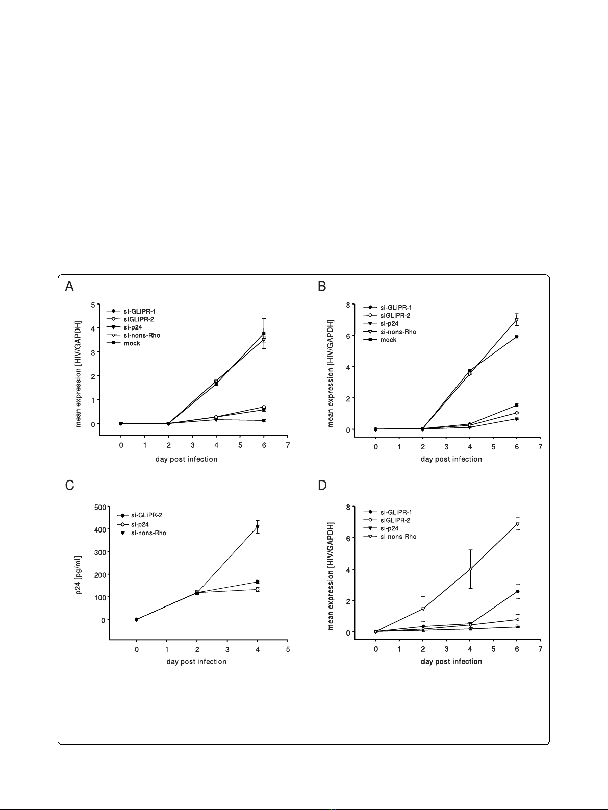
0.01 or 0.05. Sequential cell-associated HIV-1 viral
mRNA levels were determined by real-time quantitative
PCR during 6 days after infection. As expected, the
positive control siRNA (si-p24) exhibited a marked inhi-
bition in viral mRNA transcription. Similarly, the
siRNA-mediated reduction of GliPR expression was fol-
lowed by significantly reduced viral mRNA transcript
levels compared to HIV-1 infected controls, which were
mock-transfected (mock) or transfected with the non-
silencing siRNA (si-nons-Rho) at both MOI of 0.01 and
0.05 (Fig. 3a and 3b).
The effect of GliPR suppression on HIV-1 replication
was confirmed by p24 ELISA, showing a significantly
reduced p24 expression at day 4 post infection in cul-
tures with GliPR knock-down compared to controls
with non-silencing siRNA (Fig. 3c).
In order to test this phenomenon in T cells, Jurkat
cells transfected with siRNAs specific to GliPR or con-
trol siRNA were infected with HIV-1 at a MOI of 0.01.
GliPR-specific siRNAs resulted in a significant reduction
of HIV-1 replication, similar to the positive control with
siRNA against p24 (Fig 3d). Thus the T cell line results
are in line with the data in P4-CCR5 cells.
Furthermore, the effect on HIV-1 replication was
examined by the integrated HIV-1-LTR-driven reporter
vector expressing b-galactosidase in P4-CCR5 cells.
Figure 3 Effects of siRNA transfections on HIV-1 replication. P4-CCR5 cells were transfected with siRNAs directed against GliPR, viral p24 or
an unspecific sequence (non-silencing control) and subsequently infected with HIV-1
LAI
with a multiplicity of infection of 0.01 (A) and 0.05 (B),
respectively. HIV-1 RNA copy numbers were normalized per cell count by house keeping gene GAPDH. (C) P4-CCR5 cells were transfected with
siGliPR-2, si-p24 or non-silencing control siRNA and subsequently infected with HIV-1
LAI
with a multiplicity of infection of 0.01. Concentrations of
viral p24 at day 0, 2 and 4 represent mean values of triplicate samples. (D) Jurkat cells were transfected with siRNAs directed against GliPR, viral
p24 or an unspecific sequence (non-silencing control) and subsequently infected with HIV-1
LAI
with a multiplicity of infection 0.01. HIV-1 RNA
copy numbers were normalized per cell count by house keeping gene GAPDH.
Capalbo et al.Retrovirology 2010, 7:26
http://www.retrovirology.com/content/7/1/26
Page 4 of 10
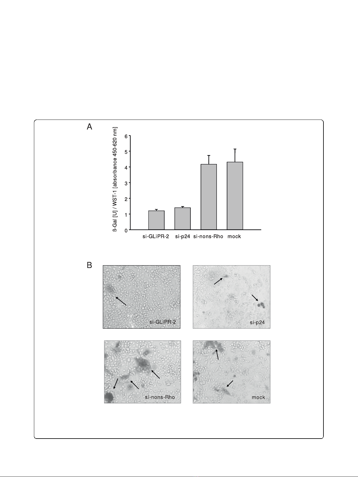
HIV-1 Tat-mediated transactivation of the LTR leads to
expression of measurable b-galactosidase activity, allow-
ing measurments of inhibitory effects on HIV-1 replica-
tion as reductions in b-galactosidase activity. The
expression of b-galactosidase was markedly decreased by
siGliPR on day four after infection, comparable to the
degree of the positive control with p24 siRNA (Fig. 4a).
The inhibition of LTR-driven transcription was
confirmed by microscopy of these cell cultures after
X-Gal staining on day six after infection (Fig. 4b).
These results demonstrated that siRNA-mediated sup-
pression of GliPR inhibited HIV-1 replication implicat-
ing that GliPR promotes HIV-1 replication. It was not
possible to employ the opposite approach by assessing
the effect of GliPR’s over-expression on HIV-1 replica-
tion, since forced expression of GliPR caused rapid
Figure 4 Expression of b-galactosidase driven by HIV-1-LTR in HIV-infected cells after siRNA transfection. P4-CCR5 cells containing a HIV-
1-LTR driven b-galactosidase reporter vector were transfected with siGliPR-2, non-silencing control siRNA or no siRNA (mock) and subsequently
infected with HIV-1
LAI
with a multiplicity of infection of 0.01. (A) b-galactosidase units at day 4 normalized by relative WST-1 values represent
mean values from triplicate samples. (B) Photomicrograph of b-gal stained P4-CCR5 cells infected with HIV-1
LAI
(MOI 0.01) after transfection with
mock, GliPR-siRNA, HIV-1 p24-siRNA or non-silencing control siRNA at day 6 post infection.
Capalbo et al.Retrovirology 2010, 7:26
http://www.retrovirology.com/content/7/1/26
Page 5 of 10


![Vaccine và ứng dụng: Bài tiểu luận [chuẩn SEO]](https://cdn.tailieu.vn/images/document/thumbnail/2016/20160519/3008140018/135x160/652005293.jpg)


%20--%3e%3cdefs%3e%3cstyle%3e%20.st0%20{%20fill:%20%23fff;%20}%20.st1%20{%20fill:%20%237800fa;%20}%20%3c/style%3e%3c/defs%3e%3cpath%20class='st1'%20d='M117.78,12.18H43.11c2.9,3.47,4.65,7.94,4.65,12.82,0,5.6-2.3,10.66-6.01,14.29h76.02l7.22-13.56-7.22-13.56Z'/%3e%3cg%3e%3cpath%20class='st0'%20d='M53.58,26.17h-.59v-1.46h.59v-4.96h2.83c1.78,0,2.67.94,2.67,2.82v5.76c0,1.87-.89,2.81-2.67,2.81h-2.83v-4.96ZM55.36,21.37v3.34h1.1v1.46h-1.1v3.34h1.01c.61,0,.91-.37.91-1.1v-5.93c0-.74-.3-1.1-.91-1.1h-1.01Z'/%3e%3cpath%20class='st0'%20d='M65.99,31.14h-1.8l-.31-2.07h-2.19l-.31,2.07h-1.64l1.82-11.39h2.62l1.82,11.39ZM65.28,18.04c-.25.46-.51.77-.75.94-.21.15-.47.22-.79.22-.26,0-.57-.07-.92-.22l-.38-.15c-.14-.05-.26-.07-.37-.07-.3,0-.53.18-.71.54l-.91-.68c.25-.46.51-.77.75-.94.21-.14.48-.21.79-.21.26,0,.57.07.92.21l.38.15c.14.05.26.07.37.07.3,0,.53-.18.71-.54l.91.68ZM61.91,27.52h1.73l-.87-5.76-.87,5.76Z'/%3e%3cpath%20class='st0'%20d='M74.53,26.89v1.52c0,1.91-.89,2.86-2.67,2.86s-2.67-.95-2.67-2.86v-5.93c0-1.91.89-2.86,2.67-2.86s2.67.95,2.67,2.86v1.11h-1.69v-1.22c0-.75-.31-1.12-.93-1.12s-.93.37-.93,1.12v6.15c0,.74.31,1.11.93,1.11s.93-.37.93-1.11v-1.63h1.69Z'/%3e%3cpath%20class='st0'%20d='M81.4,31.14h-1.8l-.31-2.07h-2.19l-.31,2.07h-1.64l1.82-11.39h2.62l1.82,11.39ZM75.9,19.2l1.52-1.91h1.71l1.51,1.91h-1.61l-.76-.95-.75.95h-1.61ZM77.32,27.52h1.73l-.87-5.76-.87,5.76ZM83.1,15.99l-1.76,1.91h-1.26l1.17-1.91h1.86Z'/%3e%3cpath%20class='st0'%20d='M84.86,19.75c1.78,0,2.67.94,2.67,2.82v1.48c0,1.87-.89,2.81-2.67,2.81h-.85v4.28h-1.79v-11.39h2.64ZM84.01,21.37v3.86h.85c.58,0,.87-.36.87-1.08v-1.71c0-.71-.29-1.07-.87-1.07h-.85Z'/%3e%3cpath%20class='st0'%20d='M93.51,19.75c1.78,0,2.67.94,2.67,2.82v1.48c0,1.87-.89,2.81-2.67,2.81h-.85v4.28h-1.79v-11.39h2.64ZM92.66,21.37v3.86h.85c.58,0,.87-.36.87-1.08v-1.71c0-.71-.29-1.07-.87-1.07h-.85Z'/%3e%3cpath%20class='st0'%20d='M98.8,31.14h-1.79v-11.39h1.79v4.88h2.03v-4.88h1.83v11.39h-1.83v-4.88h-2.03v4.88Z'/%3e%3cpath%20class='st0'%20d='M105.36,24.55h2.46v1.62h-2.46v3.34h3.09v1.63h-4.88v-11.39h4.88v1.63h-3.09v3.18ZM108.17,17.29l-1.76,1.91h-1.26l1.17-1.91h1.86Z'/%3e%3cpath%20class='st0'%20d='M112.2,19.75c1.78,0,2.67.94,2.67,2.82v1.48c0,1.87-.89,2.81-2.67,2.81h-.85v4.28h-1.79v-11.39h2.64ZM111.35,21.37v3.86h.85c.58,0,.87-.36.87-1.08v-1.71c0-.71-.29-1.07-.87-1.07h-.85Z'/%3e%3c/g%3e%3ccircle%20class='st1'%20cx='25'%20cy='25'%20r='20'/%3e%3cpath%20class='st0'%20d='M32.78,19.27c2.92,0,4.43,2.55,5.28,5.33l.71,2.17c.14.38-.33.75-.71.75h-5.61c.19-.33.24-.71.09-1.08l-.75-2.45c-.43-1.32-.99-2.64-1.79-3.77.75-.57,1.65-.94,2.78-.94h0ZM25,18.38c3.25,0,4.9,2.78,5.89,5.89l.76,2.45c.14.42-.33.8-.8.8h-11.69c-.42,0-.94-.38-.8-.8l.75-2.45c.99-3.11,2.64-5.89,5.89-5.89h0ZM25,11.35c1.74,0,3.11,1.37,3.11,3.11s-1.37,3.11-3.11,3.11-3.11-1.41-3.11-3.11,1.41-3.11,3.11-3.11h0ZM17.27,19.27c1.08,0,1.98.38,2.73.94-.8,1.13-1.37,2.45-1.74,3.77l-.8,2.45c-.14.38-.05.75.09,1.08h-5.56c-.42,0-.9-.38-.75-.75l.71-2.17c.9-2.78,2.41-5.33,5.33-5.33h0ZM17.27,12.91c1.51,0,2.78,1.27,2.78,2.83s-1.27,2.83-2.78,2.83-2.83-1.27-2.83-2.83,1.27-2.83,2.83-2.83h0ZM32.78,12.91c1.56,0,2.78,1.27,2.78,2.83s-1.23,2.83-2.78,2.83-2.83-1.27-2.83-2.83,1.27-2.83,2.83-2.83h0ZM27.07,28.56v.09c0,.57-.24,1.08-.61,1.46h0v.05c-.38.33-.9.57-1.46.57s-1.08-.24-1.46-.61h0c-.38-.38-.61-.9-.61-1.46v-.09h1.41v.09c0,.19.05.38.19.47v.05c.09.09.28.19.47.19s.38-.09.47-.19v-.05c.14-.09.24-.28.24-.47t-.05-.09h1.41ZM30.99,28.56v.09c0,1.65-.66,3.16-1.74,4.24-1.08,1.08-2.59,1.79-4.24,1.79s-3.16-.71-4.24-1.79l-.05-.05c-1.04-1.08-1.7-2.55-1.7-4.2v-.09h1.41v.09c0,1.27.47,2.4,1.27,3.25h.05c.85.85,1.98,1.37,3.25,1.37s2.4-.52,3.25-1.37c.85-.8,1.37-1.98,1.37-3.25v-.09h1.37ZM34.99,28.56v.09c0,2.78-1.13,5.28-2.92,7.07-1.79,1.79-4.29,2.92-7.07,2.92s-5.23-1.13-7.07-2.92c-1.79-1.79-2.92-4.29-2.92-7.07v-.09h1.41v.09c0,2.4.94,4.53,2.5,6.08,1.56,1.56,3.72,2.5,6.08,2.5s4.52-.94,6.08-2.5c1.56-1.56,2.5-3.68,2.5-6.08v-.09h1.41Z'/%3e%3c/svg%3e)