


YOMEDIA
ADSENSE
Báo cáo y học: "Intra-abdominal Pressures during Voluntary and Reflex Cough"
49
lượt xem 2
download
lượt xem 2
download
 Download
Vui lòng tải xuống để xem tài liệu đầy đủ
Download
Vui lòng tải xuống để xem tài liệu đầy đủ
Tuyển tập các báo cáo nghiên cứu về y học được đăng trên tạp chí y học Critical Care giúp cho các bạn có thêm kiến thức về ngành y học đề tài: Intra-abdominal Pressures during Voluntary and Reflex Cough...
AMBIENT/
Chủ đề:
Bình luận(0) Đăng nhập để gửi bình luận!
Lưu
Nội dung Text: Báo cáo y học: "Intra-abdominal Pressures during Voluntary and Reflex Cough"
- Cough BioMed Central Open Access Research Intra-abdominal Pressures during Voluntary and Reflex Cough W Robert Addington*1, Robert E Stephens2, Michael M Phelipa3, John G Widdicombe4 and Robin R Ockey5 Address: 1W. Robert Addington, D.O., 101 E. Florida Avenue, Melbourne, FL, 32901, 321-984-4628, USA, 2Department of Anatomy, Kansas City University of Medicine and Biosciences, Kansas City, MO, USA, 3Melbourne, FL, USA, 4116 Pepys Road, London SW208NY, UK and 5Orem, UT, USA Email: W Robert Addington* - wraddington@cfl.rr.com; Robert E Stephens - rstephens@kcumb.edu; Michael M Phelipa - MPHELIPA@cfl.rr.com; John G Widdicombe - JohnWiddicombeJ@aol.com; Robin R Ockey - robinockey@gmail.com * Corresponding author Published: 30 April 2008 Received: 22 January 2008 Accepted: 30 April 2008 Cough 2008, 4:2 doi:10.1186/1745-9974-4-2 This article is available from: http://www.coughjournal.com/content/4/1/2 © 2008 Addington et al; licensee BioMed Central Ltd. This is an Open Access article distributed under the terms of the Creative Commons Attribution License (http://creativecommons.org/licenses/by/2.0), which permits unrestricted use, distribution, and reproduction in any medium, provided the original work is properly cited. Abstract Background: Involuntary coughing such as that evoked from the larynx, the laryngeal cough reflex (LCR), triggers a coordinated contraction of the thoracic, abdominal and pelvic muscles, which increases intra-abdominal pressure (IAP), displaces the diaphragm upwards and generates the expiratory force for cough and airway clearance. Changes in the IAP during voluntary cough (VC) and the LCR can be measured via a pressure catheter in the bladder. This study evaluated the physiological characteristics of IAP generated during VC and the LCR including peak and mean pressures and calculations of the area under the curve (AUC) values during the time of the cough event or epoch. Methods: Eleven female subjects between the ages of 18 and 75 underwent standard urodynamic assessment with placement of an intravesicular catheter with a fiberoptic strain gauge pressure transducer. The bladder was filled with 200 ml of sterile water and IAP recordings were obtained with VC and the induced reflex cough test (RCT) using nebulized inhaled 20% tartaric acid to induce the LCR. IAP values were used to calculate the area under the curve (AUC) by the numerical integration of intravesicular pressure over time (cm H2O·s). Results: The mean (± SEM) AUC values for VC and the LCR were 349.6 ± 55.2 and 986.6 ± 116.8 cm H2O·s (p < 0.01). The mean IAP values were 45.6 ± 4.65 and 44.5 ± 9.31 cm H2O (NS = .052), and the peak IAP values were 139.5 ± 14.2 and 164.9 ± 15.8 cm H2O (p = 0.07) for VC and LCR, respectively. Conclusion: The induced LCR is the involuntary rapid and repeated synchronous expiratory muscle activation that causes and sustains an elevated IAP over time, sufficient for airway protection. VC and LCR have different neurophysiological functions. Quantification of the LCR using AUC values and mean or peak IAP values may be useful as a clinical tool for determining neurophysiological airway protection status and provide a quantitative assessment of changes in a patient's functional recovery or decline. Page 1 of 9 (page number not for citation purposes)
- Cough 2008, 4:2 http://www.coughjournal.com/content/4/1/2 jects had complaints of mild stress urinary incontinence Introduction Neurophysiological protection of the upper airway is a without any neurological history. One subject (subject critical function of the laryngeal cough reflex (LCR). 10) had multiple sclerosis (MS) and was non-ambulatory Coughing involves coordinated contractions of the tho- with internuclear ophthalmoplegia and neurological def- racic, abdominal and pelvic muscles. On videofluoros- icits associated with cranial nerves II, III, IV and VI, but no copy, reflex cough (RC) caused increased upward history of pneumonia. A further subject (subject 11) was displacement of the diaphragm as compared with volun- tested 8 weeks after sustaining a T4 complete spinal cord tary cough (VC) [1]. This diaphragmatic displacement is a injury (SCI) and therefore had serious loss of control of result of the contraction of the external abdominal her expiratory muscles; her results are mentioned briefly obliques, intercostals and associated expiratory muscles. but are not included in the statistical analyses. The force of these contractions compresses the abdominal viscera and proportionately displaces the diaphragm Evaluations were performed with a multi-channel urody- superiorly, almost to mid-sternal levels in reflex cough, namic (UD) system that used a fiber-optic, disposable but not for VC. These contractions cause an increase in strain gauge pressure transurethral bladder catheter and a intra-abdominal pressure (IAP), which is synchronized rectal catheter. With sterile technique, the calibrated blad- with urethral and rectal closure to prevent incontinence. der catheter was placed and secured to the subject's thigh. With continuous dual-channel recording, the subject's Although different patterns of "cough" have been bladder was filled slowly with sterile water until 200 ml described; the "classical" definition of cough starts with had been introduced. an inspiration, which is followed by compressive and expulsive phases; and is either a brainstem reflex or a cor- Subjects were asked to deeply inhale and perform strong tically mediated response characteristic of VC. VC appears voluntary coughs, which were recorded on the UD system. to play a role in clearing the vocal cords during speech [2]. Tartaric acid-induced reflex cough test (RCT) was used to However, the expiration reflex is a brainstem mediated elicit a LCR in all subjects [2,5,9-18]. The RCT used a jet reflex that initiates an immediate series of expiratory nebulized concentration of 20% L-(+)-tartaric acid dis- efforts without an inspiratory phase precedes the noxious solved in 0.15 mM sterile NaCl solution (Nephron Phar- stimulus. This type of cough is characterized by a synchro- maceuticals, Orlando, FL). The jet nebulizer was activated nous series of expiratory reflex coughs with a short latency with 50 psi from a tank that produced an average droplet [3-5], and has a role in clearing the upper airway of poten- diameter of 1–2 microns or less. During the RCT, the sub- tial aspirants during inhalation and swallowing [6]. ject was asked to exhale completely, the nostrils were Increased IAP provides the expiratory force for the protec- pinched closed, the nebulizer mouthpiece was placed in tive airway clearing function of the LCR and producing a the mouth and subjects sealed the mouthpiece with their VC). This distinction is physiologically important because lips during the brisk inhalation. The RCT normally causes the two types of reflex differ in neurophysiological and an immediate episode of several coughs. During VC and pharmacological mechanisms [6-8]. LCR, the intravesicular (bladder) pressure, rectal pressure and urethral EMG were also recorded for all subjects. (Fig. Previously, it has not been possible to reliably analyze the 1A) [19]. quantitative changes in the IAP associated with VC and the LCR. The changes in IAP during cough may be meas- Analysis of the IAP ured using pressure catheters in the bladder and/or rec- Graphs from the original urodynamic assessment were tum. Since quantitative measurement of changes in IAP digitized and the IAPs generated during the cough were during VC and reflex cough may be useful in the clinical quantified (Fig. 1B). Each cough epoch was analyzed setting, this investigation was designed to assess VC and throughout its duration. Deviation from baseline intra- LCR IAPs using intravesicular pressure catheters and uro- abdominal pressure defined the start of the cough epi- dynamic analysis of pressure changes. sode. The end of the cough epoch could be noted on the UD tracing as the IAP returned to nearly baseline levels. This study evaluated changes in the IAP during VC and the An analysis of the IAP rate of change indicated that an LCR as indicated by the measurements of the mean and effective sampling rate of 30 samples/sec was appropriate peak IAPs, and mathematical calculations of the area for further analysis. The IAP was measured at this rate for under the curve (AUC, pressure·time) values during VC each subject from the continuous UD recording. A graphic and LCR cough epochs. recording of pressure with vertical time lines was used to determine the peak IAPs (maximum intravesicular pres- sure during each expiratory cough effort), the mean IAP Materials and methods Following informed consent, eleven female subjects (over the period of the expiratory cough efforts), the dura- between the ages of 18 and 75 were enrolled. Nine sub- tions of the cough epochs, the number of IAP peaks and Page 2 of 9 (page number not for citation purposes)
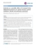
- Cough 2008, 4:2 http://www.coughjournal.com/content/4/1/2 Voluntary F igur e 1 A RCT Cough Urethral F igur e 1 B Figure (i.e., LCR) (UD) tracing (on a compressed timeline) of a subject demonstrating voluntary cough and an episode of RCT coughsurodynamic triggered by the RCT A. An 1 A. An urodynamic (UD) tracing (on a compressed timeline) of a subject demonstrating voluntary cough and an episode of RCT coughs (i.e., LCR) triggered by the RCT. A pressure sensor catheter was inserted into the subject's bladder and rectum, and the bladder was filled to 200 ml using sterile saline. Intravesicular bladder pressure was recorded at 30 samples per second. Subject was asked to voluntarily cough and the RCT was performed. Each cough episode was traced and the coordinates cor- responding to a particular bladder pressure measurement (Pves) and the IAP at that time (Tsec) were recorded for each peak, valley and slope change of the pressure tracing. B. A record was made of the complete cough episode timeline. As a part of this process, maximal IAP for each cough event was determined. Interpolation was used to fill in the remaining Pves between each annotated point. The average Pves was then calculated for each second of the timeline, and plotted as a pressure versus time graph of the cough episode. the peak values for each cough epoch, and to derive the The UD tracing for each cough epoch was quantified and AUC values during each cough epoch. In this study, AUC the coordinates corresponding to a particular IAP meas- is a product of pressure and time, expressed as cm H2O·s. urement and the IAP at that time were recorded for each peak, valley and slope change of the pressure tracing. A Page 3 of 9 (page number not for citation purposes)
- Cough 2008, 4:2 http://www.coughjournal.com/content/4/1/2 record was made of the complete cough epoch timeline Discussion (Fig. 2 and Fig. 3). Each second of the timeline was The greater AUC value with the RCT, which triggers the divided into 30 equal parts, i.e., 30 samples/s. The laryngeal cough reflex [5,21], could be due to the contin- remaining pressures were interpolated between each ual and simultaneous activation of cough-associated annotated point. The mean IAP was then calculated for expiratory muscles with rapid and repeated glottal clo- each second of the timeline, and plotted as a pressure ver- sure, compared with VC with its brief and often single sus time graph of the cough epoch (Fig. 2 and Fig. 3). event of brief glottal closure (Addington et al. cited in [22]) [1,3]. The differences in the AUC between the two From the mean IAP values, AUC values were then calcu- types of cough provide a new perspective to study the neu- lated by the numerical integration of intravesicular pres- rophysiological differences between these two events. Vol- sure over time using Boole's rule [20]. Due to the untary cough appears useful in clearing the vocal cords for diminished cough response and data points available for speech and clearing the airways once material is present in analysis, Simpson's 3/8 rule was the appropriate formula the tracheobronchial tree; it seems similar to reflex cough for the subject 11, who had a T4 complete spinal cord from the tracheobronchial tree, which starts with an inspi- injury (SCI) and an abnormal LCR [20]. A paired t-test ration to increase lung volume. The LCR does not have an was used to compare the AUC values, mean IAP and peak initial inspiration and is essentially a series of 'expiration IAP values for VC and LCR responses using SPSS statistical reflexes' with intervening inspirations; it is for involuntary software (version 10.0.5). airway protection in response to a threatening stimulus [2]. The term "cough reflex" is often used generically to include both types of "cough" and also cough bouts or Results Table 1 gives pressure values for each of the ten subjects epochs. analyzed, and summary statistics are given in Table 2. VC and LCR mean IAP values were 45.6 ± 4.65 and 44.5 ± The UD tracings indicated that the IAP appeared to be 9.31 cm H2O, respectively (p = 0.05). Although the peak greater when there was no expiratory flow and the glottis (maximum) IAP values for the LCR (164.9 ± 15.8) was adducted. During VC and RCT cough, the IAP appeared greater than the VC peak IAP (139.5 ± 14.2 cm appeared to decrease when the glottis was abducted. How- H2O), the difference was not significant (p = 0.07) (Table ever, during the coughing associated with the RCT epi- 2). sodes, the tracings showed a continuous increase in IAP above the initial baseline in all subjects, regardless of the The number of peak pressures, duration of cough events, duration of the cough episode and despite the subject hav- and AUC values were all significantly greater with the RCT ing fully exhaled before initiating the LCR, which pre- relative to voluntary cough (Fig. 2 and Fig. 3; Table 2). The vented any subsequent effective deep inhalation to assist number of peak IAPs was greater for the LCR than for VC the coughs. Although the LCR episodes may have had (6.00 ± 0.94 vs. 1.78 ± 0.28, p < 0.01), as was the episode some brief inspiratory activity late in the epoch, the IAP duration (27.0 ± 0.74 s vs. 10.2 ± 1.36 s, p < 0.01). The remained elevated above the initial baseline throughout mean (± SEM) AUC values for VC and the RCT were 349.6 the entire event – this was a consistent finding irrespective ± 55.2 cm H2O·s and 986.6 ± 116.8 cm H2O·s (p < 0.01; of the number of expiratory efforts or the duration of the Table 2), respectively. cough episode. Regarding neurological airway protection, we suggest that the main components of the LCR are pri- In the subjects with neurological impairment (Fig. 4), marily a continuous series of expiratory cough reflexes [6- subject 10 had VC and RCT AUC values of 201 and 964 8] with the possibility of some inspiratory efforts later in cm H2O·s, respectively (Table 1), and these normal val- the epoch – what may resemble the initial stages of "true" ues are included within the statistical analysis of Table 2. cough. Thus, the continuously increased IAP during the Subject 11, who had a T4 complete SCI, had VC and RCT duration of the LCR provides the sustained expiratory AUC values of 22 and 111 cm H2O·s, respectively. When force for the protective airway function of the LCR. The compared with responses in subjects without any history neurophysiological status of airway protection appears to of neurological impairment, all of these parameters were be appropriately assessed by the ability to measure the ele- decreased in the SCI subject (Fig. 4), but were similar to vated intra-abdominal pressures over time. normal values in the non-ambulatory MS subject (subject 10). The fact that peak IAP was usually greater for the LCR than for VC was surprising. The LCR was preceded by a forced The data from the SCI subject was not included in the sta- exhalation before the RCT, and the VC was preceded by a tistical analysis due to their low magnitude. There were no forced deep inhalation before producing the VC. Vide- adverse events experienced by the 11 subjects in this ofluoroscopy clearly demonstrated the changes in the size study. of the thoracic cavity by the upward displacement of the Page 4 of 9 (page number not for citation purposes)
- Cough 2008, 4:2 http://www.coughjournal.com/content/4/1/2 Patient #1 IRCT (AUC=125) Patient #1 VC (AUC=92) 180 180 160 160 140 140 120 120 Pressure Pressure 100 100 Series1 Series1 80 80 60 60 40 40 20 20 0 0 1 2 3 1 2 3 Time (sec) Time (sec) Patient #2 VC (AUC=290) Patient #2 IRCT (AUC=781) 180 180 160 160 140 140 120 120 Pressure Pressure 100 100 Series1 Series1 80 80 60 60 40 40 20 20 0 0 1 3 5 7 9 11 13 15 17 19 21 23 1 3 5 7 9 11 13 15 17 19 21 23 Time (sec) Time (sec) Patient #3 VC (AUC=326) Patient #3 IRCT (AUC=1063) 180 180 160 160 140 140 120 120 Pressure Pressure 100 100 Series1 Series1 80 80 60 60 40 40 20 20 0 0 1 3 5 7 9 11 13 15 17 19 21 23 25 27 29 1 3 5 7 9 11 13 15 17 19 21 23 25 27 Time (sec) Time (sec) Patient #4 IRCT (AUC=1214) Patient #4 VC (AUC=375) 180 180 160 160 140 140 120 120 Pressure Pressure 100 100 Series1 Series1 80 80 60 60 40 40 20 20 0 0 1 4 7 10 13 16 19 22 25 28 31 34 37 40 1 4 7 10 13 16 19 22 25 28 31 34 37 40 Time (sec) Time (sec) Patient #5 VC (AUC=612) Patient #5 IRCT (AUC=1308) 180 180 160 160 140 140 120 120 Pressure Pressure 100 100 Series1 Series1 80 80 60 60 40 40 20 20 0 0 1 3 5 7 9 11 13 15 17 19 21 23 25 27 1 3 5 7 9 11 13 15 17 19 21 23 25 27 Time (sec) Time (sec) Figure 2 Area under the Curve Graphs for Subjects 1–5 Area under the Curve Graphs for Subjects 1–5. Page 5 of 9 (page number not for citation purposes)
- Cough 2008, 4:2 http://www.coughjournal.com/content/4/1/2 Patient #6 VC (AUC=483) Patient #6 IRCT (AUC=1148) 180 180 160 160 140 140 120 120 Pressure Pressure 100 100 Series1 Series1 80 80 60 60 40 40 20 20 0 0 1 2345 67 8 9 10 11 12 13 14 15 16 1 2 3 4 5 6 7 8 9 10 11 12 13 14 15 16 17 Time (sec) Time (sec) Patient #7 VC (AUC=505) Patient #7 IRCT (AUC=1239) 180 180 160 160 140 140 120 120 Pressure Pressure 100 100 Series1 Series1 80 80 60 60 40 40 20 20 0 0 1 3 5 7 9 11 13 15 17 19 1 3 5 7 9 11 13 15 17 19 21 23 Time (sec) Time (sec) Patient #8 IRCT (AUC=1321) Patient #8 VC (AUC=488) 180 180 160 160 140 140 120 120 Pressure Pressure 100 100 Series1 Series1 80 80 60 60 40 40 20 20 0 0 1 3 5 7 9 11 13 15 17 19 21 23 25 27 29 31 33 1 3 5 7 9 11 13 15 17 19 21 23 25 27 29 31 33 35 Time (sec) Time (sec) Patient #9 VC (AUC=124) Patient #9 IRCT (AUC=703) 180 180 160 160 140 140 120 120 Pressure Pressure 100 100 Series1 Series1 80 80 60 60 40 40 20 20 0 0 1 3 5 7 9 11 13 15 17 19 21 23 1 3 5 7 9 11 13 15 17 19 21 23 Time (sec) Time (sec) Patient #10 IRCT (AUC=964) Patient #10 VC (AUC=201) 180 180 160 160 140 140 120 120 Pressure Pressure 100 100 Series1 Series1 80 80 60 60 40 40 20 20 0 0 1 3 5 7 9 11 13 15 17 19 21 23 25 1 3 5 7 9 11 13 15 17 19 21 23 25 Time (sec) Time (sec) Figure 3 Area under the Curve Graphs for Subjects 6–10 Area under the Curve Graphs for Subjects 6–10. Subject 10 had SUI and multiple sclerosis. Page 6 of 9 (page number not for citation purposes)
- Cough 2008, 4:2 http://www.coughjournal.com/content/4/1/2 Table 1: AUC Values for Voluntary and Involuntary Reflex Cough. Subject VC AUC (cm RCT AUC (cm VC mean IAP (cm RCT mean IAP VC max IAP (cm RCT max IAP (cm H2O·s) H2O·s) H2O) (cm H2O) H 2O ) H 2O ) 1 92 125 35.2 48.4 87 100 2 290 781 52.5 52.4 167 175 3 326 1063 31.6 49.8 100 170 4 375 1214 37.5 28.5 165 139 5 612 1308 70.2 58.9 211 174 6 483 1148 65.5 73.5 180 194 7 505 1239 55.0 50.8 139 173 8 488 1321 42.0 73.6 165 275 9 124 703 26.1 28.5 104 149 10 201 964 39.1 38.0 77 100 Subject 10 had a T4 complete spinal cord injury, and the IAP measurements were extremely low with a markedly compromised total cough force generated for VC and involuntary reflex cough as indicated by the AUC values of 22 and 111, respectively. As such, this subject was excluded from all statistical comparisons of VC and involuntary reflex cough. diaphragm during VC and RCT (LCR) [1]. It is well estab- traction of accessory muscles occurred especially with a lished that the expiratory strength of cough is considera- stronger voluntary cough effort. Their results for peak bly greater when starting from a large lung volume cough flow rates revealed that voluntary cough flow rate compared with a small one [23,24]. The extent to whether and the maximal cough flow rate achieved in any one this difference is due to the mechanical effect of stretched effort was significantly higher for voluntary cough than expiratory muscles, or to a lung reflex activated by lung for reflex cough. The involuntary cough results suggest inflation and enhancing the expiratory effort is debatable, that the glottis remains closed except for brief bursts of although probably both mechanisms apply [1,22,25]. We expulsive efforts. This would help to maintain the did not measure lung volumes. The fact that the expected increased level of IAP found in our results and necessary greater expiratory strength of the VC compared with the for the next expiratory cough as well as to conserve lung LCR was absent, even reversed, emphasizes a significant volume until the threat to the airway has been resolved. functional difference between the LCR and voluntary The mean EMG duration of the voluntary cough effort was cough in this investigation. The AUCs were also much significantly longer than for reflex cough [4], but they did greater for LCR than for VC. Although this might be due not consider the total expiratory electromyographic activ- to greater AUCs for individual expiratory efforts, this ity that occurs throughout the epoch of the LCR. seems unlikely and the difference probably reflects the greater number and frequency of expiratory efforts for the Lasserson's findings are important regarding the motor LCR compared with the VC, with overlapping positive sequencing activation in voluntary compared with reflex pressure curves. We cannot say if these differences also cough [4]. Our experiments differ from theirs in that we apply to cough from the lower airways. may have used a stronger reflex cough stimulus, and more targeted to the larynx, with the aim of producing strong Lasserson et al. demonstrated differences in muscle activa- expiratory efforts. But it is clear from our findings that tion between voluntary and reflex cough [4]. Reflex cough reflex cough can be assessed from one result of the stimu- from irritant chemical stimulation, assessed by surface lus, specifically an elevated mean intra-abdominal pres- electromyography (EMG), revealed simultaneous activa- sure over time. This pressure is sustained probably tion of all the expiratory muscles involved in cough, both because the glottis is closed except for very brief episodes primary and accessory. However, voluntary cough acti- of abduction associated with the expiratory airflow. These vated primary expiratory muscles first and then the con- series of expiratory coughs are essential for clearing the Table 2: Statistical comparison between values for VC and for Reflex Cough. Variable Unit VC LCR P AUC cm H2O·s 349.6 ± 55.2 986.6 ± 116.8 < 0.01 Number of peaks - 1.78 ± 0.28 6.00 ± 0.94 < 0.01 Mean IAP * cm H2O 45.6 ± 4.65 44.5 ± 9.31 0.052 Peak IAP cm H2O 139.5 ± 14.2 164.9 ± 15.8 0.07 Episode duration s 10.2 ± 1.36 27.0 ± 0.74 < 0.01 * Mean IAP calculated as a singular value for each cough event. Page 7 of 9 (page number not for citation purposes)
- Cough 2008, 4:2 http://www.coughjournal.com/content/4/1/2 Patient #10 VC (AUC=201) Patient #10 IRCT (AUC=964) 180 180 160 160 140 140 120 120 Pressure Pressure 100 100 Series1 Series1 80 80 60 60 40 40 20 20 0 0 1 3 5 7 9 11 13 15 17 19 21 23 25 1 3 5 7 9 11 13 15 17 19 21 23 25 Time (sec) Time (sec) Patient #11 VC (AUC=22) Patient #11 IRCT (AUC=111) 180 180 160 160 140 140 Pressure Pressure 120 120 100 100 Series1 Series1 80 80 60 60 40 40 20 20 0 0 1 3 5 7 9 11 13 15 17 19 21 23 25 1 3 5 7 9 11 13 15 17 19 21 23 25 Time (sec) Time (sec) Figure 4 Area under the Curve Graphs for Subjects 10 and 11 Area under the Curve Graphs for Subjects 10 and 11. Subject 10 had SUI and multiple sclerosis and subject 11 had a T4 com- plete spinal cord injury. This cohort group represents a sample of AUC values obtained from neurologically impaired subjects. airway of a threatening supraglottic stimulus. The main- voluntary cough. Involuntary cough function has multi- tained closed glottis with the LCR explains our finding of ple complex synchronous neurophysiological determi- elevated AUC values, and Lasserson's decreased peak nants that cannot be obtained from the assessment of expiratory flows with reflex compared with voluntary voluntary cough. Voluntary cough is defined by deep cough. The shorter EMG burst duration with reflex cough inhalation followed by expiratory flows and expiratory is modulated to maintain the elevated pressure without pressures. Our data demonstrated that this is significantly using too much pressure or losing the vital air needed to different than expiratory (protective) cough reflex physi- clear the airway over time. Since a forceful inspiration dur- ology. ing airway clearing may result in aspiration of material into the lungs, if inspiration does occur during an episode Our definition of the LCR as it relates to neurophysiolog- of involuntary coughing, it is brief and appears weak. Vol- ical airway protection in humans is the involuntary rapid untary cough peak airflows and EMG assessments are synchronous expiratory muscle activation that causes and unreliable determinants of airway protection since their sustains an elevated intra-abdominal pressure event over role is to clear rather than to protect the airways, and time, sufficient for airway protection following a threaten- because the patient's participation can vary greatly. These ing supraglottic laryngeal stimulus [1-3,5,12,21]. The considerations may have limited application to the usual AUC values indicate that VC and the LCR are significantly cough from the lower airways, where large expiratory air- different neurophysiological events. Quantification of the flows may be necessary to remove material from the LCR using mean or peak IAP or AUC values may be useful lungs. as a clinical tool for determining the neurophysiological status of airway protection for an individual (Addington We believe that you cannot determine involuntary neuro- et al. cited in [22]). logical airway protection status from the assessment of Page 8 of 9 (page number not for citation purposes)
- Cough 2008, 4:2 http://www.coughjournal.com/content/4/1/2 Authors' contributions 19. Cobb WS, Burns JM, Kercher KW, Matthews BD, James Norton H, Todd Heniford B: Normal intraabdominal pressure in healthy WA, RS, MP, RO were involved in the study design and adults. J Surg Res 2005, 129:231-235. protocol. RO was involved in the data collection. MP per- 20. Mathews JH, Fink KD: Numerical Methods: Using Matlab Fourth edition. Upper Saddle River, NJ: Prentice-Hall Publisher; 2004. formed the statistical analysis and was assisted by JW. JW 21. Addington WR, Stephens RE, Gilliland KA, Miller SP: Tartaric acid- analyzed the data and significantly contributed to editing induced cough and the superior laryngeal nerve evoked and writing the manuscript. WA and RS wrote much of the potential. Am J Phys Med Rehabil 1998, 77:523-526. 22. Kastelik JA, Carr MJ: Poster discussion: summary. Pulm Pharmacol manuscript and were actively engaged in the interpreta- Ther 2007, 20:446-451. tion of the results as was RO and JW. 23. Leith DE: Cough. In Lung Biology in Health and Disease: Respiratory Defense Mechanisms, Part II Edited by: Brain JD, Proctor DF, Reid LM. New York: Marcel Dekker; 1977:545-593. 24. Nishino T, Sugimori K, Hiraga K, Hond Y: Influence of CPAP on References reflex responses to tracheal irritation in anesthetized humans. J Appl Physiol 1989, 67:954-958. 1. Stephens RE, Addington WR, Miller SP, Anderson JW: Videofluor- 25. Hanacek J, Tatar M, Widdicombe J: Regulation of cough by sec- oscopy of the diaphragm during voluntary and reflex cough ondary sensory inputs. Respir Physiol Neurobiol 2006. in humans. Am J Phys Med Rehabil 2003, 82:384. 2. Stephens RE, Addington WR, Widdicombe JG: Effect of acute uni- lateral middle cerebral artery infarcts on voluntary cough and the laryngeal cough reflex. Am J Phys Med Rehabil 2003, 82:379-383. 3. Addington WR, Stephens RE, Widdicombe JG, Ockey RR, Anderson JW, Miller SP: Electrophysiologic latency to the external obliques of the laryngeal cough expiration reflex in humans. Am J Phys Med Rehabil 2003, 82:370-373. 4. Lasserson D, Mills K, Arunachalam R, Polkey M, Moxham J, Kalra L: Differences in motor activation of voluntary and reflex cough in humans. Thorax 2006, 61:699-705. 5. Addington WR, Stephens RE, Goulding RE: Anesthesia of the superior laryngeal nerves and tartaric acid-induced cough. Arch Phys Med Rehabil 1999, 80:1584-1586. 6. Widdicombe J, Fontana G: Cough: what's in a name? Eur Respir J 2006, 28:10-15. 7. Korpáš J, Tomori Z: Cough and other respiratory reflexes. In Progress in respiration research Volume 12. Edited by: Herzog H. Basel; New York: S. Karger; 1979:16-118. 8. Tatar M, Hanacek J, Widdicombe J: The expiration reflex from the trachea and bronchi. Eur Respir J 2008, 31:385-390. 9. Addington WR, Stephens RE, Gilliland KA: Assessing the laryngeal cough reflex and the risk of developing pneumonia after stroke: An interhospital comparison. Stroke 1999, 30:1203-1207. 10. Addington WR, Stephens RE, Gilliland KA, Rodriguez M: Assessing the laryngeal cough reflex and the risk of developing pneu- monia after stroke. Arch Phys Med Rehabil 1999, 80:150-154. 11. Addington WR, Stephens RE, Widdicombe JG, Anderson JW, Rekab K: Effect of tartaric acid-induced cough on pulmonary func- tion in normal and asthmatic humans. Am J Phys Med Rehabil 2003, 82:374-378. 12. Addington WR, Stephens RE, Widdicombe JG, Rekab K: Effect of stroke location on the laryngeal cough reflex and pneumonia risk. Cough 2005, 1:4. 13. Fujimura M, Kamio Y, Myou S, Hashimoto T: Effect of oral mexile- tine on the cough response to capsaicin and tartaric acid. Thorax 2000, 55:126-128. 14. Fujimura M, Sakamoto S, Kamio Y, Matsuda T: Sex difference in the inhaled tartaric acid cough threshold in non-atopic healthy subjects. Thorax 1990, 45:633-634. 15. Fujimura M, Sakamoto S, Kamio Y, Matsuda T: Effects of metha- Publish with Bio Med Central and every choline induced bronchoconstriction and procaterol induced bronchodilation on cough receptor sensitivity to inhaled cap- scientist can read your work free of charge saicin and tartaric acid. Thorax 1992, 47:441-445. 16. Fujimura M, Sakamoto S, Kamio Y, Matsuda T: Cough receptor "BioMed Central will be the most significant development for sensitivity and bronchial responsiveness in normal and asth- disseminating the results of biomedical researc h in our lifetime." matic subjects. Eur Respir J 1992, 5:291-295. Sir Paul Nurse, Cancer Research UK 17. Fujimura M, Sakamoto S, Kamio Y, Saito M, Miyake Y, Yasui M, Mat- suda T: Cough threshold to inhaled tartaric acid and bron- Your research papers will be: chial responsiveness to methacholine in patients with available free of charge to the entire biomedical community asthma and sino-bronchial syndrome. Intern Med 1992, 31:17-21. peer reviewed and published immediately upon acceptance 18. Sakamoto S, Fujimura M, Kamio Y, Saito M, Yasui M, Miyake Y, Mat- cited in PubMed and archived on PubMed Central suda T: [Relationship between cough threshold to inhaled tar- taric acid and sex, smoking and atopy in humans]. Nihon yours — you keep the copyright Kyobu Shikkan Gakkai Zasshi 1990, 28:1478-1481. BioMedcentral Submit your manuscript here: http://www.biomedcentral.com/info/publishing_adv.asp Page 9 of 9 (page number not for citation purposes)
ADSENSE


Thêm tài liệu vào bộ sưu tập có sẵn:

Báo xấu
LAVA
AANETWORK
TRỢ GIÚP
HỖ TRỢ KHÁCH HÀNG
Theo dõi chúng tôi


Chịu trách nhiệm nội dung:
Nguyễn Công Hà - Giám đốc Công ty TNHH TÀI LIỆU TRỰC TUYẾN VI NA
LIÊN HỆ
Địa chỉ: P402, 54A Nơ Trang Long, Phường 14, Q.Bình Thạnh, TP.HCM
Hotline: 093 303 0098
Email: support@tailieu.vn
Giấy phép Mạng Xã Hội số: 670/GP-BTTTT cấp ngày 30/11/2015 Copyright © 2022-2032 TaiLieu.VN. All rights reserved.

