


Báo cáo y học: "Light chain deposition disease presenting as paroxysmal atrial fibrillation: a case report"
lượt xem 3
download
 Download
Vui lòng tải xuống để xem tài liệu đầy đủ
Download
Vui lòng tải xuống để xem tài liệu đầy đủ
Tuyển tập báo cáo các nghiên cứu khoa học quốc tế ngành y học dành cho các bạn tham khảo đề tài: Light chain deposition disease presenting as paroxysmal atrial fibrillation: a case report...
Bình luận(0) Đăng nhập để gửi bình luận!
Nội dung Text: Báo cáo y học: "Light chain deposition disease presenting as paroxysmal atrial fibrillation: a case report"
- Journal of Medical Case Reports BioMed Central Open Access Case report Light chain deposition disease presenting as paroxysmal atrial fibrillation: a case report Fabio Fabbian*1, Nevio Stabellini1, Sergio Sartori2, Paola Tombesi2, Arrigo Aleotti3, Maurizio Bergami1, Simona Uggeri1, Adriana Galdi1, Christian Molino2 and Luigi Catizone1 Address: 1Renal Unit, St. Anna Hospital, Ferrara, Italy, 2Internal Medicine, University of Ferrara, Ferrara, Italy and 3Electron Microscopy Service§ University of Ferrara, Ferrara, Italy Email: Fabio Fabbian* - hrfabbia@tin.it; Nevio Stabellini - sbn@unife.it; Sergio Sartori - srs@unife.it; Paola Tombesi - srs@unife.it; Arrigo Aleotti - cme@unife.it; Maurizio Bergami - m.bergami@ospfe.it; Simona Uggeri - simonauggeri@lycos.it; Adriana Galdi - nefrologia.ferrara@email.it; Christian Molino - clinicamedica@unife.it; Luigi Catizone - l.catizone@ospfe.it * Corresponding author Published: 29 December 2007 Received: 21 June 2007 Accepted: 29 December 2007 Journal of Medical Case Reports 2007, 1:187 doi:10.1186/1752-1947-1-187 This article is available from: http://www.jmedicalcasereports.com/content/1/1/187 © 2007 Fabbian et al; licensee BioMed Central Ltd. This is an Open Access article distributed under the terms of the Creative Commons Attribution License (http://creativecommons.org/licenses/by/2.0), which permits unrestricted use, distribution, and reproduction in any medium, provided the original work is properly cited. Abstract Introduction: Light chain deposition disease (LCDD) can involve the heart and cause severe heart failure. Cardiac involvement is usually described in the advanced stages of the disease. We report the case of a woman in whom restrictive cardiomyopathy due to LCDD presented with paroxysmal atrial fibrillation. Case presentation: A 55-year-old woman was admitted to our emergency department because of palpitations. In a recent blood test, serum creatinine was 1.4 mg/dl. She was found to have high blood pressure, left ventricular hypertrophy and paroxysmal atrial fibrillation. An ACE-inhibitor was prescribed but her renal function rapidly worsened and she was admitted to our nephrology unit. On admission serum creatinine was 9.4 mg/dl, potassium 6.8 mmol/l, haemoglobin 7.7 g/dl, N- terminal pro-brain natriuretic peptide 29894 pg/ml. A central venous catheter was inserted and haemodialysis was started. She underwent a renal biopsy which showed kappa LCDD. Bone marrow aspiration and bone biopsy demonstrated kappa light chain multiple myeloma. Echocardiographic findings were consistent with restrictive cardiomyopathy. Thalidomide and dexamethasone were prescribed, and a peritoneal catheter was inserted. Peritoneal dialysis has now been performed for 15 months without complications. Discussion: Despite the predominant tubular deposition of kappa light chain, in our patient the first clinical manifestation of LCDD was cardiac disease manifesting as atrial fibrillation and the correct diagnosis was delayed. The clinical management initially addressed the cardiovascular symptoms without paying sufficient attention to the pre-existing slight increase in our patient's serum creatinine. However cardiac involvement is a quite uncommon presentation of LCDD, and this unusual case suggests that the onset of acute arrhythmias associated with restrictive cardiomyopathy and impaired renal function might be related to LCDD. Page 1 of 4 (page number not for citation purposes)
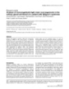
- Journal of Medical Case Reports 2007, 1:187 http://www.jmedicalcasereports.com/content/1/1/187 tion showed monoclonal kappa light chain in the urine. Introduction Light chain deposition disease (LCCD) is a systemic dis- Echocardiography detected substantial thickening of the ease involving several organs. Kidney impairment usually left wall in the septum and posterior wall and diastolic dominates the clinical picture, and proteinuria and renal ventricular dysfunction, findings suggestive of restrictive failure are the most common clinical manifestations [1]. cardiomyopathy. Heart involvement plays a crucial role in the prognosis of the disease; signs and symptoms of cardiac dysfunction The patient underwent US-guided biopsy of the lower are related to the degree of myocardial deposition of light pole of the right kidney, and two specimens were chains, and generally occur in the advanced stages of the obtained for light and electron microscopy examination. disease. We report a case of LCCD in which paroxysmal Light microscopy examination showed smooth and con- atrial fibrillation was the first clinical manifestation. tinuous deposition of eosinophil material in the tubular basement membrane, mild thickening and stiffness of the glomerular basement membrane, and increase of the Case presentation In December 2006, a 55-year-old woman was admitted to mesangial matrix. Congo red stain was negative, but the emergency department of our hospital because of pal- immunofluorescence revealed linear deposits of kappa pitations. She had a history of cholecystectomy because of light chains within the tubular basement membranes. biliary stone, hysterectomy and hypothyroidism treated Electron microscopy examination displayed coarse granu- with thyroxine 50 µg/day. In a laboratory test performed lar electron-dense deposits in the outer surface of the in October 2006, serum creatinine was 1.4 mg/dl and tubular basement membranes (Figure 1), and nonfibrillar urine analysis was normal. Clinical examination showed electron dense material along the glomerular basement hypertension and atrial fibrillation. Echocardiography membrane and in the mesangium (Figure 2). Bone mar- demonstrated left ventricular hypertrophy and diastolic row aspiration and bone biopsy were performed, and his- dysfunction, with normal ejection fraction. The atrial tologic examination of the specimens confirmed the fibrillation resolved spontaneously, and treatment with diagnosis of monoclonal immunoglobulin deposition an angiotensin converting enzyme (ACE)-inhibitor was disease associated to kappa light chain multiple myeloma. started. In January 2007, a further episode of atrial fibril- Treatment with thalidomide 100 mg/day and dexametha- lation occurred. It resolved after intravenous propaf- sone 40 mg on days 1–4 every 28 days was started, a peri- enone, but serum laboratory tests showed an increase in toneal catheter was inserted, and the patient was changed creatinine (3 mg/dl) and potassium levels (5.8 mmol/l), from haemodialysis to peritoneal dialysis. At the time of and the ACE-inhibitor was stopped. Thyroid function was normal. Three weeks later, serum creatinine was found to have further increased up to 5 mg/dl, whereas both kid- neys appeared normal on sonography (US) examination. The patient was admitted to our nephrology unit. On admission serum creatinine was 9.4 mg/dl (normal refer- ence values 0.7–1.3), potassium 6.8 mmol/l (normal ref- erence values 3.7–5.3), and haemoglobin 7.7 g/dl (normal reference values 11.5–16.5). Two units of packed red cells were transfused, a central venous catheter was inserted, and haemodialysis was started. Proteinuria was 1 g/day and urine sediment analysis showed haematuria. Serum glucose was 85 mg/dl (normal reference values 70– 110), sodium 140 mmol/l (normal reference values 136– 146), calcium 2.4 mmol/l (normal reference values 2.15– 2.55), proteins 7.1 g/dl (normal reference values 6.6–8.7), and albumin 43 g/L (normal reference values 35–46); protein electrophoresis did not show any monoclonal spike, IgG was 630 mg/dl (normal reference values 600– 1600), IgA 71 mg/dl (normal reference values 70–400), IgM 44 mg/dl (normal reference values 40–230), C3 132 mg/dl (normal reference values 90–180), C4 54 mg/dl Figure lar basement membrane electron-dense deposits along the outer surface of granular Electron1microscopy photograph showing coarsely the tubu- (normal reference values 16–38), autoantibodies were Electron microscopy photograph showing coarsely granular negative and N-terminal pro-brain natriuretic peptide electron-dense deposits along the outer surface of the tubu- (NT-proBNP) was 29894 pg/ml (Roche, Indianapolis, IN, lar basement membrane. USA; normal reference values
- Journal of Medical Case Reports 2007, 1:187 http://www.jmedicalcasereports.com/content/1/1/187 proteins, the stabilization of fibrils, and their protection from degradation [5]. In the kidneys, LCDD is often asso- ciated with deposits in the tubular basement membranes and Bowman's capsule, which may be more prominent than those deposits seen in the glomeruli. Clinical presen- tation depends in part on the site of deposition: it follows that patients with predominant glomerular deposition develop nephrotic syndrome, while those with predomi- nant tubular deposition develop renal failure and mild proteinuria [2]. In most cases, renal function declines rap- idly, with the clinical features of subacute tubulointersti- tial nephritis or rapidly progressive glomerulonephritis [3]. In our patient, the clinical presentation showed simi- lar characteristics, but it was initially misunderstood. An ACE-inhibitor was prescribed without paying sufficient attention to the slight increase in creatinine levels, and the subsequent worsening of renal function was ascribed to that medication. Such a misunderstanding delayed the correct diagnosis, and the clinical management was erro- Figure basement membrane tron-dense deposits photograph showing of the glomerular Electron2microscopyin the endothelial sidenonfibrillar elec- neously focussed on the cardiac manifestations. Indeed, Electron microscopy photograph showing nonfibrillar elec- atrial fibrillation associated with restrictive cardiomyopa- tron-dense deposits in the endothelial side of the glomerular basement membrane. thy is a quite uncommon presentation of LCDD, and the patient was referred to the nephrology unit only when the renal disease impairment became severe. writing the patient has been dialysing for 15 months and no major complications have been recorded. Endomyocardial biopsy is the gold standard to demon- strate heart involvement in LCDD. Histologic examina- The study has been conducted according to the Declara- tion of deep-frozen specimens shows amorphous Congo tion of Helsinky. red-negative deposits that are positive for light chains on immunofluorescence [6,7]. Clinical manifestations of heart involvement are variable and similar to those Discussion The pathogenesis of LCDD is similar to that of primary observed in restrictive cardiomyopathy induced by amy- amyloidosis. Both are monoclonal plasma cell prolifera- loidosis. They can range from congestive heart failure to tive disorders characterized by tissue deposition of light arrhythmias and conduction disorders; myocardial infarc- chain fragments, leading to organ impairment. Myeloma tion has also been reported [8,9]. Recently Toor et al. [10] is diagnosed in about 40% of patients with LCDD [2]. In described cardiac nonamyloidotic immunoglobulin dep- LCDD light chain fragments do not form amyloid fibrils; osition disease in 8 patients who underwent endomyocar- they are defined as non-amyloid immunoglobulin light dial biopsy. The median age was 49.5 years, none of them chains, and are mostly kappa chains. They cause renal fail- were symptomatic and on echocardiography six patients ure and extra-renal manifestations usually secondary to had concentric left ventricular hypertrophy with diastolic heart, liver and peripheral nerve involvement [3]. Survival dysfunction. One of them developed atrial fibrillation is quite variable, ranging from 1 month to 10 years from during chemotherapy and responded to therapy with dig- the onset of symptoms, and mortality is mainly due to oxin. heart or liver failure, or progression to multiple myeloma [2]. Serum creatinine at the time of diagnostic renal In our patient left ventricular morphology and the trans- biopsy seems to be the only predictive factor of renal func- mitral inflow pattern demonstrated by doppler echocardi- tion and patient survival [1,3]. ography were consistent with diastolic ventricular dysfunction due to restrictive cardiomyopathy [11]. In plasma cell disorders with dysproteinemia, the aggrega- Response to treatment may differ between amyloidosis- tion of non-amyloid immunoglobulin light chains forms induced and LCDD-induced cardiomyopathy, and the lat- granular deposits, diffusely distributed in systemic base- ter could resolve after successful treatment of the underly- ment membranes, which suggest a mechanism of aggrega- ing plasma cell disorder [7]. However, whatever signs and tion and deposition different from primary amyloidosis symptoms may reveal heart impairment in LCDD, they [4]. Several cofactors have been suggested to play a role in usually occur in the advanced stages of the disease. promoting fibrilogenesis, such as the binding to subunit Page 3 of 4 (page number not for citation purposes)
- Journal of Medical Case Reports 2007, 1:187 http://www.jmedicalcasereports.com/content/1/1/187 To our knowledge, atrial fibrillation associated with 2. Ronco PM, Aucouturier P, Mougenot B: Kidney involvement in plasma cell dyscrasias. In Oxford Textbook of Clinical Nephrology 3rd restrictive cardiomyopathy has never been previously edition. Edited by: Davison AM, Cameron JS, Grünfeld J-P, Ponticelli reported in the medical literature as a first clinical mani- C, Ritz E, Winearls CG, van Ypersele C, Barratt M, Ritter JM. Oxford (UK); Oxford University Press; 2005. festation of restrictive cardiomyopathy due to LCDD. 3. Lin J, Markowitz GS, Valeri AM, Kambham N, Sherman WH, Appel Although Palladini et al. [12] demonstrated that NT- GB, D'Agati VD: Renal monoclonal immunoglobulin deposi- proBNP assay can be useful in detecting cardiac involve- tion disease: the disease spectrum. J Am Soc Nephrol 2001, 12:1482-1492. ment in amyloidosis, we observed that natriuretic peptide 4. Kaplan B, Livneh A, Gallo G: Charge differences between in vivo levels were no more effective than echocardiography in deposits in immunoglobulin light chain amyloidosis and non- amyloid light chain deposition disease. Br J Haematol 2007, evaluating heart disease in a patient with primary amy- 136:723-728. loidosis and uraemia [13]. However, the very high levels 5. Tennents GA, Lovat LB, Pepys MB: Serum amyloid P-component of NT-proBNP observed in the patient in this present case prevents proteolysis of the amyloid fibrils of Alzheimer's dis- ease and systemic amyloidosis. Proc Natl Acad Sci USA 1995, report could have been the result of the combination of 92:4299-4303. heart involvement and impaired renal function, as renal 6. Jego P, Paillard F, Ramée MP, Grosbois B: Congestive heart failure: failure can influence NT-proBNP assay performed by revealing light chain deposition disease. European J Intern Med 2000, 11:101-103. Roche method [14]. 7. Nakamura M, Satoh M, Kowada S, Satoh H, Tashiro A, Sato F, Masuda T, Hiramori K: Reversibile restrictive cardiomyopathy due to light-chain deposition disease. Mayo Clin Proc 2002, 77:193-196. In conclusion, this unusual case suggests that the onset of 8. Garton MJ, Walton S, Ewen SW: Systemic lambda light-chain acute arrhythmias associated with restrictive cardiomyop- deposition presenting with predominant cardiac involve- athy and impaired renal function might be related to ment. Postgrad Med J 1993, 69:588-591. 9. Staros E, Katz MS: Myocardial necrosis in light chain deposi- LCDD. tion. Am heart J 1985, 110:1295-1296. 10. Toor AA, Ramdane BA, Joseph J, Thomas M, O'Hara C, Barlogie B, Walker P, Joseph L: Cardiac nonamyloidotic immunoglobulin Competing interests deposition disease. Modern Pathology 2006, 19:233-237. The author(s) declare that they have no competing inter- 11. Klein AL, Hatle LK, Taliercio CP, et al.: Serial doppler echocardi- ests. ographic follow-up of left ventricular diastolic function in cardiac amyloidosis. J Am Coll Cardiol 1990, 16:1135-1141. 12. Palladini G, Campana C, Klersy C, Balduini A, Vadacca G, Perfetti V, Authors' contributions Perlini S, Obici L, Ascari E, Melzi d'Eril G, Moratti R, Merlini G: FF, NS, MB, SU, AG, CM, LC performed the clinical work Serum N-terminal pro-brain natriuretic peptide is a sensi- tive marker of myocardial dysfunction in AL amyloidosis. Cir- and made the diagnosis, acquired, analyzed and inter- culation 2003, 107:2440-2445. preted the data. 13. Fabbian F, Stabellini N, Sartori S, Molino C, Russo G, Russo M, Can- telli S, Catizone L: Role of B-type natriuretic peptide in cardio- vascular state monitoring in a hemodialysis patient with FF and SS drafted the manuscript. primary amyloidosis. Int J Artif Organs 2006, 29(8):745-749. 14. Sykes E, Karcher RE, Eisenstadt J, Tushman DA, Balasubramaniam M, Gusway J, Perason VJ: Analytical relationships among Biosite, SS, PT performed the investigations. Bayer, and Roche methods for BNP and NTproBNP. Am J Clin Pathol 2005, 123:584-590. AA performed the electron microscopy work. Every author reviewed and approved the manuscript which is not under consideration for publication else- where in a similar form, in any language. Consent The patient was informed about the article and hypothet- ical beneficial effects of its publication on clinical practice. Written informed consent was obtained from the patient Publish with Bio Med Central and every for publication of this Case report and any accompanying scientist can read your work free of charge images. A copy of the written consent is available for "BioMed Central will be the most significant development for review by the Editor-in-Chief of this journal. disseminating the results of biomedical researc h in our lifetime." Sir Paul Nurse, Cancer Research UK Acknowledgements Your research papers will be: We gratefully acknowledge the work of the nurses of the renal unit of the available free of charge to the entire biomedical community St. Anna Hospital (Ferrara, Italy). peer reviewed and published immediately upon acceptance References cited in PubMed and archived on PubMed Central 1. Pozzi C, D'amico M, Fogazzi GB, Curioni S, Ferrario F, Pasquali S, yours — you keep the copyright Quattrocchio G, Rollino C, Segagni S, Locatelli F: Light chain dep- BioMedcentral osition disease with renal involvement: clinical characteris- Submit your manuscript here: tics and prognostic factors. Am J Kidney Dis 2003, 42:1154-1163. http://www.biomedcentral.com/info/publishing_adv.asp Page 4 of 4 (page number not for citation purposes)
CÓ THỂ BẠN MUỐN DOWNLOAD

-

Báo cáo Y học: Phosphorylation of calmodulin
 13 p |
  48
|
48
|  6
6
-

báo cáo khoa học: " Successful management of refractory pleural effusion due to systemic immunoglobulin light chain amyloidosis by vincristine adriamycin dexamethasone chemotherapy: a case report"
5 p | 52
| 5
-

Báo cáo y học: "Light chain (AL) amyloidosis: update on diagnosis and management"
17 p | 40
| 4
-
Báo cáo y học: "Analysis of immunoglobulin light chain rearrangements in the salivary gland and blood of a patient with Sjögren’s syndrome"
12 p | 51
| 3
-

Báo cáo y học: "Resolution of cast nephropathy following free light chain removal by haemodialysis in a patient with multiple myeloma: a case report"
5 p | 36
| 3
-
Báo cáo Y học: ER–resident chaperone interactions with recombinant antibodies in transgenic plants
10 p | 43
| 3
-
Báo cáo Y học: Convulxin induces platelet shape change through myosin light chain kinase and Rho kinase
7 p | 39
| 3
-
Báo cáo Y học: The inhibitory region of troponin-I alters the ability of F-actin to interact with different segments of myosin
13 p | 38
| 2


Chịu trách nhiệm nội dung:
Nguyễn Công Hà - Giám đốc Công ty TNHH TÀI LIỆU TRỰC TUYẾN VI NA
LIÊN HỆ
Địa chỉ: P402, 54A Nơ Trang Long, Phường 14, Q.Bình Thạnh, TP.HCM
Hotline: 093 303 0098
Email: support@tailieu.vn
Giấy phép Mạng Xã Hội số: 670/GP-BTTTT cấp ngày 30/11/2015 Copyright © 2022-2032 TaiLieu.VN. All rights reserved.

