RESEA R C H Open Access
Suppression of LPS-induced matrix-
metalloproteinase responses in macrophages
exposed to phenytoin and its metabolite,
5-(p-hydroxyphenyl-), 5-phenylhydantoin
Ryan Serra
1
, Abdel-ghany Al-saidi
1
, Nikola Angelov
2
, Salvador Nares
1*
Abstract
Background: Phenytoin (PHT) has been reported to induce gingival (gum) overgrowth (GO) in approximately 50%
of patients taking this medication. While most studies have focused on the effects of PHT on the fibroblast in the
pathophysiology underlying GO, few studies have investigated the potential regulatory role of macrophages in
extracellular matrix (ECM) turnover and secretion of proinflammatory mediators. The aim of this study was to
evaluate the effects of PHT and its metabolite, 5-(p-hydroxyphenyl-), 5-phenylhydantoin (HPPH) on LPS-elicited
MMP, TIMP, TNF-aand IL-6 levels in macrophages.
Methods: Human primary monocyte-derived macrophages (n= 6 independent donors) were pretreated with 15-
50 μg/mL PHT-Na
+
or 15-50 μg/mL HPPH for 1 hour. Cells were then challenged with 100 ng/ml purified LPS from
the periodontal pathogen, Aggregatibacter actinomycetemcomitans. Supernatants were collected after 24 hours and
levels of MMP-1, MMP-2, MMP-3, MMP-9, MMP-12, TIMP-1, TIMP-2, TIMP-3, TIMP-4, TNF-aand IL-6 determined by
multiplex analysis or enzyme-linked immunoadsorbent assay.
Results: A dose-dependent inhibition of MMP-1, MMP-3, MMP-9, TIMP-1 but not MMP-2 was noted in culture
supernatants pretreated with PHT or HPPH prior to LPS challenge. MMP-12, TIMP-2, TIMP-3 and TIMP-2 were not
detected in culture supernatants. High concentrations of PHT but not HPPH, blunted LPS-induced TNF-a
production although neither significantly affected IL-6 levels.
Conclusion: The ability of macrophages to mediate turnover of ECM via the production of metalloproteinases is
compromised not only by PHT, but its metabolite, HPPH in a dose-dependent fashion. Further, the preferential
dysregulation of macrophage-derived TNF-abut not IL-6 in response to bacterial challenge may provide an
inflammatory environment facilitating collagen accumulation without the counteracting production of MMPs.
Background
Drug-induced gingival (gum) overgrowth (DIGO) is
widely recognized as a common unwanted sequelae
associated with a variety of medications. Among these,
the antiepileptic agent, PHT (Dilantin®), has been
reported to induce gingival overgrowth (GO) in approxi-
mately 50% of patients taking this medication [1,2]. PHT
is a hydantoin-derivative anticonvulsant that exerts its
anticonvulsant properties by stabilizing neuronal cell
membranes to the action of sodium, potassium, and cal-
cium. The drug also affects the transport of calcium
across cell membranes and decreases the influx of cal-
cium ions across membranes by decreasing membrane
permeability and blocking intracellular uptake [3]. PHT
is primarily metabolized by liver cytochrome P450
enzymes, particularly CYP2C9 and CYP2C19 [4] to form
enantiomers of 5-(4-hydroxyphenyl-),5-phenylhydantoin
(HPPH) which in addition to PHT, have been implicated
in the pathogenesis of DIGO [5,6].
* Correspondence: Salvador_Nares@dentistry.unc.edu
1
Department of Periodontology, School of Dentistry, University of North
Carolina at Chapel Hill, Chapel Hill, North Carolina, USA
Full list of author information is available at the end of the article
Serra et al.Journal of Inflammation 2010, 7:48
http://www.journal-inflammation.com/content/7/1/48
© 2010 Serra et al; licensee BioMed Central Ltd. This is an Open Access article distributed under the terms of the Creative Commons
Attribution License (http://creativecommons.org/licenses/by/2.0), which permits unrestricted use, distribution, and reproduction in
any medium, provided the original work is properly cited.

While most studies have focused on the role of the
fibroblast [7-10], it is likely that other cells contribute to
the pathogenesis of DIGO. In particular, tissue macro-
phages, present in elevated numbers within gingival tis-
sues, possibly in response to accumulation of the plaque
biofilm[2,11],mayplayaroleinpathogenesis.These
long-lived, multifaceted cells, strategically poised along
portals of entry, perform numerous functions of vital
importance to the host. In addition to their key role in
immunity [12], the macrophage is recognized as the
major mediator of normal connective tissue turnover
and maintenance, as well as for orchestrating repair dur-
ing wound healing [13-18]. It has a dualistic role to
receive, amplify, and transmit signals to fibroblasts,
endothelial cells, and vascular smooth muscle cells by
producing pro-inflammatory and catabolic cytokines.
However, during tissue turnover and wound healing it
secretes anabolic peptide growth factors [12]. Given this
duality of function, any perturbation can lead to patho-
logical processes. We have demonstrated that the clini-
cal presentation of PHT-induced gingival overgrowth is
associated with a specific macrophage phenotype char-
acterized by high expression levels of IL-1band PDGF-
B [11,19] suggesting that this drug-induced macrophage
phenotype could contribute to the pathogenesis of
DIGO. These cellular attributes might explain the
dichotomy of the lesion where there is both periodontal
inflammation typically associated with connective tissue
catabolism paradoxically juxtaposed with gingival over-
growth,- a clear anabolic signal of wound repair and
regeneration.
As tissue homeostasis requires the proper balance of
metabolism and catabolism, it is possible that macro-
phage-derived cytokines, MMPs and TIMP levels are
altered in response to PHT and HPPH. Here we investi-
gated the effects of these agents on production of
MMPs, TIMPs, and pro-inflammatory cytokines in
human monocyte-derived macrophages and report that
indeed, PHT and HPPH significantly modulate macro-
phage MMP and cytokine protein levels in response to
purified LPS from the periodontal pathogen, Aggregati-
bacter actinomycetemcomitans.
Methods
Monocyte isolation and macrophage differentiation
Peripheral blood mononuclear cells were obtained from
commercially-available buffy coats (Oklahoma Blood
Institute, Oklahoma City, OK, USA) derived from
healthy donors by density gradient centrifugation using
Ficoll-paque (Amersham, Uppsala,Sweden).Sixinde-
pendent cultures were obtained from 6 independent
donors. Monocytes were isolated using CD14 MicroBe-
ads (Miltenyi Biotec, Auburn, CA, USA) according to
manufacturer’s instructions and cultured as previously
described [12,20,21]. Briefly, isolated monocytes were
plated onto duplicate 12-well tissue culture-treated
plates (BD Biosciences, San Jose, CA, USA) at a density
of 5 × 10
5
cells/cm
2
in serum-free DMEM with L-gluta-
mine (Cellgro, Manassas, VA, USA) containing 50 μg/
mL gentamicin (Sigma, St. Louis, MO, USA) at 37 C,
5% CO
2
to promote monocyte attachment. After 2
hours, heat-inactivated fetal bovine serum (FBS, Invitro-
gen, Carlsbad, CA, USA) was added to a final concentra-
tion of 10%. Cells were >95% CD14+ as determined by
FACS analysis (data not shown) prior to culture.
Macrophage stimulation
After 5 days, the media and non-adhered cells were
removed and replaced with complete media (DMEM,
10% FBS, gentamicin) and incubated at 37 C, 5% CO
2
.
Media was replaced every 2 days. Experiments were
initiated upon confirmation of macrophage differentia-
tion after 7 days in culture [12,20,21]. Macrophages
were used between day 7 and 10 and pretreated with
either: 1) 15 μg/mL of PHT-Na+ (Sigma), (serum levels,
[22-24]), 2) 50 μg/mL PHT-Na+ (high dose), 3) 15 μg/
mL PHT metabolite (Sigma), (5-(4’-hydroxyphenyl),5-
phenylhydantoin, HPPH), or 4) 50 μg/mL HPPH for 1
hour. Untreated cells served as control cultures. Stock
solutions of PHT-Na+ (150 mg/mL) were made in ster-
ile deionized water while HPPH (150 mg/mL) solutions
were made in DMSO. Each stock solution was further
diluted prior to use. The total concentration of DMSO
in cultures was always less than 0.05%. DMSO concen-
trations less than 0.1% have been reported not to affect
cellular viability and function [25,26]. Nevertheless, we
confirmed these findings in preliminary studies exposing
macrophage cultures to 0.05% DMSO (data not shown).
To induce production of MMPs and proinflammatory
cytokines, macrophages were challenged with 100 ng/
mL purified LPS from the Gram-negative, periodontal
pathogen, Aggregatibacter actinomycetemcomitans
(A. actinomycetemcomitans (Aa), serotype b, strain Y4, a
kind gift from K. L. Kirkwood, University of South Car-
olina, USA) for 24 hours. Isolation and purification of
Aa LPS has been previously described [27]. Previous stu-
dies have demonstrated that LPS from this organism is
capable of inducing MMP and TIMP production [28-30]
and our preliminary studies determined that this con-
centration of LPS was capable of significantly inducing
TNF-alevels in human primary macrophages and THP-
1 cells induced for macrophage differentiation (data not
shown).
MMP, TIMP protein assays
After 24 hours, the media was collected, spun at 12,000 ×
g, transferred to fresh tubes and stored at -80 C until
further use. Quantification of supernatant MMP and
Serra et al.Journal of Inflammation 2010, 7:48
http://www.journal-inflammation.com/content/7/1/48
Page 2 of 10

TIMP levels were determined using the Luminex 100
System (Luminex Co., Austin, TX, USA) and the Fluoro-
kine MAP Multiplex Human MMP Panel and the Fluoro-
kine MAP Human TIMP Multiplex Kit, respectively
according to the manufacturer’s instructions (both from
R&D,Minneapolis,MN,USA).Thesekitsmeasure
levels of pro-, mature, and TIMP complexed MMPs. Six
independent experiments were performed from cells
derivedfrom6differentdonors. The assays were per-
formed in 96-well plates, as previously described [20].
For MMP determination, microsphere beads coated with
monoclonal antibodies against MMP-1, MMP-2, MMP-
3, MMP-9, MMP-12 were added to the wells. For TIMP
determination, microsphere beads coated with monoclo-
nal antibodies against TIMP-1, TIMP-2, TIMP-3, and
TIMP-4 were added to the wells of a separate plate. To
remain below the upper level of quantitation, samples
containing LPS were diluted 10-fold prior to analysis.
This dilution factor was based on our preliminary studies.
Samples and standards were pipetted into wells, incu-
bated for 2 hours with the beads then washed using a
vacuum manifold (Millipore Corporation, Billerica, MA
USA). Biotinylated secondary antibodies were added and
incubation for 1 h. The beads were then washed and
incubated for an additional 30 minutes with streptavidin
conjugated to the fluorescent protein, R-phycoerythrin
(streptavidin/R-phycoerythrin). The beads were washed
andanalyzed(aminimumof50peranalyte)usingthe
Luminex 100 system. The Luminex 100 measures the
amount of fluorescence associated with R-phycoerythrin,
reported as median fluorescence intensity of each spec-
tral-specific bead allowing it to distinguish the different
analytes in each well. The concentrations of the unknown
samples (antigens in macrophage supernatants) were esti-
mated from the standard curve using a third-order poly-
nomial equation and expressed as pg/mL after adjusting
for the dilution factor. Samples below the detection limit
of the assay were recorded as zero. The minimum detect-
able concentrations for the assays were as follows: MMP-
1: 4.4 pg/mL, MMP-2: 25.4 pg/mL, MMP-3: 1.3 pg/mL,
MMP-9: 7.4 pg/mL, TIMP-1: 1.54 pg/mL, TIMP-2: 14.7
pg/mL, TIMP-3: 86 pg/mL and TIMP-4: 1.29 pg/mL. All
values were standardized for total protein using the Brad-
ford assay (Pierce, Thermo Scientific, Rockford, IL, USA)
according to manufacturer’s instructions. Briefly, culture
supernatants were mixed with assay reagent and incu-
batedfor10minutesatroomtemperaturein96well
plates. Bovine serum albumin (BSA, Invitrogen) was used
as a standard. The absorbance at 595 nm was read using
a SpectraMax M2 microplate reader (Molecular Devices,
Sunnyvale, CA, USA). Values obtained from untreated
control cultures were arbitrarily used as a baseline mea-
sure. The ratio, (control)/(supernatant protein value) was
used to normalize each sample based on total protein.
Cytokine assays
After 24 hours, supernatants (n= 6 independent
donors) were collected and levels of TNF-aand IL-6
determined by ELISA (RayBiotech, Norcross, GA, USA)
according to manufacturer’s instructions. The absor-
bance at 450 nm was read using a SpectraMax M2
microplate reader (Molecular Devices) with the wave-
length correction set at 550 nm. The rated sensitivities
of the commercial ELISA kits was 15 pg/mL for TNF-a
and 6 pg/mL for IL-6. Values were standardized for
total protein using the Bradford assay as described
above.
Cell viability assays
Viability of macrophages was evaluated using the CellTi-
ter 96 AQueous One Solution Cell Proliferation Assay
[3-(4,5-diethylthiazol-2-yl)-5-(3-carboxymethoxyphenyl)-
2-(4-sulfophenyl)-2H-tetrazolium, inner salt, MTS] assay
according to the manufacturer’s protocol (Promega,
Madison, WI, USA). This colorimetric method can be
used to determine the number of viable cells in prolif-
eration or to evaluate cytotoxicity. Briefly, macrophages
were cultured in triplicate in 96-well plates and treated
with PHT, HPPH and LPS as described above. Unstimu-
lated cells served as control cultures. After 24 h, the
cells were incubated with MTS for 2 h at 37 C, 5% CO
2
.
The absorbance was read at 490 nm using a microplate
reader.
Statistical analysis
Data were analyzed using a hierarchical multiple regres-
sion approach relative to LPS, drug and dose. The first
tier sought to establish the validity of the positive con-
trol, LPS vs the negative control group. The second tier
of this analysis was aimed at determining whether PHT
or HPPH have an effect on MMP, TIMP, TNF-aand
IL-6 levels. Finally, the third tier sought to contrast dose
and compare one drug with another. Data were
expressed as mean ± SEM and compared using a two-
tailed Student’sttest for correlated samples (GraphPad
Prism, GraphPad Software, La Jolla, CA, USA). Results
were considered statistically significant at p< 0.05.
Results
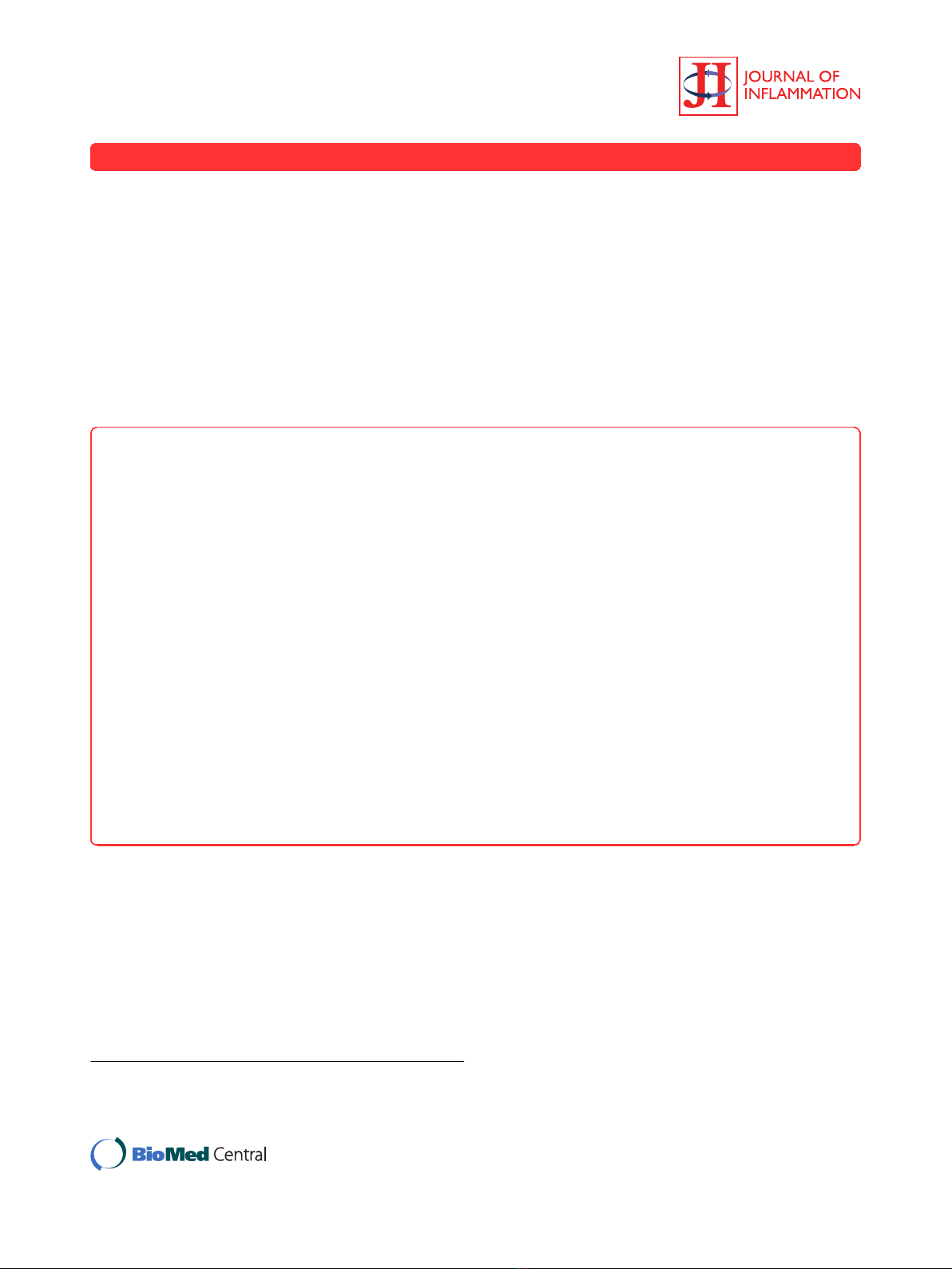
PHT and HPPH inhibit LPS-induced supernatant levels of
MMP-1, MMP-3, MMP-9, and TIMP-1 in a dose dependent
manner
To evaluate the effects of PHT and its metabolite,
HPPH on macrophage MMP and TIMP levels, human
monocyte-derived macrophages were pretreated for 1
hour with either 15 μg/mL or 50 μg/mL of these agents
prior to challenge with LPS. Previous studies have deter-
mined that PHT plasma levels of 10-20 μg/mL are
necessary to effectively maintain effective seizure control
Serra et al.Journal of Inflammation 2010, 7:48
http://www.journal-inflammation.com/content/7/1/48
Page 3 of 10
[22-24]. Thus, the concentrations used in our study
represent therapeutic as well as elevated levels of PHT
permitting the evaluation of dose on MMP and TIMP
production. To rule out the possibility that differences
in supernatant levels of these readouts were due to
decreased cell viability, we performed a viability assay
on cells cultured in each condition. No significant differ-
ences were noted in the viability of cells exposed to LPS
and either dose of PHT, HPPH, PHT/LPS or HPPH/
LPS as determined by MTS assay. Further, we standar-
dized the results of each analyte to total protein concen-
tration for each condition using a Bradford assay. No
differences were noted for any analyte examined in con-
ditioned media from macrophage cultures treated with
PHT or HPPH alone compared to control cultures (p>
0.05). As expected, LPS markedly induced supernatant
MMP-1, MMP-3, MMP-9, TIMP-1 but not MMP-2
levels in our 6 independent cultures after a 24 hour
exposure (Fig. 1A-D). Compared to untreated control
cultures, LPS significantly increased secretion of MMP-1
despite the presence of either PHT or HPPH at any
dose. This was similarly observed for MMP-3 levels with
the exception of cultures pretreated with 15 μg/mL
HPPH which despite elevated levels, did not reach sta-
tistical significance (p> 0.05). In contrast, exposure of
macrophages to 50 μg/mL of either PHT or HPPH prior
to LPS stimulation prevented a significant increase in
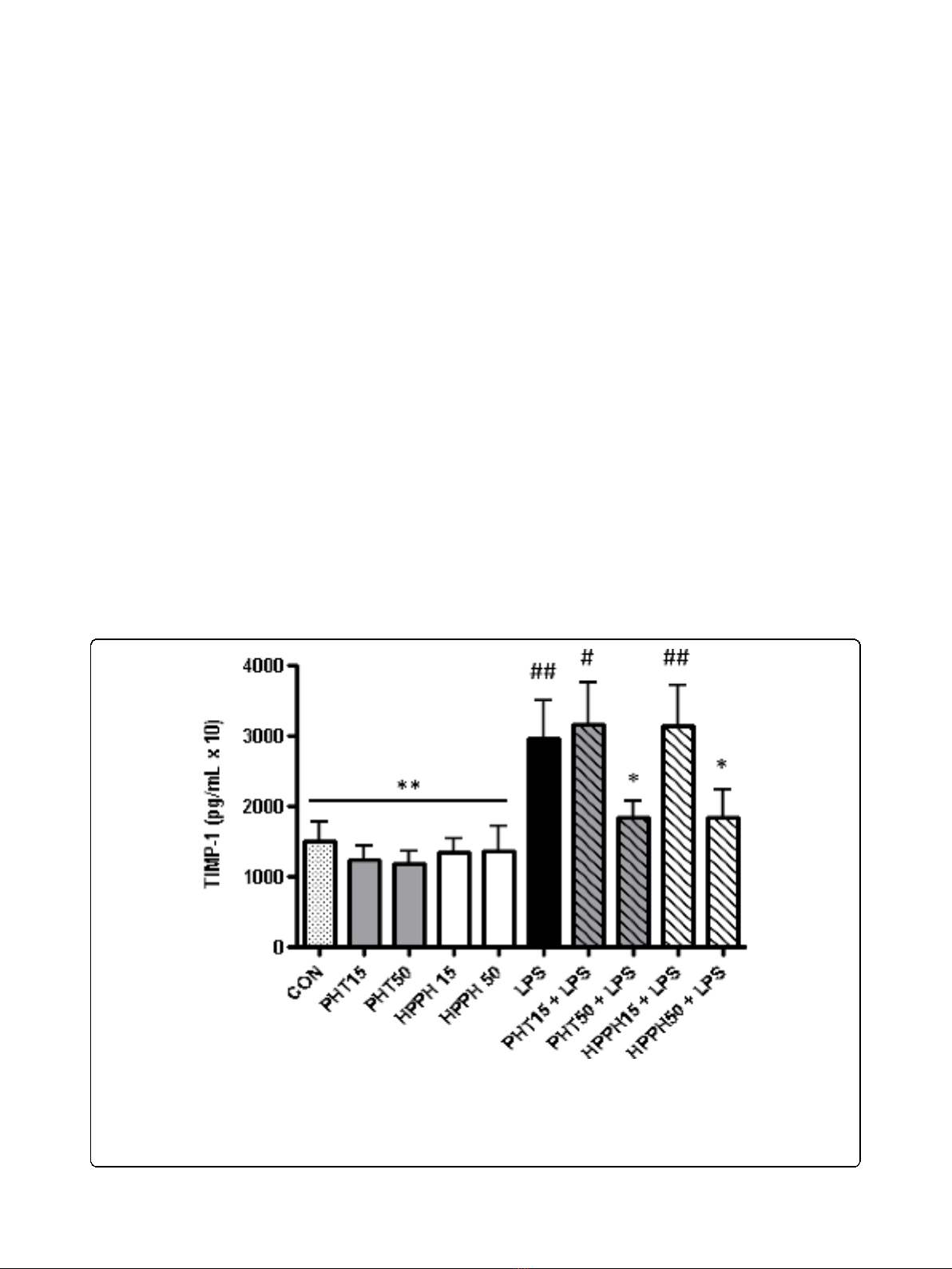
MMP-9andTIMP-1(Fig.1DandFig.2).Levelsof
Figure 1 The effect of phenytoin, HPPH and LPS on levels of (A) matrix metalloproteinase-1, (B) matrix metalloproteinase-2, (C) matrix
metalloproteinase-3, and (D) matrix metalloproteinase-9 in conditioned medium from macrophage cultures. Primary human monocyte-
derived macrophages (n=6 independent cultures) were pretreated with phenytoin or HPPH (15 μg/mL and 50 μg/mL) for 1 hour prior to
challenge with 100 ng/mL A. actinomycetemcomitans LPS and the levels of matrix metalloproteinase-1, matrix metalloproteinase-2, matrix
metalloproteinase-3, and matrix metalloproteinase-9 measured after 24 hours in conditioned media by multiplex analysis. MMP-1, matrix
metalloproteinase-1; MMP-2, matrix metalloproteinase-2; MMP-3, matrix metalloproteinase-3; MMP-9, matrix metalloproteinase-9; CON, control;
PHT, phenytoin; HPPH, 5-(4-hydroxyphenyl-),5- phenylhydantoin; LPS, lipopolysaccharide. Compared to CON, # p<0.05, ## p<0.01, ### p<0.001,
compared to LPS, * p<0.05, ** p<0.01, *** p<0.001. Student t-test, n=6 independent donors.
Serra et al.Journal of Inflammation 2010, 7:48
http://www.journal-inflammation.com/content/7/1/48
Page 4 of 10
MMP-9 and TIMP-1 remained near control levels
despite the potent proinflammatory challenge thus
demonstrating the ability of these agents to alter macro-
phage function. Compared to LPS alone, pretreatment
with 50 μg/mL PHT significantly blunted LPS-induced
levels of MMP-1 (p< 0.05). In cultures pretreated with
50 μg/mL HPPH, MMP-3 levels were not significantly
different compared to LPS-only treated cultures (p>
0.05) although the trend for reduced supernatant levels
of MMP3 was evident. However, exposure of macro-
phages to either 15 μg/mL or 50 μg/mL PHT prior to
LPS stimulation significantly blunted supernatant MMP-
3levels(p<0.01andp< 0.001, respectively, Fig. 1C)
compared to LPS-only treated cultures. Interestingly, a
trend for higher levels of MMP-1 were noted in cultures
treated with HPPH while MMP-3 levels were slightly
elevated in cultures treated with either PHT and HPPH
although neither reached statistical significance (p>
0.05) (Fig. 1, A, C).
Elevated levels (50 μg/mL) of PHT or HPPH signifi-
cantly reduced MMP-9 and TIMP-1 levels compared to
LPS-only treated cells (Fig. 1D and Fig. 2). The levels of
these analytes remained near control values despite LPS
challenge. Interestingly, HPPH but not PHT was
associated with reduced levels of MMP-2 compared to
LPS only, but this relationship was not statistically sig-
nificant. MMP-12 and TIMPs-2-4 remained below levels
of detection in all groups and cultures.
Supernatant levels of TNF-abut not IL-6, is decreased in
response to PHT
At 24 hours, supernatant levels of TNF-aand IL-6
were significantly increased by LPS compared to
untreated controls (p< 0.001). Similar to MMP and
TIMP levels, no significant differences in TNF-aand
IL-6 levels were observed in supernatants exposed to
either 15 or 50 μg/mL PHT and HPPH alone com-
pared to untreated cultures although a trend for
decreased levels of TNF-awas evident (Fig 3A). How-
ever, macrophage cultures pretreated with 50 μg/mL
PHT prior to challenge with LPS showed a significant
(p<0.05)decreaseinTNF-alevels compared to LPS
only treated cultures. No difference was noted for 15
μg/mL of PHT or HPPH at either concentration (Fig.
3A). Regardless of dosage, pretreatment with PHT or
HPPH prior to LPS challenge had no significant effect
(p> 0.05) on IL-6 secretion when compared to LPS
only treated cultures.
Figure 2 The effect of phenytoin, HPPH and LPS on levels of tissue inhibitor of matrix metalloproteinase-1 in conditioned medium
from macrophage cultures. Primary human monocyte-derived macrophages (n= 6 independent cultures) were pretreated with phenytoin or
HPPH (15 μg/mL and 50 μg/mL) for 1 hour prior to challenge with 100 ng/mL A. actinomycetemcomitans LPS and the levels of tissue inhibitor of
matrix metalloproteinase-1 measured after 24 hours in conditioned media by multiplex analysis. TIMP-1, tissue inhibitor of matrix
metalloproteinase-1; CON, control; PHT, phenytoin; HPPH, 5-(4-hydroxyphenyl-),5-phenylhydantoin; LPS, lipopolysaccharide. Compared to CON, #
p< 0.05, ## p< 0.01, ### p< 0.001, compared to LPS, * p< 0.05, ** p< 0.01, *** p< 0.001. Student t-test, n= 6 independent donors.
Serra et al.Journal of Inflammation 2010, 7:48
http://www.journal-inflammation.com/content/7/1/48
Page 5 of 10


![Vaccine và ứng dụng: Bài tiểu luận [chuẩn SEO]](https://cdn.tailieu.vn/images/document/thumbnail/2016/20160519/3008140018/135x160/652005293.jpg)


%20--%3e%3cdefs%3e%3cstyle%3e%20.st0%20{%20fill:%20%23fff;%20}%20.st1%20{%20fill:%20%237800fa;%20}%20%3c/style%3e%3c/defs%3e%3cpath%20class='st1'%20d='M117.78,12.18H43.11c2.9,3.47,4.65,7.94,4.65,12.82,0,5.6-2.3,10.66-6.01,14.29h76.02l7.22-13.56-7.22-13.56Z'/%3e%3cg%3e%3cpath%20class='st0'%20d='M53.58,26.17h-.59v-1.46h.59v-4.96h2.83c1.78,0,2.67.94,2.67,2.82v5.76c0,1.87-.89,2.81-2.67,2.81h-2.83v-4.96ZM55.36,21.37v3.34h1.1v1.46h-1.1v3.34h1.01c.61,0,.91-.37.91-1.1v-5.93c0-.74-.3-1.1-.91-1.1h-1.01Z'/%3e%3cpath%20class='st0'%20d='M65.99,31.14h-1.8l-.31-2.07h-2.19l-.31,2.07h-1.64l1.82-11.39h2.62l1.82,11.39ZM65.28,18.04c-.25.46-.51.77-.75.94-.21.15-.47.22-.79.22-.26,0-.57-.07-.92-.22l-.38-.15c-.14-.05-.26-.07-.37-.07-.3,0-.53.18-.71.54l-.91-.68c.25-.46.51-.77.75-.94.21-.14.48-.21.79-.21.26,0,.57.07.92.21l.38.15c.14.05.26.07.37.07.3,0,.53-.18.71-.54l.91.68ZM61.91,27.52h1.73l-.87-5.76-.87,5.76Z'/%3e%3cpath%20class='st0'%20d='M74.53,26.89v1.52c0,1.91-.89,2.86-2.67,2.86s-2.67-.95-2.67-2.86v-5.93c0-1.91.89-2.86,2.67-2.86s2.67.95,2.67,2.86v1.11h-1.69v-1.22c0-.75-.31-1.12-.93-1.12s-.93.37-.93,1.12v6.15c0,.74.31,1.11.93,1.11s.93-.37.93-1.11v-1.63h1.69Z'/%3e%3cpath%20class='st0'%20d='M81.4,31.14h-1.8l-.31-2.07h-2.19l-.31,2.07h-1.64l1.82-11.39h2.62l1.82,11.39ZM75.9,19.2l1.52-1.91h1.71l1.51,1.91h-1.61l-.76-.95-.75.95h-1.61ZM77.32,27.52h1.73l-.87-5.76-.87,5.76ZM83.1,15.99l-1.76,1.91h-1.26l1.17-1.91h1.86Z'/%3e%3cpath%20class='st0'%20d='M84.86,19.75c1.78,0,2.67.94,2.67,2.82v1.48c0,1.87-.89,2.81-2.67,2.81h-.85v4.28h-1.79v-11.39h2.64ZM84.01,21.37v3.86h.85c.58,0,.87-.36.87-1.08v-1.71c0-.71-.29-1.07-.87-1.07h-.85Z'/%3e%3cpath%20class='st0'%20d='M93.51,19.75c1.78,0,2.67.94,2.67,2.82v1.48c0,1.87-.89,2.81-2.67,2.81h-.85v4.28h-1.79v-11.39h2.64ZM92.66,21.37v3.86h.85c.58,0,.87-.36.87-1.08v-1.71c0-.71-.29-1.07-.87-1.07h-.85Z'/%3e%3cpath%20class='st0'%20d='M98.8,31.14h-1.79v-11.39h1.79v4.88h2.03v-4.88h1.83v11.39h-1.83v-4.88h-2.03v4.88Z'/%3e%3cpath%20class='st0'%20d='M105.36,24.55h2.46v1.62h-2.46v3.34h3.09v1.63h-4.88v-11.39h4.88v1.63h-3.09v3.18ZM108.17,17.29l-1.76,1.91h-1.26l1.17-1.91h1.86Z'/%3e%3cpath%20class='st0'%20d='M112.2,19.75c1.78,0,2.67.94,2.67,2.82v1.48c0,1.87-.89,2.81-2.67,2.81h-.85v4.28h-1.79v-11.39h2.64ZM111.35,21.37v3.86h.85c.58,0,.87-.36.87-1.08v-1.71c0-.71-.29-1.07-.87-1.07h-.85Z'/%3e%3c/g%3e%3ccircle%20class='st1'%20cx='25'%20cy='25'%20r='20'/%3e%3cpath%20class='st0'%20d='M32.78,19.27c2.92,0,4.43,2.55,5.28,5.33l.71,2.17c.14.38-.33.75-.71.75h-5.61c.19-.33.24-.71.09-1.08l-.75-2.45c-.43-1.32-.99-2.64-1.79-3.77.75-.57,1.65-.94,2.78-.94h0ZM25,18.38c3.25,0,4.9,2.78,5.89,5.89l.76,2.45c.14.42-.33.8-.8.8h-11.69c-.42,0-.94-.38-.8-.8l.75-2.45c.99-3.11,2.64-5.89,5.89-5.89h0ZM25,11.35c1.74,0,3.11,1.37,3.11,3.11s-1.37,3.11-3.11,3.11-3.11-1.41-3.11-3.11,1.41-3.11,3.11-3.11h0ZM17.27,19.27c1.08,0,1.98.38,2.73.94-.8,1.13-1.37,2.45-1.74,3.77l-.8,2.45c-.14.38-.05.75.09,1.08h-5.56c-.42,0-.9-.38-.75-.75l.71-2.17c.9-2.78,2.41-5.33,5.33-5.33h0ZM17.27,12.91c1.51,0,2.78,1.27,2.78,2.83s-1.27,2.83-2.78,2.83-2.83-1.27-2.83-2.83,1.27-2.83,2.83-2.83h0ZM32.78,12.91c1.56,0,2.78,1.27,2.78,2.83s-1.23,2.83-2.78,2.83-2.83-1.27-2.83-2.83,1.27-2.83,2.83-2.83h0ZM27.07,28.56v.09c0,.57-.24,1.08-.61,1.46h0v.05c-.38.33-.9.57-1.46.57s-1.08-.24-1.46-.61h0c-.38-.38-.61-.9-.61-1.46v-.09h1.41v.09c0,.19.05.38.19.47v.05c.09.09.28.19.47.19s.38-.09.47-.19v-.05c.14-.09.24-.28.24-.47t-.05-.09h1.41ZM30.99,28.56v.09c0,1.65-.66,3.16-1.74,4.24-1.08,1.08-2.59,1.79-4.24,1.79s-3.16-.71-4.24-1.79l-.05-.05c-1.04-1.08-1.7-2.55-1.7-4.2v-.09h1.41v.09c0,1.27.47,2.4,1.27,3.25h.05c.85.85,1.98,1.37,3.25,1.37s2.4-.52,3.25-1.37c.85-.8,1.37-1.98,1.37-3.25v-.09h1.37ZM34.99,28.56v.09c0,2.78-1.13,5.28-2.92,7.07-1.79,1.79-4.29,2.92-7.07,2.92s-5.23-1.13-7.07-2.92c-1.79-1.79-2.92-4.29-2.92-7.07v-.09h1.41v.09c0,2.4.94,4.53,2.5,6.08,1.56,1.56,3.72,2.5,6.08,2.5s4.52-.94,6.08-2.5c1.56-1.56,2.5-3.68,2.5-6.08v-.09h1.41Z'/%3e%3c/svg%3e)