
BioMed Central
Page 1 of 16
(page number not for citation purposes)
Retrovirology
Open Access
Research
The formation of cysteine-linked dimers of BST-2/tetherin is
important for inhibition of HIV-1 virus release but not for sensitivity
to Vpu
Amy J Andrew†, Eri Miyagi†, Sandra Kao and Klaus Strebel*
Address: Laboratory of Molecular Microbiology, Viral Biochemistry Section, National Institute of Allergy and Infectious Diseases, NIH, Bethesda,
Maryland, 20892-0460, USA
Email: Amy J Andrew - andrewa@niaid.nih.gov; Eri Miyagi - emiyagi@niaid.nih.gov; Sandra Kao - skao@niaid.nih.gov;
Klaus Strebel* - kstrebel@niaid.nih.gov
* Corresponding author †Equal contributors
Abstract
Background: The Human Immunodeficiency virus type 1 (HIV-1) Vpu protein enhances virus
release from infected cells and induces proteasomal degradation of CD4. Recent work identified
BST-2/CD317 as a host factor that inhibits HIV-1 virus release in a Vpu sensitive manner. A current
working model proposes that BST-2 inhibits virus release by tethering viral particles to the cell
surface thereby triggering their subsequent endocytosis.
Results: Here we defined structural properties of BST-2 required for inhibition of virus release
and for sensitivity to Vpu. We found that BST-2 is modified by N-linked glycosylation at two sites
in the extracellular domain. However, N-linked glycosylation was not important for inhibition of
HIV-1 virus release nor did it affect surface expression or sensitivity to Vpu. Rodent BST-2 was
previously found to form cysteine-linked dimers. Analysis of single, double, or triple cysteine
mutants revealed that any one of three cysteine residues present in the BST-2 extracellular domain
was sufficient for BST-2 dimerization, for inhibition of virus release, and sensitivity to Vpu. In
contrast, BST-2 lacking all three cysteines in its ectodomain was unable to inhibit release of wild
type or Vpu-deficient HIV-1 virions. This defect was not caused by a gross defect in BST-2
trafficking as the mutant protein was expressed at the cell surface of transfected 293T cells and was
down-modulated by Vpu similar to wild type BST-2.
Conclusion: While BST-2 glycosylation was functionally irrelevant, formation of cysteine-linked
dimers appeared to be important for inhibition of virus release. However lack of dimerization did
not prevent surface expression or Vpu sensitivity of BST-2, suggesting Vpu sensitivity and inhibition
of virus release are separable properties of BST-2.
Background
Vpu is an 81 amino acid type 1 integral membrane protein
[1,2] that has been shown to cause proteasomal degrada-
tion of CD4 [3,4] but also enhances the release of virions
from infected cells [5-7]. These two biological activities of
Vpu are mechanistically distinct and involve different
structural domains in Vpu. In particular, two conserved
phosphoserine residues in the cytoplasmic domain of Vpu
Published: 8 September 2009
Retrovirology 2009, 6:80 doi:10.1186/1742-4690-6-80
Received: 21 July 2009
Accepted: 8 September 2009
This article is available from: http://www.retrovirology.com/content/6/1/80
© 2009 Andrew et al; licensee BioMed Central Ltd.
This is an Open Access article distributed under the terms of the Creative Commons Attribution License (http://creativecommons.org/licenses/by/2.0),
which permits unrestricted use, distribution, and reproduction in any medium, provided the original work is properly cited.

Retrovirology 2009, 6:80 http://www.retrovirology.com/content/6/1/80
Page 2 of 16
(page number not for citation purposes)
(S52, S56) are crucial for CD4 degradation but have no or
only a partial effect on virus release [8-11]. On the other
hand, Vpu's transmembrane (TM) domain is critical for
enhancement of particle release but it can be substituted
by other membrane anchors without effect on CD4 degra-
dation [12,13]
Previous data suggested that Vpu regulates the detach-
ment of otherwise complete virions from the cell surface
[5,14]. Subsequently, several mechanisms of Vpu medi-
ated virus release have been proposed. First, a Vpu-associ-
ated ion channel activity was implicated in the regulation
of virus release. Vpu has the ability to assemble into a
monovalent cation-specific ion channel [15-19]. Rand-
omization of Vpu's TM domain did not affect membrane
association but inhibited Vpu's ion channel activity and,
at the same time, impaired its ability to regulate virus
release [12,17]. These observations established a correla-
tion between Vpu ion channel activity and increased virus
release activity. A second alternative model suggested that
Vpu might interfere with the activity of Task-1, a cellular
ion channel, through the formation of hetero-oligomeric
complexes [20]. Overexpression of a dominant-negative
fragment of Task-1 inhibited Task-1 ion channel activity
and increased release of vpu-deficient particles thus creat-
ing a functional correlation between Task-1 ion channel
activity and reduced HIV-1 particle release [20]. It is not
known, however, if expression of Task-1 is tissue specific
and it remains unclear, exactly how either Vpu or Task-1
ion channel activities might regulate detachment of parti-
cles from the cell surface.
A third model invokes the inactivation of a cellular inhib-
itor of virus release. This model is based on the observa-
tion that Vpu-dependent virus release is host cell-
dependent [21]. Indeed, in addition to Task-1, several
other host factors have been identified whose overexpres-
sion was associated with reduced virus release. These
include the Vpu binding protein UBP [22], the recently
identified host factors BST-2 (also referred to as tetherin,
CD317, or HM1.24 [23,24]), and CAML [25]. Among
those, BST-2 is of particular interest since its cell type-spe-
cific expression most closely matches that of Vpu-depend-
ent cell types and silencing of BST-2 expression in HeLa
cells by siRNA or shRNA rendered virus release from these
cells Vpu-independent [23,24].
A functional Vpu-BST-2 interaction was first reported in a
quantitative membrane proteomics study where Vpu
expressed from an adenovirus vector was found to reduce
cellular expression of BST-2 in HeLa cells [26]. Intrigu-
ingly, subsequent reports found that BST-2 expression var-
ied in a cell type dependent manner; BST-2 mRNA was
constitutively expressed in cell types such as HeLa, Jurkat,
or CD4+ T cells but not 293T or HT1080 cells and thus
corresponded to cell types known to depend on Vpu for
efficient virus release [23,24]. Also, BST-2 expression was
induced by interferon treatment in 293T and HT1080
cells [24] consistent with the previous observation that
interferon treatment of various cell lines that did not nor-
mally require Vpu for efficient virus release became Vpu-
dependent [27]. Additionally, ectopic expression of BST-2
in 293T or HT1080 cells rendered these cells Vpu depend-
ent. This strongly suggested that BST-2 was indeed a host
factor whose inhibitory effect on virus release was coun-
teracted by Vpu [23,24].
BST-2 was originally identified as a membrane protein in
terminally differentiated human B cells of patients with
multiple myeloma [28,29] BST-2 is a 30-36 kDa type II
transmembrane protein, consisting of 180 amino acids
[30]. The protein has both an N-terminal transmembrane
domain and a C-terminal glycosyl-phosphatidylinositol
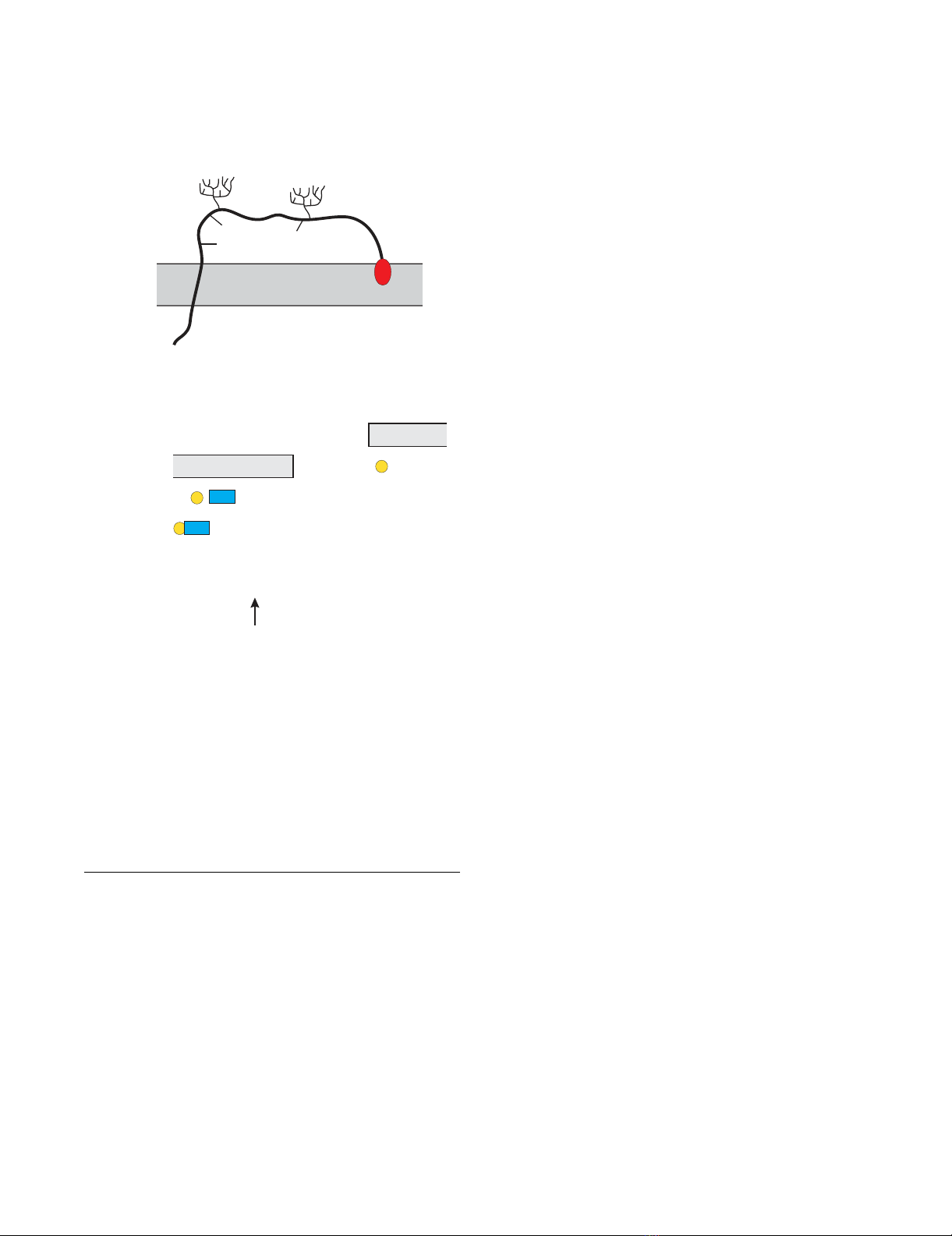
(GPI) anchor (Fig. 1) [31]. BST-2 protein associates with
lipid rafts at the cell surface and on internal membranes,
presumably the TGN [31]. Also, BST-2 forms stable
cysteine-linked dimers [29] and is modified by N-linked
glycosylation [29,31]. However, the precise function of
these BST-2 modifications remains unknown. N-linked
glycosylation was dispensable for inhibition of Lassa and
Marburg virus release, but the significance of BST-2 glyco-
sylation has not been examined in relation to HIV-1 [32].
Recent data suggest that the BST-2 TM domain is critical
for interference by Vpu [33-35]. consistent with previous
observations of the importance of the Vpu TM domain for
the regulation of virus release [12,13,36]. Furthermore,
antagonism of BST-2 was reported to involve intracellular
reduction of BST-2 levels by Vpu [37-40]. and was shown
to encompass a β-TrCP-dependent endo-lysosomal path-
way [38].
Here we analyzed the functional importance of various
structural properties of BST-2. We show that both pre-
dicted N-linked glycosylation sites are utilized in the
human protein. Interestingly, while endogenous BST-2 in
HeLa cells and other cell types contained almost exclu-
sively complex carbohydrate modifications, a large pro-
portion of transiently expressed BST-2 was modified by
high-mannose carbohydrates, a modification common to
endoplasmic reticulum (ER) resident glycoproteins.
Intriguingly, mutation of both glycosylation sites did not
inhibit cell surface expression of BST-2 and neither abol-
ished sensitivity to Vpu nor eliminated BST-2's inhibitory
effect on HIV-1 particle release. Thus, carbohydrate mod-
ification of BST-2 does not appear to have any functional
significance as far as HIV-1 virus release is concerned. In
contrast, the formation of cysteine linked dimers of BST-2
appeared to be functionally important. We confirmed that
BST-2 forms cysteine-linked dimers involving three
cysteine residues in the extracellular domain. Mutation of
Retrovirology 2009, 6:80 http://www.retrovirology.com/content/6/1/80
Page 3 of 16
(page number not for citation purposes)
individual cysteine residues or of any two of the three
cysteine residues in combination failed to affect BST-2
dimerization and had no effect on BST-2's inhibition of
virus release. In contrast, BST-2 mutated in all three
cysteine residues was unable to inhibit HIV-1 virus
release. Interestingly, this mutant was still expressed at the
cell surface and remained sensitive to Vpu. The inability of
the triple cysteine mutant to inhibit virus release was
therefore not due to gross mislocalization or misfolding
of the protein. Our results suggest that the formation of
cysteine-linked dimers is a critical requirement for the
inhibition of virus release by BST-2.
Methods
Plasmids
The full length infectious HIV-1 molecular clone pNL4-3
and the Vpu deletion mutant pNL4-3/Udel have been
described [5,41] For transient expression of Vpu, the
codon-optimized vector pcDNA-Vphu [42] was
employed. Plasmid pcDNA-BST-2 is a vector for the
expression of human BST-2 under the control of the
cytomegalovirus immediate-early promoter. BST-2 was
amplified by RT-PCR from HeLa mRNA using the primers
5' ATAAC TCGAG GTGGA ATTCA TGGCA TCTAC TTCGT
ATGAC TATTGC and 3' AAGCT TGGTA CCTCA CTGCA
GCAGA GCGCT GAGGC CCAGC AGCAC. The resulting
PCR product was cleaved with XhoI and KpnI and cloned
into the XhoI/KpnI sites of pcDNA3.1(-) (Invitrogen
Corp., Carlsbad CA). Mutation of cysteine residues C53,
C63, and C91 in human BST-2, either alone or in combi-
nation, to alanine was accomplished by PCR-based muta-
genesis of pcDNA-BST-2 and resulted in pcDNA-BST-2
C53A, pcDNA-BST-2 C63A, pcDNA-BST-2 C91A, pcDNA-
BST-2 C12 (C53,63A), pcDNA-BST-2 C13 (C53,91A),
pcDNA-BST-2 C23 (C63,91A), and pcDNA-BST-2 C3A
(C53,63,91A). Mutation of two potential N-linked glyco-
sylation sites was similarly accomplished by PCR-based
mutagenesis and resulted in the change of asparagine res-
idues N65 and N92 to glutamine in human BST-2 either
alone or in combination. PCR products were cloned into
pcDNA-BST-2 to obtain pcDNA-BST-2 N1 (N65Q),
pcDNA-BST-2 N2 (N92Q), and pcDNA-BST-2 N1/N2
(N65,92Q). The presence of the desired mutations and
the absence of additional mutations were verified for each
construct by sequence analysis.
Antisera
Anti-BST-2 antiserum was elicited in rabbits by using a
bacterially expressed MS2-BST-2 fusion protein composed
of amino acids 1 to 91 of the MS2 replicase and amino
acids 41 to 162 of BST-2 generating a polyclonal antibody
against the extracellular portion of BST-2. Polyclonal anti-
Vpu serum (rabbit), directed against the hydrophilic C-
terminal cytoplasmic domain of Vpu expressed in
Escherichia coli [43] was used for detection of Vpu. Serum
from an HIV-positive patient was used to detect HIV-1-
specific capsid (CA) and Pr55gag precursor proteins.
Tubulin was identified using a monoclonal antibody to α-
tubulin (Sigma-Aldrich, Inc., St. Louis MO).
Tissue culture and transfections
HeLa and 293T cells were propagated in Dulbecco's mod-
ified Eagles medium (DMEM) containing 10% fetal
bovine serum (FBS). For transfection, cells were grown in
25 cm2 flasks to about 80% confluency. Cells were trans-
fected using LipofectAMINE PLUS™ (Invitrogen Corp,
Carlsbad CA) following the manufacturer's recommenda-
tions. A total of 5 μg of plasmid DNA per 25 cm2 flask was
Schematic of the BST-2 structureFigure 1
Schematic of the BST-2 structure. (A) BST-2 is a type 2
integral membrane protein. The N-terminus localizes to the
cytoplasm. The BST-2 ectodomain contains three cysteine
residues (C53, C63, C91) and two potential sites for N-
linked glycosylation (N65, N92). The C-terminus of BST-2 is
modified by the addition of a glycosyl-phosphatidylinositol
(gpi) anchor. (B) Predicted amino acid sequence of human
BST-2. The predicted transmembrane region is indicated by a
box. Signals for N-linked glycosylation are marked in blue;
cysteine residues in the BST-2 extracellular domain are high-
lighted in yellow. The arrow points to the predicted site of
cleavage for the addition of the gpi anchor [31].
1 mastsydycr vpmedgdkrc klllgigilv
31 lliivilgvp liiftikans eacrdglrav
61 mecrnvthll qqelteaqkg fqdveaqaat
91 cnhtvmalma sldaekaqgq kkveelegei
121 ttlnhklqda saeverlrre nqvlsvriad
151 kkyypssqds ssaaapqlli vllglsallq
N1
N2
A
B
N65 (N1) N92 (N2)
C53
C63
C91
gpi
NH2

Retrovirology 2009, 6:80 http://www.retrovirology.com/content/6/1/80
Page 4 of 16
(page number not for citation purposes)
used. Total amounts of transfected DNA was kept con-
stant in all samples of any given experiment by adding
empty vector DNA as appropriate. Cells were harvested 24
h post transfection.
Immunoblotting
For immunoblot analysis of intracellular proteins, whole
cell lysates were prepared as follows: Cells were washed
once with PBS, suspended in PBS (400 μl/107 cells), and
mixed with an equal volume of sample buffer (4%
sodium dodecyl sulfate, 125 mM Tris-HCl, pH 6.8, 10%
2-mercaptoethanol, 10% glycerol, and 0.002%
bromophenol blue). For analysis of cysteine mutants
under non-reducing conditions, cells were suspended in
PBS and mixed with an equal volume of sample buffer
that did not contain 2-mercaptoethanol. Proteins were
solubilized by boiling for 10 to 15 min at 95°C with occa-
sional vortexing of the samples to shear cellular DNA.
Residual insoluble material was removed by centrifuga-
tion (2 min, 15,000 rpm in an Eppendorf Minifuge). Cell
lysates were subjected to SDS-PAGE; proteins were trans-
ferred to PVDF membranes and reacted with appropriate
antibodies as described in the text. Membranes were then
incubated with horseradish peroxidase-conjugated sec-
ondary antibodies (Amersham Biosciences, Piscataway
NJ) and visualized by enhanced chemiluminescence
(ECL, Amersham Biosciences).
Metabolic labeling and immunoprecipitations
Cells were transfected as described in the text with con-
stant amounts of proviral vectors and increasing amounts
of BST-2. Twenty-four hours later, cells were washed with
PBS, scraped and resuspended in 3 ml labeling media
lacking methionine (Millipore Corp., Billerica MA). Cells
were then incubated for 15 minutes at 37°C to deplete the
endogenous methionine pool. After starvation cells were
pelleted again, resuspended in 400 μl of labeling medium,
and 150 μCi of [35S]-methionine was added to each sam-
ple. Cells were labeled for 90 minutes at 37°C. Then, cells
were pelleted (20 sec, 10,000 × g). The virus-containing
supernatant was removed and filtered through 0.45 μm
cellulose acetate spin filters (Corning Costar Corp., Cam-
bridge MA). Virions were lysed in 0.1% Triton-X100, 0.1%
bovine serum albumin (BSA) in PBS. Cells were lysed in
200 μl of Triton lysis buffer (50 mM Tris pH 7.5, 150 mM
NaCl, 0.5% Triton-X100) and incubated on ice for 5 min-
utes. After lysis, the cells were pelleted at 13,000 × g for 2
minutes to remove insoluble material. The supernatants
and the virus lysates were incubated on a rotating wheel
for 1 hr at 4°C with protein A-Sepharose coupled with an
HIV-positive patient serum. Beads were washed twice with
wash buffer (50 mM Tris pH 7.4, 300 mM NaCl, 0.1% Tri-
ton X-100). Bound proteins were eluted by heating in
sample buffer for 10 min at 95°C, separated by SDS-
PAGE, and visualized by fluorography.
Concanavalin A (ConA) and datura stramonium lectin (DS
lectin) binding assays
For glycoprotein analysis of BST-2, cell lysates were pre-
pared as follows: Cells were washed once with PBS and
lysed in 300 μl of lysis buffer (50 mM Tris pH 8.0, 100
mM NaCl, 5 mM ethylenediaminetetraacetic acid, 0.5%
CHAPS) and 40 μl of DOC (2% deoxycholate in lysis
buffer). The cell extracts were clarified at 13,000 × g for 2
min and the supernatant was incubated on a rotating
wheel for 1-3 h at 4°C with ConA or DS lectin resin (Vec-
tor Laboratories, Burlingame CA) in 0.1% BSA-PBS. Com-
plexes were washed three times with 50 mM Tris, 300 mM
NaCl, and 0.1% Triton X100, pH 7.4. Bound proteins
were eluted from beads by heating in sample buffer for 5
- 10 min at 95°C and analyzed by immunoblotting.
Glycoprotein analysis
All digestions were performed directly on BST-2 bound to
either ConA or DS lectin resin. Control reactions con-
ducted in parallel did not contain enzyme. For endogly-
cosidase H (Endo H) and Peptide: N-Glycosidase F
analysis (PNGase) beads were washed with denaturing
buffer (New England BioLabs, Ipswich MA), then resus-
pended in denaturing buffer and boiled at 95°C for 10
min. The supplied reaction buffer was added along with
0.1% NP-40 according to the manufacturer's suggestion.
An excess of enzyme, 2500 units of Endo H (New England
BioLabs, Ipswich MA) or PNGase (New England BioLabs,
Ipswich MA), was added and samples were digested at
37°C for 3 h. Bound proteins were eluted from beads by
heating in an equal volume of sample buffer for 10 min at
95°C and analyzed by immunoblotting. For endo-β-
galactosidase (Endo B) analysis, beads were first washed
with 50 mM sodium acetate (pH 5.8), then resuspended
in the same buffer supplemented with BSA to 0.1%. 5 mU
of Endo B (Associates of Cape Cod, East Falmouth MA)
was added and digested at 37°C for 16 h along with a con-
trol lacking the enzyme.
FACS analysis
Cells were washed twice with ice-cold 20 mM EDTA-PBS,
followed by 2 washes in ice-cold 1% BSA-PBS. Cells were
treated for 10 min with 50 μg of mouse IgG (Millipore,
Temecula CA) to block non-specific binding sites. Cells
were incubated with primary antibody (α-BST-2) for 30
min at room temperature. Cells were then washed twice
with ice-cold 1% BSA-PBS followed by addition of allo-
phycocyanin (APC)-conjugated anti rabbit IgG secondary
antibody (Jackson Immuno Research Lab Inc., West Grove
PA) in 1% BSA-PBS. Incubation was for 30 min at room
temperature in the dark. Cells were then washed twice
with ice-cold 1% BSA-PBS and fixed with 1% paraformal-
dehyde in PBS. Finally, cells were analyzed on a FACS Cal-
ibur (BD Biosciences Immunocytometry Systems,
Mountain View CA). Data analysis was performed using

Retrovirology 2009, 6:80 http://www.retrovirology.com/content/6/1/80
Page 5 of 16
(page number not for citation purposes)
Flow Jo (Tree Star, San Carlos CA). For gating of trans-
fected cells, pEGFP-N1 (Clontech, Mountain View CA)
was cotransfected.
Results
Endogenous and exogenously expressed BST-2 have
distinct glycosylation profiles
The biochemical characterization of BST-2 necessitates
exogenous expression of the protein. For that purpose, the
BST-2 gene was PCR-amplified from HeLa cell mRNA and
cloned under the control of the cytomegalovirus immedi-
ate-early promoter as described in Methods. Ectopic
expression of BST-2 from pcDNA-BST-2 was analyzed by
immunoblot analysis of transiently transfected 293T cells
using a BST-2-specific antibody (Fig. 2A, lane 3). HeLa
cells expressing endogenous BST-2 (Fig. 2A, lane 1) and
mock-transfected 293T cells (Fig. 2A, lane 2) were ana-
lyzed in parallel. Endogenous BST-2 in HeLa cells
appeared as a smear of multiple bands with an apparent
Mr of 30-40 kDa, presumably due to N-linked glycosyla-
tion. Consistent with a previous report [24], untransfected
293T cells did not reveal BST-2 expression (Fig. 2A, lane
2). Of note, the bulk of transiently expressed BST-2 in
293T cells exhibited faster mobility in the gel than the
endogenous protein and had an apparent Mr of 28-29
kDa (Fig. 2A, lane 3, arrow). Only a minor fraction of the
exogenously expressed BST-2 protein exhibited an electro-
phoretic mobility comparable to that of the endogenous
protein in HeLa cells. The appearance of faster migrating
forms of exogenously expressed BST-2 is not cell type-spe-
cific and was observed in transiently transfected HeLa cells
as well (data not shown). Titrating transfected BST-2 DNA
to the limit of detection in 293T cells did not prevent the
appearance of the faster migrating forms of BST-2 (data
not shown). Thus, the appearance of faster migrating
forms of BST-2 in transiently transfected 293T cells are
presumably a result of transient expression rather than
protein overexpression per se.
Exogenously expressed BST-2 contains high-mannose as
well as complex carbohydrate modifications
It has been previously reported that rodent BST-2 is glyco-
sylated, however the type of glycosylation has not been
examined [29,31] Smeared protein patterns similar to
BST-2 were previously observed for proteins with complex
carbohydrate modifications [44] and it was likely that the
30-40 kDa and 28-29 kDa forms of BST-2 detected in
transfected 293T cells above represented various carbohy-
drate modifications. We therefore performed an endogly-
cosidase analysis of transiently expressed BST-2.
Enzymatic reactions were performed on BST-2 that was
previously enriched by adsorption to either Concanavalin
A (ConA) or datura stramonium lectin (DS lectin) resin.
ConA recognizes α-linked high-mannose oligosaccha-
rides, which are intermediates in N-linked glycosylation
and are typically found on proteins that have not yet
exited the endoplasmic reticulum (ER); DS lectin on the
other hand binds β1-4 linked N-acetylglucosamine or N-
acetyllactosamine repeats, which are characteristic of fully
processed oligosaccharides and produce the smear pattern
on protein gels noted above [44]. The latter modifications
are typically found on glycoproteins that have exited the
ER (for review see [45]). Peptide:N-Glycosidase F
(PNGase) cleaves glycoproteins between the innermost
GlcNAc and asparagine residues of all oligosaccharides
from N-linked glycoproteins [46]. Therefore all N-linked
oligosaccharides will be sensitive to PNGase treatment.
Endo-β-N-acetylglucosaminidase H (EndoH), on the
other hand, is more selective than PNGase and cleaves the
chitobiose core of high-mannose from N-linked glycopro-
teins [46]. Because of that, ER associated proteins are gen-
erally sensitive to EndoH treatment. Proteins exiting the
ER to the Golgi typically undergo additional sugar modi-
fications and, as a result, become EndoH resistant. A third
type of endoglycosidase, endo-β-galactosidase (EndoB),
cleaves glycoproteins after β-galactosidic linkages [47]
typically observed on glycoproteins that have exited the
ER. Accordingly, glycoproteins residing in the ER are gen-
erally EndoB resistant.
As expected, PNGase treatment removed all oligosaccha-
rides (Fig. 2B &2C, lane 3) resulting in deglycosylated pro-
teins with a Mr of 17-19 kDa and appeared as a doublet.
The reason why deglycosylated BST-2 runs as a doublet is
not clear but could be due to other modifications such as
phosphorylation or the presence and absence of the GPI
anchor. PNGase-treated BST-2 samples adsorbed to DS
lectin columns revealed an additional protein doublet of
38-40 kDa (Fig. 2B, lane 3). The precise nature of this pro-
tein species is unclear but its mobility in the gel is consist-
ent with that predicted for a dimer form of BST-2. BST-2
enriched by DS lectin columns was largely resistant to
EndoH treatment (Fig. 2B, compare lanes 1 & 2). EndoH
resistance indicated that the 30-40 kDa population of
BST-2 contained complex sugar modifications and there-
fore has likely exited the ER. This was confirmed by their
sensitivity to EndoB treatment (Fig. 2B, lane 5). In con-
trast, the 28-29 kDa population of BST-2 enriched by
ConA was highly sensitive to EndoH (Fig. 2C, lane 2) but
was resistant to EndoB treatment (Fig. 2C, lane 5). These
results suggest that the 28-29 kDa protein population
observed in transfected 293T cells represents a high-man-
nose form of BST-2. Therefore, exogenously expressed
BST-2 consists of two populations: a 30-40 kDa form con-
taining complex sugar modifications (referred to as "post-
ER form" for the remainder of the text) and a predomi-
nant 28-29 kDa population containing high-mannose oli-
gosaccharide modifications (referred to as "high-mannose
form" in the following). HeLa cells expressing endog-
enous BST-2 were not entirely devoid of the high-man-


![Hình ảnh học bệnh não mạch máu nhỏ: Báo cáo [Năm]](https://cdn.tailieu.vn/images/document/thumbnail/2024/20240705/sanhobien01/135x160/1985290001.jpg)


