
Open Access
Available online http://ccforum.com/content/11/6/R132
Page 1 of 9
(page number not for citation purposes)
Vol 11 No 6
Research
The relationship between gastric emptying, plasma
cholecystokinin, and peptide YY in critically ill patients
Nam Q Nguyen1,2, Robert J Fraser2,3, Laura K Bryant3, Marianne J Chapman4, Judith Wishart2,
Richard H Holloway1,2, Ross Butler5 and Michael Horowitz2
1Department of Gastroenterology and Hepatology, Royal Adelaide Hospital, North Terrace, Adelaide, South Australia, 5000
2Discipline of Medicine, University of Adelaide, Royal Adelaide Hospital, Adelaide, South Australia, 5000
3Investigation and Procedures Unit, Repatriation General Hospital, Daw Road, Adelaide, South Australia, 5000
4Department of Anaesthesia and Intensive Care, Royal Adelaide Hospital, Adelaide, South Australia, 5000
5Centre for Paediatric and Adolescent Gastroenterology, Children, Youth and Women's Health Service, Adelaide, South Australia, 5000
Corresponding author: Nam Q Nguyen, quoc.nguyen@health.sa.gov.au
Received: 10 Oct 2007 Revisions requested: 15 Nov 2007 Revisions received: 23 Nov 2007 Accepted: 21 Dec 2007 Published: 21 Dec 2007
Critical Care 2007, 11:R132 (doi:10.1186/cc6205)
This article is online at: http://ccforum.com/content/11/6/R132
© 2007 Nguyen et al.; licensee BioMed Central Ltd.
This is an open access article distributed under the terms of the Creative Commons Attribution License (http://creativecommons.org/licenses/by/2.0),
which permits unrestricted use, distribution, and reproduction in any medium, provided the original work is properly cited.
Abstract
Introduction Cholecystokinin (CCK) and peptide YY (PYY) are
released in response to intestinal nutrients and play an important
physiological role in regulation of gastric emptying (GE). Plasma
CCK and PYY concentrations are elevated in critically ill
patients, particularly in those with a history of feed intolerance.
This study aimed to evaluate the relationship between CCK and
PYY concentrations and GE in critical illness.
Methods GE of 100 mL of Ensure® meal (106 kcal, 21% fat)
was measured using a 13C-octanoate breath test in 39
mechanically ventilated, critically ill patients (24 males; 55.8 ±
2.7 years old). Breath samples for 13CO2 levels were collected
over the course of 4 hours, and the GE coefficient (GEC)
(normal = 3.2 to 3.8) was calculated. Measurements of plasma
CCK, PYY, and glucose concentrations were obtained
immediately before and at 60 and 120 minutes after
administration of Ensure.
Results GE was delayed in 64% (25/39) of the patients.
Baseline plasma CCK (8.5 ± 1.0 versus 6.1 ± 0.4 pmol/L; P =
0.045) and PYY (22.8 ± 2.2 versus 15.6 ± 1.3 pmol/L; P =
0.03) concentrations were higher in patients with delayed GE
and were inversely correlated with GEC (CCK: r = -0.33, P =
0.04, and PYY: r = -0.36, P = 0.02). After gastric Ensure, while
both plasma CCK (P = 0.03) and PYY (P = 0.02)
concentrations were higher in patients with delayed GE, there
was a direct relationship between the rise in plasma CCK (r =
0.40, P = 0.01) and PYY (r = 0.42, P < 0.01) from baseline at
60 minutes after the meal and the GEC.
Conclusion In critical illness, there is a complex interaction
between plasma CCK, PYY, and GE. Whilst plasma CCK and
PYY correlated moderately with impaired GE, the pathogenetic
role of these gut hormones in delayed GE requires further
evaluation with specific antagonists.
Introduction
In health, cholecystokinin (CCK) and peptide YY (PYY) are
important humoral mediators of nutrient-induced small intesti-
nal feedback, which regulates gastric emptying (GE) and
energy intake [1-5]. In response to the presence of nutrients
(particularly fat and protein) in the small intestine, CCK and
PYY are released in a load-dependent manner from enteroen-
docrine cells, predominantly in the proximal small intestine for
CCK and the distal small intestine for PYY [5-8]. CCK has also
been reported to mediate the initial postprandial release of
PYY [9,10]. In healthy humans, exogenous administration of
CCK and PYY is associated with relaxation of the proximal
stomach, inhibition of antral motor activity, stimulation of con-
tractions localised to the pylorus, slowing of GE
[1,2,4,7,11,12], and a reduction in energy intake [3,4,13-16].
CCK antagonists have been shown to increase GE and
energy intake in humans [17-19]. The effects of PYY antago-
nism on GE in humans, however, are unknown. Furthermore,
both plasma CCK and PYY concentrations are elevated in
patients with chronic nutrient deprivation, malnutrition, and
ANOVA = analysis of variance; APACHE = Acute Physiology and Chronic Health Evaluation; AUC0–120 min = area under curve over the course of 120
minutes; BMI = body mass index; CCK = cholecystokinin; GE = gastric emptying; GEC = gastric emptying coefficient; ICU = intensive care unit;
PYY = peptide YY.

Critical Care Vol 11 No 6 Nguyen et al.
Page 2 of 9
(page number not for citation purposes)
anorexia nervosa [20-22], conditions that are known to be
associated with a high prevalence of delayed GE [23,24].
Impaired gastric motor function and associated feed intoler-
ance occur in up to 50% of critically ill patients and can
adversely affect both morbidity and mortality [25,26]. Whilst
the mechanisms underlying delayed GE in critical illness
remain poorly defined, exaggerated inhibitory feedback on GE
arising from the interaction of nutrients with the small intestine
is likely to be important [27]. For example, in response to duo-
denal nutrient, there is a greater degree of antral hypo-motility,
pyloric hyperactivity [27], and exaggerated release of both
CCK and PYY in critically ill patients [28,29]. Furthermore, the
CCK and PYY responses are substantially greater in those
patients who have feed intolerance [28,29]. In the fasted state,
there is an increase in plasma concentrations of hormones that
slow GE, such as CCK and PYY, and a decrease in hormones
that may accelerate GE, such as ghrelin [28-30]. The effects
of exogenous CCK and PYY on gastric motility are also com-
parable to the motor disturbances in both the proximal and dis-
tal stomach observed in critically ill patients [27,31,32].
Whereas the above evidence supports a potential role for both
CCK and PYY in the mediation of enhanced nutrient-induced
enterogastric feedback during critical illness, the relationships
between plasma CCK and PYY concentrations and GE in crit-
ical illness have hitherto not been evaluated. This study was
designed to examine the following hypotheses: (a) slow GE is
associated with elevated plasma concentrations of CCK and
PYY, and (b) GE is a determinant of postprandial concentra-
tions of CCK and PYY in the critically ill.
Materials and methods
Subjects
Studies were performed prospectively in 39 unselected criti-
cally ill patients (24 males; 55.8 ± 2.7 years old) who were
admitted to a level-3 intensive care unit (ICU) between May
2005 and November 2006. Any patient at least 17 years old
was eligible for inclusion if he or she was sedated, mechani-
cally ventilated, and able to receive enteral nutrition. Exclusion
criteria included any contraindication to passage of an enteral
tube; a history of gastric, oesophageal, or intestinal surgery;
recent major abdominal surgery; evidence of liver dysfunction;
administration of prokinetic therapy within 24 hours prior to the
study; and a history of diabetes mellitus. All patients were
receiving an insulin infusion according to a standard protocol,
which was designed to maintain the blood glucose concentra-
tion between 5.0 and 7.9 mmol/L [27-29,31]. Written
informed consent was obtained from the next of kin for all
patients prior to enrolment into the study. The study was
approved by the Human Research Ethics Committee of the
Royal Adelaide Hospital and performed according to the
National Health and Medical Research Council guidelines for
the conduct of research on unconscious patients.
Study protocol and techniques
Critically ill patients were studied in the morning, after a mini-
mum 8-hour fast. All patients were sedated, with either propo-
fol or a combination of morphine and midazolam, throughout a
minimum of 24 hours prior to the study. The type of sedation
was determined by the intensivist in charge of the patient and
did not influence patient selection. In all patients, a 14- to 16-
French gauge Levin nasogastric feeding tube (Pharma-Plast,
Lynge, Denmark) was already in situ in the stomach, as part of
clinical care, and the correct position of the feeding tube was
confirmed radiologically prior to commencing the study.
GE was measured by a 13C-octanoate breath test, with the
patient in the supine position and the head of the bed elevated
to 30°. Gastric contents were initially aspirated and discarded,
and then 100 mL of liquid nutrient meal (Ensure™; Abbott Aus-
tralia, Kurnell, Australia) containing 106 kcal with 21% of fat
and labelled with 100 μL of13C-octanoate (100 mg/mL; Cam-
bridge Isotope Laboratories, Inc., Andover, MA, USA) was
infused slowly over the course of 5 minutes into the stomach
via the nasogastric tube. End-expiratory breath samples were
obtained from the ventilation tube using a T-adapter (Datex-
Engström, now part of GE Healthcare, Little Chalfont, Buck-
inghamshire, UK) and holder for vacutainers (blood needle
holder; Reko Pty Ltd, Lisarow, Australia) containing a needle
(VenoJect®; Terumo Corporation, Tokyo, Japan). Samples
were collected at baseline, every 5 minutes for the first hour,
and every 15 minutes thereafter, for a subsequent 3 hours
after meal administration [33]. Time (t) = 0 minutes was
defined as the time when all of the Ensure had been infused
into the stomach. To avoid sampling other than end-expiratory
air, sampling was timed to the end-expiratory phase by obser-
vation of the patient and the time-flow curve on the ventilation
monitor.
Blood samples (5 mL) for the measurement of plasma CCK
and PYY were collected into chilled EDTA (ethylenediamine-
tetraacetic acid) tubes immediately before and at 60 and 120
minutes after the delivery of the intragastric meal. Blood sam-
ples were centrifuged at 4°C within 30 minutes of collection
and stored at -70°C for subsequent analysis. Blood samples
for the measurement of blood glucose were also collected at
baseline, every 15 minutes for the first hour, and every 30 min-
utes for the subsequent 3 hours.
Measurements
Gastric emptying
GE was assessed indirectly by using 13C-octanoate breath
tests. This non-invasive technique has been validated against
gastric scintigraphy, using both solid and liquid meals, in
healthy subjects and non-critically ill patients [34-39]. In criti-
cally ill patients, the breath test has a sensitivity of 71% and a
specificity of 100% in detecting delayed GE, with a modest
correlation between gastric half-emptying time determined by
breath test and scintigraphy [40].

Available online http://ccforum.com/content/11/6/R132
Page 3 of 9
(page number not for citation purposes)
The concentration of CO2 and the percentage of 13CO2 were
measured in each sample by means of an isotope ratio mass
spectrometer (ABCA model 20/20; Europa Scientific, Crewe,
UK). Samples containing less than 1% CO2 were regarded as
being non-end-expiratory and were excluded from further anal-
ysis. The 13CO2 concentration over time was plotted, and the
resultant curves were used to calculate a GE coefficient
(GEC) [41], using non-linear regression formulae: GEC =
ln(y)) and y = atbe -et, where y is the percentage of 13CO2
excretion in breaths per hour, t is time in hours, and a, b, and c
are regression estimated constants [36,38,42]. GEC is a glo-
bal index for the GE rate, and the normal range for normal GE
has been established previously in a group of 28 healthy vol-
unteers (normal GEC = 3.2 and 3.8) [33].
Plasma cholecystokinin, peptide YY, and blood glucose
Plasma CCK concentrations were measured by radioimmu-
noassay using an adaptation of the method of Santangelo and
colleagues [43]. A commercially available antibody (C2581,
lot 105H4852; Sigma-Aldrich, St. Louis, MO, USA) raised in
rabbits against synthetic sulphated CCK-8 was used. This
antibody binds to all CCK peptides containing the sulphated
tyrosine residue in position 7 and has 26% cross-reactivity
with un-sulphated CCK-8, less than 2% cross-reactivity with
human gastrin 1, and no cross-reactivity with structurally unre-
lated peptides. Antibody was added at a dilution of 1:17,500,
and iodine-125-labeled sulphated CCK-8 with Bolton-Hunter
reagent (74 TBq/mmol; Amersham International, now part of
GE Healthcare) was used as a tracer. Incubation proceeded
for 7 days at 4°C. The antibody-bound fraction was separated
by the addition of dextran-coated charcoal containing gelatin
(0.015 g gelatin, 0.09 g dextran, and 0.15 g charcoal in 30 mL
of assay buffer). The detection limit was 1 pmol/L, and the
intra-assay coefficient of variation at 50 pmol/L was 9.5%.
Plasma PYY concentrations were measured by radioimmu-
noassay using an antiserum raised in rabbits against human
PYY (1–36) (Sigma-Aldrich) [43]. This antiserum showed less
than 0.001% cross-reactivity with human pancreatic polypep-
tide and sulphated CCK-8 and 0.0025% cross-reactivity with
human neuropeptide Y. Tracer (Prosearch International, Mal-
vern, Australia) was prepared by radio-labeling synthetic
human PYY (1–36) (Auspep Pty Ltd, Parkville, Australia) using
the lactoperoxidase method. Mono-iodo-tyrosine-PYY was
separated from free iodine-125, diiodo-PYY, and unlabeled
PYY by reverse-phase high-performance liquid chromatogra-
phy (Phenomenex Jupiter C4 300A 5u column catalogue
number 00B-4167-EO 250 _ 4.6 mm; Phenomenex, Inc., Tor-
rance, CA, USA). Standards (1.6 to 50 fmol/tube) or samples
(200 μL of plasma) were incubated in assay buffer with 100
μL of antiserum at a final dilution of 1:10,000 for 20 to 24
hours at 4°C, 100 μL of iodinated PYY (10,000 cpm) was then
added, and the incubation continued for another 20 to 24
hours. Separation of the antibody-bound tracer from free
tracer was achieved by the addition of 200 μL of dextran-
coated charcoal containing gelatin (0.015 g of gelatin, 0.09 g
of dextran, and 0.15 g of charcoal per 30 mL of assay buffer)
and the mixture was incubated at 4°C for 20 minutes and then
centrifuged at 4°C for 25 minutes. Radioactivity of the bound
fraction was determined by counting the supernatants in a
gamma counter. The intra- and inter-assay coefficients of vari-
ation were 12.3% and 16.6%, respectively. The minimum
detectable concentration was 4 pmol/L [43]. Blood glucose
concentrations were measured by means of a portable gluco-
meter (Precision Plus; Abbott Laboratories, Abbott Park, IL,
USA).
Statistical analysis
Data are presented as mean ± standard error of the mean. The
integrated changes in plasma concentrations of CCK and PYY
were calculated and expressed as areas under the curve over
the 120 minutes (AUC0–120 min) after the Ensure meal. Differ-
ences in demographic characteristics, in baseline blood glu-
cose, CCK, and PYY concentrations, and in AUC0–120 min for
plasma CCK and PYY between critically ill groups were com-
pared using the Student unpaired t test and the chi-square
test. Changes in plasma concentrations of CCK and PYY over
time were determined by one-way repeated measures analysis
of variance (ANOVA). Potential differences between patients
with normal versus delayed GE with respect to the plasma
CCK, PYY, and blood glucose responses to the meal were
evaluated using two-way ANOVA with post hoc analyses. The
relationships between GE with baseline plasma CCK and
PYY, changes in plasma CCK and PYY (from baseline to t =
60 minutes and t = 120 minutes), and demographic factors
(age, body mass index [BMI], Acute Physiology and Chronic
Health Evaluation [APACHE] II score [41], and serum creati-
nine) were assessed using the Pearson correlation. Signifi-
cance was accepted at a P value of less than 0.05.
Results
The duration of ICU stay prior to the study was 4.60 ± 0.34
days. The admission diagnoses included multi-trauma (n =
12), head injury (n = 12), sepsis (n = 11), respiratory failure (n
= 9), cardiac failure (n = 3), aortic dissection (n = 3), pancre-
atitis (n = 1), and retroperitoneal bleed (n = 1). The mean
APACHE II score on the study day was 22.4 ± 0.9. Twenty-
five patients (64%) were sedated with morphine and mida-
zolam, and 14 patients (36%) with propofol. Nineteen patients
(48%) required inotropic support with either adrenaline or
noradrenalin. Acid suppression therapy (ranitidine or panto-
prazole) was given to 32 (82%) of the 39 patients. Renal func-
tion was normal in the majority of patients (82%; 32/39) at the
time of study, with a serum creatinine of 0.10 ± 0.01 mmol/L.
None of the 7 patients with renal impairment (mean serum cre-
atinine = 0.23 ± 0.04 mmol/L) required haemodialysis. Before
enrolment into the study, 24 (66%) patients had received
enteral feeds for a mean duration of 3.52 ± 0.36 days, and 15
(34%) patients had not received any nutritional support prior
to the study. Ten patients (42%) who received prior enteral

Critical Care Vol 11 No 6 Nguyen et al.
Page 4 of 9
(page number not for citation purposes)
nutrition had feed intolerance, defined as aspirates of greater
than 250 mL during gastric enteral feeding [44]. The mean
duration of ICU stay prior to the study did not differ between
the two groups (fed: 4.9 ± 0.5 days versus not fed: 4.2 ± 0.4
days; P = 0.78).
Gastric emptying
GE was delayed in 64% (25/39) of the patients, with a mean
GEC of 2.8 ± 0.1. The demographic data and characteristics
of patients who had normal and delayed GE are summarised
in Table 1. There was no relationship between the GEC and
age (P = 0.23), gender (P = 0.82), BMI (P = 0.86), APACHE
II score at time of study (P = 0.68), type of sedation, use of ino-
tropes or acid suppression, presence of sepsis, or prior enteral
feeding. The mean fasting blood glucose concentration was
7.14 ± 0.24 mmol/L, which increased slightly after the meal to
a peak of 8.13 ± 0.28 mmol/L (P < 0.01). There were no dif-
ferences in either fasting or postprandial blood glucose con-
centrations between patients with delayed and normal GE (P
> 0.05).
Table 1
Demographic data and characteristics of critically ill patients, classified according to their rate of gastric emptying
Normal GE (n = 14) Delayed GE (n = 25) P value
Age, years 57.5 ± 3.8 56.3 ± 2.8 0.87
Gender, male/female 7/7 17/8 0.41
Body mass index, kg/m228.3 ± 1.3 27.7 ± 1.2 0.78
APACHE II score on study day 22.6 ± 1.1 22.1 ± 1.0 0.86
Serum creatinine, mmol/L 0.08 ± 0.01 0.11 ± 0.02 0.14
Baseline blood glucose, mmol/L 7.1 ± 0.2 7.1 ± 0.2 0.99
Admission diagnosisa, percentage (number)
Sepsis 36% (5) 19% (5) 0.28
Respiratory failure 43% (6) 15% (4) 0.13
Multi-trauma 21% (3) 32% (8) 0.48
Head injuryb21% (3) 34% (9) 0.48
Aortic dissection 7% (1) 8% (2) 0.99
Pancreatitis 0% (0) 4% (1) 0.99
Retroperitoneal bleed 7% (1) 0% (0) 0.35
Medication, percentage (number)
Morphine ± midazolam 57% (8) 68% (17) 0.44
Propofol 43% (6) 31% (8) 0.44
Inotropes (adrenaline/noradrenalin) 57% (8) 46% (12) 0.51
Plasma CCK concentration, pmol/L
Fasting 6.1 ± 0.4 8.5 ± 1.0 0.045
Postprandial
At 60 minutes 8.2 ± 0.7 10.1 ± 0.8 0.03
At 120 minutes 7.1 ± 0.7 9.8 ± 0.8 0.03
Plasma PYY concentration, pmol/L
Fasting 15.6 ± 1.3 22.8 ± 2.2 0.03
Postprandial
At 60 minutes 21.0 ± 1.8 25.0 ± 2.2 0.02
At 120 minutes 18.9 ± 1.9 24.9 ± 2.0 0.02
Data are mean ± standard error of the mean. aOne patient may have one or more admission diagnoses. bIncluding sub-arachnoid haemorrhage
and massive cerebral ischemic event. APACHE II, Acute Physiology and Chronic Health Evaluation II; CCK, cholecystokinin; GE, gastric
emptying; PYY, peptide YY.
Available online http://ccforum.com/content/11/6/R132
Page 5 of 9
(page number not for citation purposes)
Plasma cholecystokinin and peptide YY concentrations
Baseline plasma CCK concentration was 7.74 ± 0.87 pmol/L
and PYY was 20.4 ± 2.0 pmol/L. Baseline plasma PYY, but
not CCK, was positively related to age (r = 0.37; P = 0.01)
and BMI (r = 0.50; P < 0.01). Baseline plasma concentrations
of both CCK and PYY were not related to gender (P = 0.82),
the APACHE II score on the study day (P = 0.40), serum cre-
atinine (P = 0.28), the type of sedation, the use of inotropes or
acid suppression, the presence of sepsis, or prior enteral nutri-
tion. There was no relationship between baseline plasma CCK
and PYY (P = 0.80).
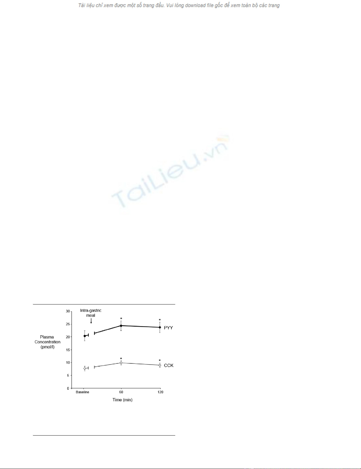
In response to the gastric meal, there was a small but signifi-
cant rise in plasma CCK and PYY (P = 0.01) (Figure 1). The
integrated changes in plasma CCK (r = 0.45; P < 0.001), but
not PYY, from baseline to 120 minutes were positively corre-
lated with age. There was no relationship between integrated
plasma CCK or PYY with gender, BMI, APACHE II scores on
study day, serum creatinine, the type of sedation, the use of
inotropes and acid suppression, presence of sepsis, or prior
history of receiving enteral nutrition. Both plasma CCK and
PYY remained above baseline at 120 minutes (Figure 1), par-
ticularly in patients with delayed GE (P < 0.05) (Table 1).
There was a positive correlation between the magnitude of the
increase in plasma PYY and CCK concentrations at 60 min-
utes (r = 0.33; P = 0.03).
Relationship between gastric emptying, plasma
cholecystokinin, and peptide YY
Baseline plasma CCK (8.5 ± 1.0 versus 6.1 ± 0.4 pmol/L; P
= 0.045) and PYY (22.8 ± 2.2 versus 15.6 ± 1.3 pmol/L; P =
0.03) concentrations were higher in patients with delayed GE
compared with those with normal GE. The GEC was inversely
related to both baseline plasma CCK (r = -0.33; P = 0.04) and
PYY (r = -0.36; P = 0.02) (Figure 2). Similarly, plasma CCK (P
= 0.03) and PYY (P = 0.02) concentrations were higher at 60
and 120 minutes in patients with delayed GE. The GEC was
inversely related to plasma CCK (r = -0.32; P = 0.049) and
PYY (r = -0.30; P = 0.06) at 120 minutes, but not at 60 min-
utes. The absolute changes in plasma CCK (r = 0.40; P =
0.01) and PYY (r = 0.42; P < 0.01) at 60 minutes, as well as
the integrated changes in plasma CCK (r = 0.36; P = 0.03)
and PYY over 120 minutes (r = 0.38; P = 0.02), were directly
related to the GEC (Figure 3). The integrated changes in
plasma CCK and PYY, however, were not significantly differ-
ent in patients with delayed versus normal GE (CCK: AUC 0–
120 min: 130 ± 42 versus 160 ± 38 pmol/L-minutes, P = 0.61;
PYY: AUC 0–120 min: 174 ± 98 versus 414 ± 155 pmol/L-min-
utes, P = 0.16).
Discussion
Whilst we have shown previously that plasma CCK and PYY
levels are increased in critically ill patients [28-30] and that
CCK and PYY are known to slow GE, the present study is the
first to directly demonstrate a relationship between GE and
plasma concentrations of CCK and PYY in critical illness. The
major observations are that, during critical illness, (a) GE was
inversely related to both fasting and postprandial plasma CCK
and PYY concentrations but (b) the postprandial increases in
plasma CCK and PYY were also directly related to GE.
Together with previous studies that have shown that entero-
gastric hormones [28-30] and feedback responses [27] to
small intestinal nutrients are exaggerated in the critically ill, the
relationship between enterogastric hormones and GE in the
present study supports a putative pathogenesis role of enter-
ogastric hormones in disordered GE during critical illness.
However, the weakness of the relationship in these patients
when compared with that previously reported in healthy sub-
jects [1,2,4,7,11,12] highlights the complexity of the regula-
tory mechanisms and further suggests that other factors such
as admission diagnosis and medication have a role in disor-
dered GE.
The substantially higher fasting plasma CCK and PYY concen-
trations in our critically ill patients with delayed GE are consist-
ent with our previous reports on critically ill patients with feed
intolerance [28-30]. The observation that the rate of GE is
inversely related to the fasting levels of CCK and PYY sug-
gests that they may contribute to the regulation of GE in criti-
cally ill patients. Although the strength of the correlation was
only modest, the relationship should not be regarded as weak,
as this was a cross-sectional study. The mechanisms
underlying the elevated fasting levels of these hormones are
unknown. Nutritional deprivation is likely to be relevant since
inadequate nutritional support is common in critically ill
patients, fasting slows GE even in healthy subjects, and fast-
ing CCK and PYY concentrations are higher in patients with
anorexia nervosa and malnutrition [21,22]. The lack of differ-
ences in fasting hormonal concentrations between patients
with and without nutritional support in the present study sug-
Figure 1
Plasma cholecystokinin (CCK) and peptide YY (PYY) concentrations at baseline and after intragastric Ensure (100 mL, 106 kcal with 21% lipid) in 39 critically ill patients (mean ± standard error of the mean)Plasma cholecystokinin (CCK) and peptide YY (PYY) concentrations at
baseline and after intragastric Ensure (100 mL, 106 kcal with 21%
lipid) in 39 critically ill patients (mean ± standard error of the mean). *P
< 0.05 versus baseline.


