


Summary of Chemical doctoral thesis: Chemical constituents and biological activities of two starfish Anthenea Sibogae and Anthenea Aspera collected from Vietnamese coast
lượt xem 3
download
 Download
Vui lòng tải xuống để xem tài liệu đầy đủ
Download
Vui lòng tải xuống để xem tài liệu đầy đủ
Summary of Chemical doctoral thesis: Chemical constituents and biological activities of two starfish Anthenea Sibogae and Anthenea Aspera collected from Vietnamese coast
Bình luận(0) Đăng nhập để gửi bình luận!
Nội dung Text: Summary of Chemical doctoral thesis: Chemical constituents and biological activities of two starfish Anthenea Sibogae and Anthenea Aspera collected from Vietnamese coast
- MINISTRY OF EDUCATION VIETNAM ACADEMY OF SCIENCE AND TRANING AND TECHNOLOGY GRADUATE UNIVERSITY OF SCIENCES AND TECHNOLOGY ------------------------- NGUYEN ANH HUNG CHEMICAL CONSTITUENTS AND BIOLOGICAL ACTIVITIES OF TWO STARFISH ANTHENEA SIBOGAE AND ANTHENEA ASPERA COLLECTED FROM VIETNAMESE COAST Major: Natural Products Chemistry Code: 9 44 01 17 SUMMARY OF CHEMICAL DOCTORAL THESIS HA NOI - 2020
- This dissertation was completed in: Graduate university of Sciences and Technology – Vietnam Academy of Science and Technology. Supervisor 1: Assoc. Prof. Dr. Tran Thi Thu Thuy Supervisor 2: Dr.Sc. Alla Anatolievna Kicha Reviewer 1: Reviewer 2: Reviewer 3: The thesis will be defended in front of the Academy Thesis Evaluation Council at the: Graduate university of Sciences and Technology - Vietnam Academy of Science and Technology. At the time..… hour…., ………, 2020 Thesis can be found at the: - Library of Graduate university of Sciences and Technology - Vietnam National Library
- PREAMBLE 1. The necessary of the thesis Vietnam is endowed with more than 1 million km2 of sea area, tropical monsoon climate, densely estuarine which are ideal conditions for a diverse and rich marine organison. Since the 1970s there have been a few studies on natural compounds from marine organison. However, comparing to the potential source of marine life in our country, up to now, the research in this field is still too few and scattered, especially the studies in echinoderms. Starfish are invertebrates, belonging to echinoderms and have long been known as a nutritious source. About 1800 species of starfish occur in all the world’s oceans, from tropics to frigid polar water. However, only about 80 species have been studied for their chemical composition and biological activity. The Oreasterdae family includes 20 genera: Acheronaster, Anthaster, Anthenea, Astrosarkus, Bothriaster, Choriaster, Culcita, Goniodiscaster…. Currently, there are only 9 species of the Oreasterdae family which were studied in the world, of which 2 species have been studied in Vietnam, namely Anthenea chinensis and Culcita novaeguineae. The results of those studies showed that substances isolated from the starfish of the Oreasterdae family have anti-inflammatory, analgesic, hypotensive, cytotoxic, antibacterial and antifungal actioity. In the aim to find active ingredients for use in medicine and pharmacy from Vietnamese medicinal remedies, we have selected 2 starfish species of the genus Anthenea. "Chemical constituents and biological activities of two starfish Anthenea sibogae and Anthenea aspera collected from Vietnamese coast”. 2. Research objectives of the thesis - Chemical investigation of two starfish Anthenea sibogae and Anthenea aspera. - Biological evaluation of isolated compounds from 2 starfish Anthenea sibogae and Anthenea aspera. 3. The main research content of the thesis 1
- To achieve the above objectives, the thesis has implemented the following contents: • Isolation of pure compounds from 2 starfish Anthenea aspera and Anthenea sibogae collected from Vietnam’s coast. • Structural elucidation of the chemical structure of isolated compounds. • Biological evaluation of isolated compounds: anticancer activities. CHAPTER 1. INTRODUCTION The literature review is a collection of national and international researchs on: 1.1. Introduction about starfish 1.2. Chemical constituents of the Oreasteridae starfish 1.3. Biological activity of starfish CHAPTER 2. MATERIALS AND METHODS 2.1. Materials Starfish samples were collected at Van Boi island, Quang Ninh province in June 2012 and identified by Assoc. Do Cong Thung, Institute of Marine Resources and Environment, Vietnam Academy of Science and Technology. The voucher specimens are deposited at Anthenea sibogae: DG02-BTL, Anthenea aspera: SBĐ 12 2.2. Methods for chemical and biological studies 2.2.1. Isolation methods The chromatographic methods are used to isolate pure from starfish compounds including: thin - layer chromatography (TLC), normal or reverse - phase silica gel column chromagraphy (RP - C18), Polychrome1, High Performance Liquid Chromatography (HPLC). 2.2.2. Structural determination The structures of isolated compounds are determined by the combination of physical parameters with modern spectroscopic methods such as melting point (Mp), optical rotation ([α]D), Mass Gas Chromatography Spectroscopy (GC-MS), Electrospray Ionization Mass Spectrometry (ESI-MS), High Resolution Electrospray Ionization Mass spectrometry (HR-ESI-MS), 2
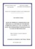
- Nuclear Magnetic Resonance Spectroscopy including one-dimensional spectrum (1H, 13C and DEPT, TOCSY 1D) and two-dimensional spectrum (COSY, HSQC, HMBC and NOESY) recorded on Bruker Avance III 500 MHz or Bruker Avance III 700 MHz using TMS as internal standard. 2.2.3. Biological evaluation methods 2.2.3.1. Cytotoxicity assay The cytotoxic activity was tested by MTS method on human breast cancer cell line T-47D. The tests were conducted at the Pacific Institute of Organic Biochemistry, Russian Federal Academy of Sciences, Vladivostok. 2.2.3.2. Cell proliferation assay The tested substances were added to the cell culture medium at concentrations (50 μM) then incubated for additional 24, 48, and 72 h at 37 0C in a 5% CO2 incubator. The tumors formed were measured in using absorbance at 490/630 nm using Fedorov method. 2.2.3.3. Soft agar clonogenic assay The tested substances were added to the cell culture medium at a concentration of 50 μM. The culture was maintained at 37 0C in 5% CO2 incubator for 2 weeks and the cells colonies visualized scored microscope and the ImageJ computer software. CHAPTER 3. EXPERIMENTAL PART 3.1. Extraction and isolation procedure for starfish Anthenea sibogae This section details how to isolate compounds from A. sibogae. The separation of compounds was summarized in the diagram in Figure 3.1. 3.1.1. Sample treatment of starfish A. sibogae 3
- Fresh A. sibogae (2 Kg) H/E 40/1, 5/1 Extraction four times MeOH (t = 45 0C) ASB1 (7 mg) Crude MeOH ASB2 (5 mg) Crude CH2Cl2 CH2Cl2 (3x500 ml) E/Me 30/1, 10/1 ASB3 (6 mg) Residue 80 g ASB4 (7 mg) Dissolved in H2O (1.0 L). Elution AgNO3; EtOH EtOH Fraction 10,5 g CH2Cl2/MeOH (9/11:5) F1…. F8 MeOH/H2O (15/15:1) F6 F6.1, F6.2, F6.5 CHCl3/EtOH (3:1Ò2:1) F6.3 (87 mg) F6.4 (25 mg) HPLC (Diasorb-130-C16T) F6.3.1 (6,9 mg) 75% EtOH,; 25 ml/min F6.3.2 (2,3 mg) F6.3.3 (3,2 mg), F6.3.4 (5,8 mg), HPLC (Diasfer-110-C18) F6.4.1 (13,5 mg), HPLC (Diasfer-110-C18) 80%MeOH; 0,5 ml/min 83%MeOH; 0,5 ml/min HPLC (Diasfer-110-C18) 85% MeOH: 0,5 ml/min ASB5 (2,4 mg; tR = 35 min) ASB7 (0,3 mg; tR = 31,7 min) ASB6 (2,1 mg; tR = 39,8 min) ASB8 (1,2mg; tR = 30,4 min) ASB9 (0,6 mg; tR = 27,6 min) ASB10 (1,0 mg; tR = 27,5 min) ASB11 (1,9 mg; tR = 58,2 min) Figure 3.1. Diagram for the isolation procedure of starfish A. sibogae 3.1.2. Physical properties and spectral data of isolated compounds 3.2. Extraction and isolation procedure for starfish A. aspera This section details how to isolate compounds from A. aspera. The separation of compounds was summarized in the diagram in Figure 3.2. 3.2.1. Sample treatment of starfish A. aspera 4
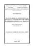
- Fresh Anthenea aspera (10 Kg) 1. Chopped 2. Extracted by EtOH (3x8L), filtered 3. Evaporated Crude EtOH 213 g EtOAc (1Lx3) n-hexane (1Lx3) MeOH (1Lx3) Crude hexane Crude MeOH Crude EtOAc (SDE) (SDH) (SDM) 68 g 45 g 96 g CHCl3/MeOH gradien CH2Cl2/ MeOH 10:0-8:2 hexane/CH2Cl2 100:0-0:100 CH2Cl2/ MeOH 100:0-0:100 SDH1 ….. SDH4 SDH6 …... SDH9 SDE1 …… SDE3 SDE7 SDE9 SDM1 …. SDM4 SDM7 …. SDM11 Hexane/EtOAc gradien RP-C18 CHCl3/MeOH/H2O 4/1/0,1 MeOH/H2O/NH4OH CH2Cl2/ MeOH 3/1 70/29/1 CHCl3/MeOH/H2O 5/1/0,1 CHCl3/MeOH/H2O AA1 AA2 CH2Cl2/ MeOH 05/1 2/1/0,1 0,17 g 0,53 g AA4 15 mg AA5 AA6 AA3 48 mg 30 mg AA9 1,02 g AA7 AA8 AA10 AA11 9 mg 17 mg 33 mg 10 mg 8 mg Figure 3.2. Diagram for the isolation procedure of starfish A. aspera 3.2.2. Physical properties and spectral data of isolated compounds. CHAPTER 4 - RESULTS AND DISCUSSION This chapter presents the results of isolation and structural elucidation of isolated compounds from 2 starfish A. aspera and A. sibogae, cytotoxic activity, tumor suppression activity on soft agar and anti-proliferative activity of some isolated compounds. 4.1. Structural elucidation of isolated compounds from the starfish A. sibogae This section details the results of the structural determination of 11 compounds isolating from A. sibogae, including 6 new compounds and 5 known compounds. ASB2. Thymine ASB1. Cholesterol 5
- ASB3. L-tyrosine ASB4. Tryptophan ASB6. Anthenoside S2 (new compound) ASB5. Anthenoside S1 (new compound) ASB7. Anthenoside S3 (new compound) ASB8. Anthenoside S4 (new compound) ASB9. Anthenoside S5 (new compound) ASB10. Anthenoside S6 (newcompound) ASB11. Mixture of Anthenoside J và Anthenoside K 6
- 4.1.5. ASB5 compound: (20R, 22E)-7-O- (6-O-methyl-β-D-galactofuranosyl) - 16-O- (3-O-methyl- β -D-glucopyranosyl) -24-nor- 5α-cholesta-8(14), 22(23) - diene-3α, 6β, 7β, 16α-tetraol (anthenoside S1, new compound) The molecular formula of ASB5 was determined to be of C40H66O14 from the [M + Na]+ sodium adduct ion peak at m/z 793,4346 in the (+)-HR-ESI-MS spectrum. The 1H- and 13 C-NMR spectroscopic data reffered to the steroidal nucleus of ASB5 revealed the chemical shifts of H- and C-atoms of two angular Me groups H3–C(18) [δ(H) 0,91 (s), δ(C) 20,3] and H3–C(19) [δ(H) 0,84 (s); δ(C) 15,5], an 8(14) double bond (δ(C) 126,6; 147,6), two HC–O groups, including H‒ C(3) [δ(H) 4,07 ‒ 4,08 (m, W = 11,8); δ(C) 67,5] and H‒C(6) [δ(H) 3,64 (t, J = 2,8); δ(C) 75,2], as well as two HC–O groups bearing Omonosaccharide residues, including H‒C(7) [δ(H) 4,23 (d, J = 2,8); δ(C) 78,5] and H‒C(16) [δ(H) 4,63 (td, J = 9,0; 5,0); δ(C) 79,2]. The width of the multiplet of H‒C(3) about 12 Hz corresponded well to the 3α-OH configuration while the width of the multiplet of H‒C(3) at the 3β-OH configuration is more than 30 Hz. The H- and C-atom resonances of the H3‒C(18), H3‒C(19), H‒C(3), H‒C(6), H‒C(7), and H‒C(16) were similar to the same signals in the NMR spectra of anthenoside Q and testified about a Δ8(14)-3α,6β,7β,16α-tetrahydroxysteroidal nucleus glycosylated at the C(7) and C(16) positions in ASB5. The NMR spectra of the aglycon side chain showed the presence of three secondary Me groups H3–C(21) [δ(H) 1,10 (d, J = 7,0); δ(C) 23,8], H3–C(26) [δ(H) 0,98 (d, J = 6,7); δ(C) 23,2], and H3–C(27) [δ(H) 0,97 (d, J = 6,7); δ(C) 23,1], and a 22(23) double bond [δ(H) 5,74 (ddd, J = 15,3; 8,8; 1,2), 5,30 (dd, J = 15,3; 6,8); δ(C) 133,9; 137,2]. Based on these data, a (22E)-Δ22-24-nor-cholestane side chain has been assumed in ASB5. A thorough analysis of the COSY, HSQC, HMBC, and ROESY spectra led to the assignment of all the H- and C-atom resonances of the steroidal moiety in ASB5 (Tables 4.6 and 4.7, Fig. 4.1.27). The H- and C-atom sequences at H-C(1) to H-C(7), H-C(9) 7
- to H-C(12) through H-C(11), H-C(15) to H-C(17), H-C(17) to H-C(20), H-C(20) to H-C(22), H-C(23) to H3-C(27) were ascertained using the COSY and HSQC experiments. The total structure of the steroidal aglycon of ASB5 was supported by the key HMBC correlations H-C(6)/C(8), C(10); H-C(15)/C(8), C(14), C(17); H3-C(18)/C(12), C(13), C(14), C(17); H3-C(19)/С(1), С(9), С(10); H3- C(21)/C(17), C(20), C(22); H-C(22)/C(25); and H-C(23)/C(20), C(25), C(26), C(27). The key ROESY cross-peaks, such as H3-C(19)/Hβ-C(2), Hβ-C(4), Hβ- C(11); H3-C(18)/Hβ-C(12), Hβ-C(15), H-C(16); H-C(5)/Hα-C(1), Hα-C(9); Hα- C(4)/Hα-C(6); Hβ-C(4)/H-C(19); and Hα‒C(7)/Hα-C(15), along with the values of the coupling constants of H-C(6), H-C(7), and H-C(16), confirmed the 3α,6β,7β,16α relative configurations of O-bearing substituents and 5α/9α/10β/13β steroidal nucleus in ASB5. The resonance of H3-C(21) at δ(H) 1,10 (δ(H) 1,10 for (20R)-Δ22- and δ(H) 1,00 for (20S)-Δ22-steroids) as well as the ROESY correlations of H3-C(18)/H-C(20), H3-C(21); H3-C(21)/Hβ-C(12); and H-C(22)/H- C(16) allowed us to assume the (20R)-configuration. As a result, we proposed the (20R,22E)-24-nor-5α-cholesta-8(14),22(23)-diene-3α,6β,7β,16α-tetraol structure as the aglycon moiety of ASB5. The 1H-NMR spectrum of ASB5 showed two resonances of anomeric H- atoms at δ(H) 4,33 and 5,02, correlated in the HSQC experiment with corresponding C-atom signals at δ(C) 102,9 and 108,4, resp. The (+)-ESI-MS/MS spectrum of the [M + Na]+ ion peak at m/z 793 exhibited fragment ion peaks at m/z 599 ([(M + Na) – С7H14O6]+) and 217 ([С7H14O6 + Na]+). The (‒)-ESI- MS/MS spectrum of the [M ‒H]- ion peak at m/z 769 displayed fragment ion peaks at m/z 575 ([(M – H) – С7H14O6]-) and 193 ([С7H13O6]-). All peaks corresponded to the loss of O-methyl-hexose residue. The chemical shifts and coupling constants of H-C(1)-H-C(6) of two O-methyl-hexose units were determined by the irradiation of anomeric H-atoms in the 1D TOCSY 8
- experiments. Moreover, the H- and C-atom signals of the monosaccharide residues of ASB5 were established using 2D-NMR experiments (Tables 4.6 and 4.7). These chemical shifts and the corresponding coupling constants coincided well with those of terminal 3-O-methyl-β-glucopyranosyl and 6-O-methyl-β- galactofuranosyl residues. Acid hydrolysis of glycoside 1 with 2M TFA was carried out to ascertain the stereochemical series of its monosaccharide units. Alcoholysis of the obtained monosaccharides by (R)-(-)-2-octanol followed by acetylation, GC analysis, and comparison of retention times of acetylated (-)-2- octyl glycoside derivatives with the corresponding derivatives of standard monosaccharides allowed us to establish the D-configuration of the 3-O-methyl- glucose and 6-O-methyl-galactose (Experimental Section). The attachment positions of the monosaccharide units to the steroidal aglycon were defined by the HMBC and ROESY spectra, where the cross-peaks between H-C(1’) of 3-OMe- Glcp and C(16), H-C(16) of the aglycon and H-C(1’’) of 6-OMe-Galf and C(7), H-C(7) of the aglycon were observed (Fig. 1.27). On the basis of all the above mentioned data, the structure of anthenoside S1 was elucidated to be (20R,22E)-7- O-(6-O-methyl-β-D-galactofuranosyl)-16-O-(3-O-methyl-β-D-glucopyranosyl)- 24-nor-5α-cholesta-8(14),22(23)-diene-3α,6β,7β,16α-tetraol (ASB5). Fig. 4.1.27. Chemical structure ASB5 9
- Table 4.6. 1H-NMR data of compounds ASB5-ASB10 Vị ASB5 ASB6 ASB7 ASB8 ASB9 ASB10 trí 1 1,51-1,55 1,51- 1,50-1,56 (m) 1,51-1,55 1,52- 1,50- (m) 1,55 (m) 1,28-1,31 (m) (m) 1,55 (m) 1,54 (m) 1,29-1,31 1,28- 1,28-1,34 1,29- 1,28- (m) 1,30 (m) (m) 1,32 (m) 1,30 (m) 2 1,61-1,63 1,60- 1,60-1,64 (m) 1,60-1,62 1,60- 1,60- (m) 1,64 (m) (m) 1,63 (m) 1,63 (m) 3 4,07-4,08 4,06- 4,06-4,08 (m) 4,06-4,08 4,07- 4,07- (m) 4,08 (m) (m) 4,09 (m) 4,09 (m) 4 1,96 (td, J 1,96 (td, 1,96 (td, J = 1,96 (td, J 1,98 (td, 1,96 (td, = 14,0; J = 14,0; 14,0, 2,8) =14,0;2,8) J= J = 14,0; 2,8) 2,8) 1,36 (br d, J = 1,36 (br d, J 13,7, 2,8) 1,37 (br d, 1,36 (td, 14,0) = 14,0) 2,8) 1,37 J = 14,0) J=14,0) 1,38 (br (brd, J = d, J = 14,0) 13,7) 5 2,12 (dt, J 2,12 (dt, 2,12 (dt, J = 2,13 (dt, J 2,16 (td, 2,13 (dt, = 14,0, J =14,0; 14,0; 2,8) =14,0, 2,8) J =13,7; J = 14,0, 2,8) 2,8) 2,8) 2,8) 6 3,64 (t, J 3,63 (t, J 3,64(t, J = 2,8) 3,64 (t, J = 3,53 (t, 3,61- = 2,8) = 2,8) 2,8) J = 2,8) 3,63 (m) 7 4,23 (d, J 4,22 (d, J 4,26 (d, J = 2,8) 4,27 (d, J = 4,25 (d, 4,23 (d, = 2,8) = 2,8) 2,8) J = 2,8) J = 2,8) 8 - - - - - - 9 2,25-2,28 2,25- 2,26-2,28 (m) 2,25-2,29 2,24- 2,25- (m) 2,28 (m) (m) 2,26 (m) 2,28 (m) 10 - - - - - - 11 1,63-1,67 1,63- 1,62-1,66 (m) 1,62-1,66 1,65- 1,63- (m) 1,66 (m) 1,50-1,56 (m) (m) 1,68 (m) 1,67 (m) 1,52-1,55 1,51- 1,51-1,56 1,50- 1,51- (m) 1,55 (m) (m) 1,56 (m) 1,57 (m) 12 1,82 (dt, J 1,81 (dt, 1,85 (dt, J = 1,87-1,89 1,80 (dt, 1,82 (dt, 10
- = J = 12,4; 12,0; 3,3) (m) J= J = 12,3; 12,5;3,3) 3,4) 1,28-1,32 (m) 1,30-1,34 12,0; 3,5) 1,24 1,23-1,27 1,23- (m) 3,4) ‒ 1,28 (m) 1,27 (m) 1,19- (m) 1,22 (m) 13 - - - - - - 14 - - - - - - 15 2,86 (ddd, 2,85 2,84 (ddd, J = 2,85 (ddd, J 2,93 2,87 J = 17,0; (ddd, J = 17,0; 8,7; 3,0) = 17,0; 8,6; (ddd, J (ddd, J = 9,0, 2,8) 17,0; 9,0, 2,68 (ddd, J = 2,9) = 16,8; 17,0; 2,64(ddd, 2,8) 17,0; 4,1; 1,8) 2,62-2,69 9,0; 3,0) 9,0, 3,1) J = 17,0; 2,64 (m) 2,38- 2,60 5,0, 2,1) (ddd, J = 2,41 (m) (ddd, J = 17,0; 5,0, 17,0; 2,1) 5,2, 1,8) 16 4,63(td, J 4,62 (td, 4,55 (td, J = 4,57 (td, J = 4,47 4,46 (td, = 9,0; 5,0) J = 9,0; 8,7; 4,1) 8,6, 4,2) (ddd, J J = 9,0; 5,0) = 9,8, 5,2) 9,0; 6,0) 17 1,53 (dd, 1,54 (dd, 1,49-1,51 (m) 1,51 (dd, J 1,46 1,48 (dd, J = 9,0; J = 9,0; = 8,6, 6,7) (dd, J = J = 9,0; 4,0) 4,0) 9,8; 2,7) 4,6) 18 0,91 (s) 0,91 (s) 0,94 (s) 0,95 (s) 0,89 (s) 0,93 (s) 19 0,84 (s) 0,84 (s) 0,85 (s) 0,85 (s) 0,83 (s) 0,85 (s) 20 2,38-2,42 2,38- 1,67-1,74 (m) 1,69-1,75 2,37- 1,66- (m) 2,42 (m) (m) 2,42 (m) 1,71 (m) 21 1,10 (d, J 1,10 (d, 1,03 (d, J = 6,8) 1,03 (d, J = 1,09 (d, 1,06 (d, = 7,0) J = 7,1) 6,8) J = 7,3) J = 6,8) 22 5,74 (ddd, 5,76 (d, J 1,71-1,76 (m) 1,85-1,89 5,65 1,79- J = 15,3; = 15,3; 1,43-1,47 (m) (m) (dd, J = 1,86 (m) 8,8, 1,2) 8,8, 1,2) 1,52-1,56 15,4, 1,43- (m) 7,3) 1,47 (m) 23 5,30 (dd, 5,31(dd, 2,07-2,12 (m) 2,19-2,23 5,37 2,21- J = 15,3, J = 15,3; 1,88-1,93 (m) (m) (dd, J = 2,24 (m) 11
- 6,8) 6,8) 1,87-1,91 15,4, 1,92- (m) 7,3) 1,97 (m) 24 - - 5,14 (br t, J - 1,92(br - =7,5) td, J =7,3, 3,3) 25 2,22-2,30 2,26- - 2,24-2,31 1,56- 2,24- (m) 2,29 (m) (m) 1,62 (m) 2,30 (m) 26 0,98 (d, J 0,98 (d, J 1,67 (s) 1,03 (d, J = 0,89 (d, 1,04 (d, = 6,7) = 6,6) 6,9) J = 6,5) J = 6,9) 27 0,97 (d, J 0,97 (d, J 1,60 (s) 1,03 (d, J = 0,89 (d, 1,04 (d, = 6,7) = 6,6) 6,9) J = 6,5) J = 6,9) 28 4,72 (d, J = 4,75 (br 1,3) s) 4,70 (br s) 4,72 (br d, J =1,3) 3-OMe-β- 4-OMe- 3-OMe-β-D- 3-OMe-β- 3-OMe- β-D- D-Glcp β-D- Glcp D-Glcp β-D- Galf Glcp Galf 1’ 4,33 (d, J 4,29 (d, J 4,32 (d, J = 7,7) 4,32 (d, J = 4,97 (br 4,95 (d, = 7,7) =7,8) 7,8) s) J = 2,4) 2’ 3,23 (dd, 3,17 (dd, 3,21(dd, J = 9,1, 3,22 (dd, J 4,00 3,96 (dd, J = 9,3; J =9,3; 7,7) =9,1, 7,8) (dd, J = J = 4,8; 7,7) 7,8) 2,5; 1,1) 2,4) 3’ 3,09 (t, J 3,46 (t, J 3,08 (t, J = 9,1) 3,08 (t, J = 3,74 4,04 (dd, = 9,3) = 9,3) 9,1) (dd, J = J = 7,2; 5,6; 2,5) 4,8) 4’ 3,36 (t, J 3,10 (t, J 3,32 (t, J = 9,1) 3,27-3,29 4.08 3,88 (dd, = 9,3) = 9,3) (m) (dd, J = J = 7,2; 5.6, 3.5) 2,4) 5’ 3,27 (ddd, 3,26 3,25 (ddd, J = 3,26 (m) 3.73- 3,70- J= (ddd, J = 9,1; 5,7; 2,5) 3.75 (m) 3,73 (m) 9,3;5,5, 9,3; 5,1, 12
- 2,5) 2,2) 6’ 3,88(dd, 3,86(dd, 3,86(dd, J=11,6, 3,86(dd, 3,65(br 3,62 (dd, J=11,6; J=11,6; 2,5) J=11,6, 2,5) d, J = 11,2; 2,5) 2,2) 3,65(dd,J=11,6, 3,63(dd, J=6,1) 7,2) 3,70(dd, 3,71(dd, 5,7) J=11,6, 5,6) 3,59 (dd, J=11,6; J=11,6; J = 11,2, 5,5) 5,1) 4,5) OMe 3,63 (s) 3,56 (s) 3,62 (s) 3,62 (s) 3,41 (s) 6-OMe-β- 6-OMe- 6-OMe-β-D- 6-OMe-β- 6-OMe- D-Galf β-D-Galf Galf D-Galf β-D- Galf 1’’ 5,02 (d, J 5,01 (d, J 5,05 (d, J = 2,0) 5.04 (d, J = 4.99 (d, = 2,0) = 2,0) 2.0) J = 2.3) 2’’ 3,90 (dd, 3,89- 3,90 (dd, J = 3.90 (dd, J 3.91 (dd, J = 3,7; 3,91 (m) 4,3, 2,0) =3.6, 2.0) J = 3.8, 2,0) 2.3) 3’’ 3,94 (dd, 3,94 (dd, 3,90-3,96 (m) 3.93 (dd, J 3.95 (dd, J = 6,2; J = 6,1; = 6.8, 3.6) J = 6.2, 3,7) 3,6) 3.8) 4’’ 3,90- 3,89- 3,91(dd, J = 5,8, 3.92 (dd, J 3.88 (dd, 3,91(m) 3,91 (m) 3,4) = 6.8, 3.5) J = 6.2, 3.9) 5’’ 3,82 (ddd, 3,82 3,82 (ddd, J = 3.82 (ddd, J 3.82 ‒ J = 7,0; (ddd, J = 7.2, 4.8, 3.4) = 7.1, 4.8, 3.85 (m) 5.2; 3,4) 7,0; 5,2, 3.5) 3,3) 6’’ 3,53 (dd, 3,53 (dd, 3,53(dd, J = 3,54 (dd, J 3,53 (d, J = 10,3; J = 10,1; 10,1, 4,8) = 10.1, 4.8) J = 6,0) 5,2) 5,2) 3,52 (dd, J = 3,52 (dd, J 3,52 (dd, J 3,52 (dd, 10,1; 7,2) = 10.1, 7,1) = J = 10,1; 10,3;7,0) 7,0) OMe 3,38 (s) 3,38 (s) 3,38 (s) 3,38(s) 3,39(s) 13
- Measured in CD3OD, 700 MHz 13 Table 4.7. C-NMR spectroscopic data of compounds ASB5-ASB10 Position ASB5 ASB6 ASB8 ASB9 ASB10 1 34.6 34.5 34.6 34.5 34.5 2 29.6 29.6 29.6 29.5 29.6 3 67.5 67.5 67.3 67.4 67.5 4 33.3 33.3 33.2 33.4 33.3 5 38.0 38.0 37.9 37.7 38.0 6 75.2 75.5 75.5 77.4 75.2 7 78.5 78.7 78.7 72.1 78.4 8 126.6 126.6 126.6 128.4 127.0 9 45.9 45.9 45.6 45.8 45.8 10 38.8 38.8 38.9 38.7 38.9 11 19.5 19.5 19.6 19.4 19.5 12 37.2 37.1 37.6 36.9 37.3 13 45.4 45.4 45.4 44.9 45.1 14 147.6 147.6 148.0 146.8 147.4 15 34.3 34.5 34.2 33.5 33.8 16 79.2 79.4 80.1 76.9 77.7 17 62.8 62.8 62.7 62.6 62.7 18 20.3 20.3 19.7 20.5 20.1 19 15.5 15.4 15.5 15.3 15.4 20 37.2 37.1 34.1 37.1 32.9 21 23.8 23.8 20.8 24.6 21.4 22 133.9 133.8 34.6 137.7 33.8 23 137.2 137.4 33.0 129.5 33.3 24 - - 158.4 43.2 157.7 25 32.3 32.3 34.9 29.9 34.9 26 23.2 23.2 22.6 22.8 22.5 27 23.1 23.1 22.4 22.3 28 108.6 107.2 3-OMe-β-D- 4-OMe- 3-OMe-β- 3-OMe- β-D- Glcp β-D- D-Glcp β-D- Galf Glcp Galf 1’ 102.9 103.0 102.6 108.2 107.6 2’ 75.2 75.2 75.1 80.9 83.7 3’ 87.9 78.2 87.8 88.9 78.3 4’ 71.5 81.2 71.7 84.3 84.4 14
- 5’ 77.8 77.2 77.9 73.2 72.4 6’ 63.2 62.7 63.4 65.1 65.4 OMe 61.0 60.8 61.0 58.1 6-OMe -β- 6-OMe- 6-OMe-β- 6- D-Galf β-D- D-Galf OMe- Galf β-D- Galf 1’’ 108.4 108.4 108.5 108.4 2’’ 83.4 83.4 83.4 83.5 3’’ 78.7 78.7 78.7 78.7 4’’ 85.0 85.0 85.0 85.0 5’’ 70.8 70.8 70.8 70.8 6’’ 75.5 75.5 75.5 75.5 OMe 59.4 59.4 59.4 59.4 Measured in CD3OD, 176 MHz 4.2. Structure elucidation of isolated compounds from the starfish A. aspera 4 sterols and 7 other compounds were isolated for the first time from hexane, ethyl acetate and methanol extracts of starfish A. aspera collected in Northeast Vietnam. AA1. Cholesterol AA2. Lathosterol AA4.Cholestan-3β,5α,6β,15α,16β-26-hexol AA3. Cholest-4-ene-3β,6β-diol AA5. Cyclo(L-glycine-L-proline) AA6. L-glycine-L-propyl 15
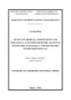
- AA7.Cyclo(L-alanine-4-hydroxyl-L-prolyl AA8. L-Phenylalanine AA9. Tyramine AA10. Thymine AA11. Uracil 4.2.1. AA1 compound: cholesterol Compound AA1 has melting point, Rf and NMR spectrum coincide with compound ASB1 4.2.2. AA2 compound: Lathosterol (Cholest-7,8-ene-3 -ol) Fig. 4.2.8. Chemical structure of AA2 Table 4.14. NMR spectrum data of AA2 and reference substance Position # C Ca,c Ha,b(mult., J, Hz) 1 37.1 37.1 1.82 m/ 1.07 m 2 31.3 31.4 1.80 m/ 1.61 m 3 70.7 71.1 3.60 m 4 37.8 38.0 1.27 m/ 1.72 m 5 40.2 40.3 1.40 m 6 29.6 29.7 1.76 m 7 117.2 117.4 5.15 m 8 139.3 139.6 - 9 49.4 49.5 1.61 m 10 34.1 34.2 - 11 21.5 21.6 1.57 m, 1.45 m 12 39.5 39.5 1.20; 2.02 m 13 43.2 43.4 - 16
- 14 54.9 55.0 1.80 overlap 15 22.9 23.0 1.40 m; 1.52 m 16 27.9 27.9 1,88 m; 1.26 m 17 56.1 56.1 1.20 m 18 11.8 11.8 0.53 s 19 12.9 13.0 0.79 s 20 36.1 36.0 1.36 m 21 18.8 18.8 0.92 d (6.5) 22 36.1 36.2 0.99 m; 1.34 m 23 23.9 23.9 1.14 m, 1.34 m 24 39.4 39.6 1.13-1.10 m 25 27.9 28.0 1.52 m 26 22.5 22.6 0.86 d (7.0) 27 22.7 22.8 0.87 d (7.0) a CD3OD, b500 MHz, c125 MHz, #δC data of [58] 4.2.3. AA3 compound: cholest-4-ene-3 ,6 -diol Fig. 4.2.13. Chemical structure of AA3 Table 4.15. NMR spectrum data of AA3 and reference substance Position Ca,c Ha,b(mult., J, Hz) C δCa,c Ha,b(mult., J, Hz) 1 37.4 1.76 (m); 1.32 (m) 15 25.2 1.64 (m); 1.40 (m) 2 29.6 1.91 (m); 1.49 (m) 16 29.2 1.88 ( m), 1.32 (m) 3 69.2 4.11-4.16 (trùng H-6) 17 57.6 1.14 (m) 4 121.4 5.67 (d, J 1.5 Hz) 18 12.4 0.75 (s) 5 149.5 - 19 19.2 1.08 (s) 6 68.6 4.11-4.16 (trùng H-3) 20 37.1 1.43 (m) 7 43.7 0.87 (m) 21 19.2 0.95 (d, J 6.5) 8 35.8 1.58 (m) 22 37.3 1.39 (m); 1.05 (m) 9 55.9 0.75 (m) 23 24.9 1.14-1.22 (m) 10 38.9 - 24 40.7 1.10-1.21 (m) 11 22.1 1.39; 1.53 (m) 25 29.1 1.55 (m) 12 41.1 2.04-2.06 (m) 26 23.2 0.89 (d, 6.5) 17
- 13 43.2 - 27 22.9 0.86 (d, 6.5) 14 57.4 1.07 (m) a CD3OD, b500 MHz, c125 MHz, #δC data of [77] 4.2.4. AA4 compound: cholestane 3 ,5,6 ,15,16 ,26-hexol Fig. 4.2.21. Chemical structure of AA4 Table 4.16. NMR spectrum data of AA4 and reference substance Position # C Cac Hab, mult (J = Hz) HMBC (HC) NOESY 1 31.7 31.7 1.79 m; 1.51 m C-5, C-19 2 33.5 33.5 1.62 m; 1.35 m C-10 3 68.4 68.3 4.03 m (5.5) 4 41.6 41.5 2.08 dd (11.5; 13.0) C-3, C-5 5 76.6 76.6 - 6 76.6 76.4 3.49 dd (2.5; 3.0) C-4, C-5, C-8, H-4, H-7 C-10 7 35.4 35.2 1.89 m C-6, C-8, C-9, C-14 8 32.2 31.1 2.01 m C-7, C-9, C-14 9 46.7 46.6 1.41 m 10 39.5 39.3 - 11 22.0 21.9 1.38 m C-13 12 42.1 42.0 1.98 m; 1.20 m C-9, C-14 13 44.9 44.7 - 14 61.2 60.9 0.98 m C-13, C-16, C-18 15 85.0 85.1 3.76 dd (2.5; 10.0) C-8, C-14, C-16 H-18 16 83.2 83.0 3.99 dd (2.5; 7.5) C-13, C-15 H-17 17 60.1 59.9 1.27 m C-13, C-18, C-20 18 15.2 15.1 0.93 s C-12, C-13, C-14, C-17 18
CÓ THỂ BẠN MUỐN DOWNLOAD

-

Summary of chemical doctoral thesis: Synthesis of hydrotalcites bearing corrosion inhibitors and fabrication of nanocomposite coatings for corrosion protection of carbon steel
 31 p |
  27
|
27
|  7
7
-
Summary of Chemical doctoral thesis: Synthesis of hydrotalcite bearing benzothiazolylthiosuccinic acid (BTS) modified by silane and applied in solventborne epoxy coating for corrosion protection of carbon steel
31 p | 34
| 7
-

Summary of chemistry doctoral thesis: Study on chemical constituents and biological activities of Tacca vietnmensis and Tacca chantrierispecies growing in Vietnam
27 p | 50
| 5
-
Summary of Chemistry Doctoral thesis: Study on chemical constituents and cytotoxic activities of Glochidion Glomerulatum and Glochidion Hirsutum growing in study on chemical constituents and cytotoxic activities of glochidion glomerulatum and glochidion hirsutum growing in Vietnam
27 p | 53
| 5
-
Summary of Chemistry Doctoral Thesis: Research on chemical constituents and biological activities of callicarpa candicans and callicarpa macrophylla growing in Vietnam
29 p | 19
| 4
-

Summary of Chemical doctoral thesis: Study on chemical composition and develop extraction technology process to make valuable products from Docynia indica (Wall.) Decne) fruits in Vietnam
27 p | 47
| 4
-
Summary of Chemistry doctoral thesis: Chemical research and biological activity of two species of tai chua (Garcinia cowa Roxb. Ex Choisy) and dang hoang (Garcinia hanburyi Hook. F) growing in Vietnam
27 p | 15
| 4
-
Summary of Chemical doctoral thesis: Study on fabrication nano Platinum modified glassy carbon electrode for application to analyze lead, cadmium in the water environment
28 p | 33
| 4
-
Summary of Chemistry doctoral thesis: Study on chemical constituents and biological activities of Vitex limonifolia Wall. Ex C.B.Clarke and Vitex trifolia L.
27 p | 33
| 4
-
Summary of Chemistry Doctoral thesis: Study on chemical constituents and biological activities from Culcita Novaeguineae Müller & Troschel, 1842 and Pentaceraster Gracilis (Lutken, 1871) in Viet Nam
28 p | 27
| 4
-
Summary of Chemical Doctoral thesis: Synthesis and characterization of Silica/Polypyrrole Nanocomposite oriented for use in organic corrosion protection coating
27 p | 53
| 4
-

Summary of Chemical doctoral thesis: Studying the chemical composition and biological activity of the Sheic species (Callicarpa candicans) and the large-leafed Tuzhou (Callicarpa macrophylla) in Vietnam
29 p | 37
| 3
-
Summary of Chemical doctoral thesis: Study on enhancement of technical characteristics for some composite rubbers with nano additive
27 p | 29
| 3
-
Summary of Chemical doctoral thesis: Study on chemical constituents and biological activities of two species Markhamia stipulata var. canaenses V.S. Dang and Stereospermum binhchaunesis V.S. Dang of Bignoniaceae
27 p | 18
| 3
-
Summary Of Chemistry Doctoral Thesis: Study on chemical constituents and biological activities from the tubers of Ophiopogon Japonicus (L.F.) KER-GAWL
27 p | 44
| 3
-
Summary of chemistry doctoral thesis: Study on chemical constituents and biological activities from the leaves of Excoecaria agallocha L. and Excoecaria cochinchinensis Lour.
27 p | 33
| 3
-

Summary of chemical doctoral thesis: Research on building a dataset for qualitative and quantitative analysis of active ingredients in F. multiflora (Thunberg) Haraldson by high performance liquid chromatography
23 p | 28
| 2


Chịu trách nhiệm nội dung:
Nguyễn Công Hà - Giám đốc Công ty TNHH TÀI LIỆU TRỰC TUYẾN VI NA
LIÊN HỆ
Địa chỉ: P402, 54A Nơ Trang Long, Phường 14, Q.Bình Thạnh, TP.HCM
Hotline: 093 303 0098
Email: support@tailieu.vn
Giấy phép Mạng Xã Hội số: 670/GP-BTTTT cấp ngày 30/11/2015 Copyright © 2022-2032 TaiLieu.VN. All rights reserved.

