NANO EXPRESS Open Access
Ag nanoparticles/PPV composite nanofibers with
high and sensitive opto-electronic response
Jinfeng Chen
1
, Peipei Yang
1
, Chunjiao Wang
2
, Sumei Zhan
1
, Lianji Zhang
1
, Zonghao Huang
1*
, Wenwen Li
1
,
Cheng Wang
1,3
, Zijiang Jiang
1
, Chen Shao
1
Abstract
The novel Ag nanoparticles/poly(p-phenylene vinylene) [PPV] composite nanofibers were prepared by
electrospinning. The transmission electron microscope image shows that the average diameter of composite fibers
is about 500 nm and Ag nanoparticles are uniformly dispersed in the PPV matrix with an average diameter of
about 25 nm. The Fourier transform infrared spectra suggest that there could be a coordination effect to a certain
extent between the Ag atom and the πsystem of PPV, which is significantly favorable for the dissociation of
photoexcitons and the charge transfer at the interface between the Ag nanoparticle and the PPV. The Au top
electrode device of the single Ag/PPV composite nanofiber exhibits high and sensitive opto-electronic responses.
Under light illumination of 5.76 mW/cm
2
and voltage of 20 V, the photocurrent is over three times larger than the
dark current under same voltage, which indicates that this kind of composite fiber is an excellent opto-electronic
nanomaterial.
Introduction
Recently,1Dopto-electronicnanomaterials,especially
the 1D organic opto-electronic nanomaterials, have
received much attention of scientists because of their dis-
tinctive geometries, novel opto-electronic properties, and
the potential application in nano/micro devices [1-5].
Electrospinning is an efficient technique for the fabri-
cation of 1D polymer-based nanomaterials. Up to now,
a lot of polymers and polymer-based composite materi-
als have been fabricated by electrospinning [5-7]. Poly
(p-phenylene vinylene) [PPV] is a typical conjugated
polymer which has good photoluminescent [PL] and
electroluminescent propertiesaswellasphotovoltaic
and nonlinear optical properties [8-10]. Our research
group has successfully fabricated the PPV nanofibers
and the PPV-based composite nanofibers by electrospin-
ning, such as TiO
2
/PPV and CdSe/PPV nanofibers, etc.,
which showed novel opto-electronic properties [11,12].
Metal nanomaterials exhibit many novel physical and
chemical characteristics which arise from their quan-
tum confinement effects and their enormously large
specific surface areas. Therefore, metal nanomaterials
are used as a kind of block to build advanced func-
tional materials or to improve the efficiency of devices
in many researches. Lee et al. [13] reported that the
incorporation of gold nanodots on the indium tin
oxide surface can obviously increase the power conver-
sion efficiency of poly(3-hexylthiophene)/[6][6]-phenyl
C61-butyric acid methyl ester solar cell. Nah et al. [14]
reported that the electrochromic absorption was mark-
edly enhanced in Ag nanoparticles embedded in MEH-
PPV composite films. The opto-electronic response of
the pristine PPV film device is relative low [10], which
makes the investigation of the opto-electronic charac-
ter of a single PPV nanofiber difficult. We expect that
incorporating Ag nano-particles in PPV nanofibers can
prepare a novel composite nanofiber with a high opto-
electronic response.
In this paper, Ag nanoparticles/PPV composite nanofi-
bers were successfully prepared by electrospinning.
Then, the Au top electrode device of a single composite
nanofiber was fabricated on a SiO
2
substratebyan
‘organic ribbon mask’technique, which showed high
and sensitive opto-electronic response.
* Correspondence: huangzh295@nenu.edu.cn
1
Faculty of Chemistry, Northeast Normal University, Changchun, 130024,
People’s Republic of China
Full list of author information is available at the end of the article
Chen et al.Nanoscale Research Letters 2011, 6:121
http://www.nanoscalereslett.com/content/6/1/121
© 2011 Chen et al; licensee Springer. This is an Open Access article distributed under the terms of the Creative Commons Attribution
License (http://creativecommons.org/licenses/by/2.0), which permits unrestricted use, distribution, and reproduction in any medium,
provided the original work is properly cited.
Experimental
Preparation of Ag nanoparticles and Ag/PPV composite
nanofibers
Sodium borohydride (NaBH
4
) was purchased from Sino-
pharm Chemical Reagent Co., Ltd. (Shanghai, China),
while ethanol and silver sulfate (Ag
2
SO
4
)werefrom
Beijing Beihua Fine Chemicals Co., Ltd. (Beijing, China).
All reagents were of analytical grade and used without
further purification.
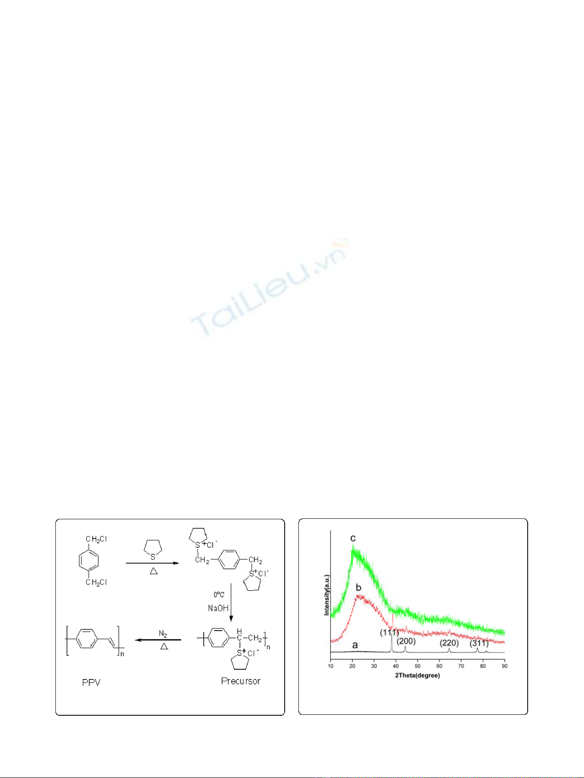
The synthesis route of PPV is presented in Figure 1,
and the PPV precursor ethanol solution (0.4 wt.%) was
prepared according to [15]. Ag nanoparticles were pre-
pared by the reduction of silver ions in Ag
2
SO
4
with
NaBH
4
.
The synthesis process of Ag nanoparticles/PPV com-
posite nanofibers is as follows: Firstly Ag
2
SO
4
(0.21 g,
0.67 mmol) was dissolved in 100 ml distilled water to
get a clear solution, and then NaBH
4
(1.40 g, 0.037 mol)
was added into the solution with vigorous stirring under
N
2
-saturated atmosphere. After filtering, drying, and tri-
turating, we obtained Ag nanoparticles. Then, 3 mg of
Ag nanoparticles was added into 0.4 wt.% PPV precur-
sor ethanol solution (2.63 g) with stirring at room tem-
perature for 24 h to obtain a new solution. Then, the
solution was electrospun at room temperature, with
positive voltage of 15 kV, humidity of 45%, and tip-to-
collector distance of 20 cm. Finally, the electrospun
fibers were heated at 180°C for 4 h in a vacuum oven
for conversion of the PPV precursor to PPV.
The pristine PPV nanofibers were also prepared in a
similar procedure as described above.
Characterization
The small-angle X-ray diffraction [SAXRD] measurements
were performed on a small-angle X-ray diffractometer
(PX13-010, Japan). Fourier transform infrared [FTIR] mea-
surements were carried out on a Fourier transform infra-
red spectrometer (Magana 560, Nicolet Corp., Madison,
WI, USA). The photoluminescence excitation [PLE] and
PL measurements were made on an Eclipse Fluorescence
Spectrophotometer (Varian Corp., Palo Alto, CA, USA).
The morphology of nanofibers was observed using a trans-
mission electron microscope [TEM] (FP 5021/20, Czech
Republic). A scanning electron microscope [SEM] (ESEM
XL-30, FEI Company, Hillsboro, OR, USA) was used to
reveal the structure of the Au top electrode device of a sin-
gle Ag/PPV composite nanofiber. The opto-electronic
response of this device was measured with a Keithley 4200
SCS and a Micromanipulator 6150 probe station in a
clean and shielded box.
Results and discussion
SAXRD patterns
The SAXRD patterns of Ag nanoparticles, Ag/PPV com-
posite nanofibers, and pure PPV nanofibers are shown
in Figure 2, using CuKaradiation (l= 0.154060 nm).
The same diffraction peaks at 38.13°, 44.23°, 64.48°, and
77.33° in the SAXRD patterns of Ag particles and Ag/
PPV composite fibers can verify the generation of highly
crystalline Ag nanoparticles with the face-centered cubic
crystal structure (JCPDS card no. 04-0783).
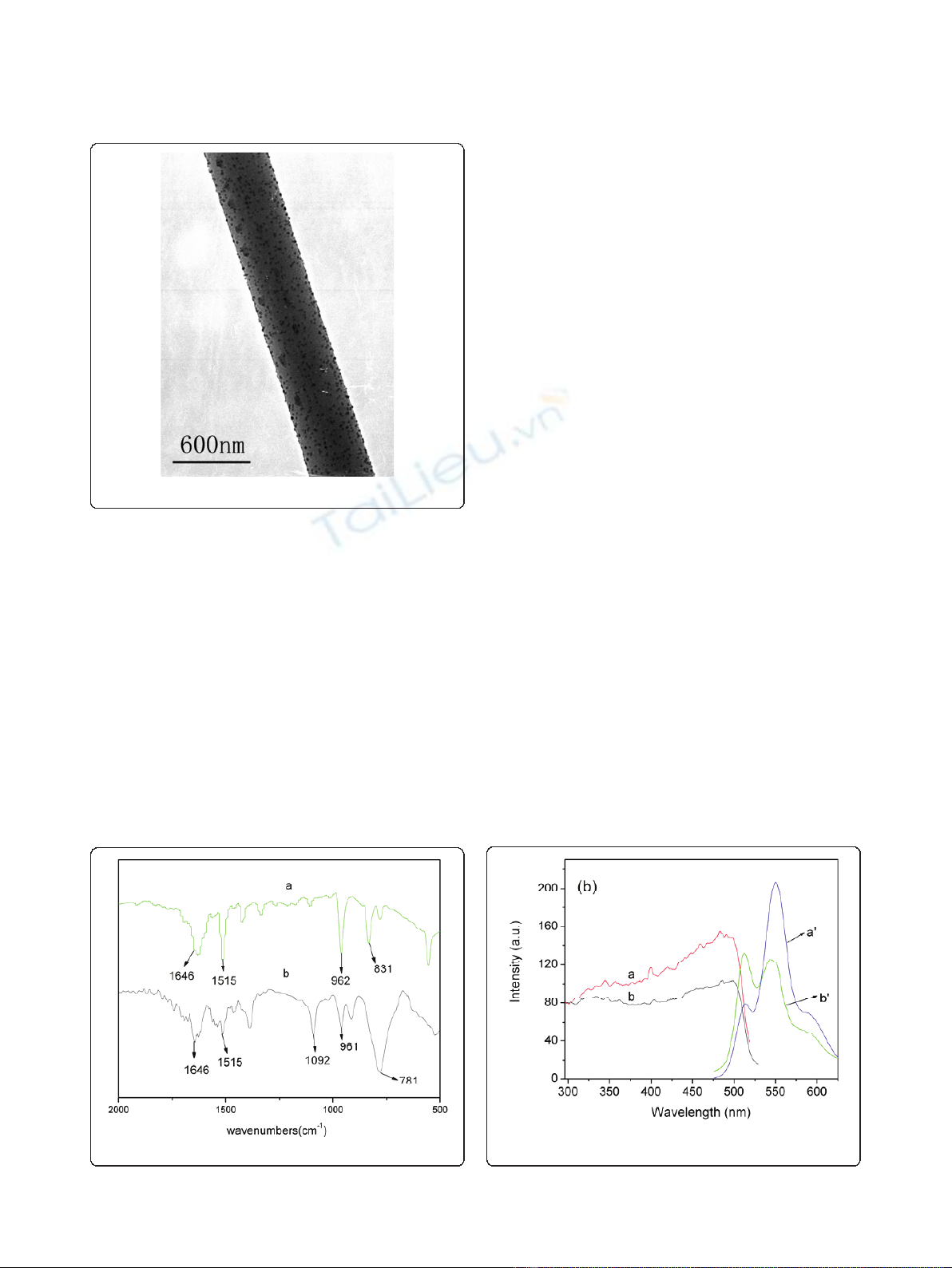
TEM image
TheTEMimageofanAg/PPVcompositefiber
(Figure 3) shows that the fiber diameter was about
500 nm and the average diameter of Ag nanoparticles
was about 25 nm. During electrospinning, the Cou-
lomb repulsion among charged Ag nanoparticles
should be the main factor making the Ag nanoparticles
uniformly dispersed.
Figure 1 The synthesis route of PPV.
Figure 2 XRD patterns.aAg nanoparticles, bAg/PPV composite
fibers, cpure PPV fibers.
Chen et al.Nanoscale Research Letters 2011, 6:121
http://www.nanoscalereslett.com/content/6/1/121
Page 2 of 5
FTIR spectra
From the FTIR spectra of pristine PPV fibers and com-
posite fibers (Figure 4), we can conclude that both pris-
tine PPV fibers and composite fibers have the three
similar characteristic absorption peaks at 1,646 cm
-1
(C = C bond stretching mode), 1,515 cm
-1
(C-C ring
stretching mode), and 962 cm
-1
(trans-vinylene C-H
out-of-plane bending mode), which implies that the con-
jugation structure of PPV is basically kept in composite
fibers. However, the characteristic absorption peak of
the pristine PPV fibers at 831 cm
-1
(p-phenylene C-H
out-of-plane bending mode) was obviously broadened in
the spectrum of composite fibers. This phenomenon
could be explained by the coordination effect to a
certain extent between the 5Sorbital of the Ag atom,
locating over the conjugation plane of PPV, and the π
system of PPV (especially the πsystem of the benzene
ring part). Therefore, the SP
3
hybrid orbital component
could be partly introduced into the C-H bond (SP
2
hybrid orbital) of the benzene ring. The occurrence of
the coordination effect should be significantly favorable
for the charge separation of photoexcitons and the
charge transfer at the interface between Ag nanoparti-
cles and PPV so as to obviously improve the opto-
electronic response of the composite materials.
PL spectra
Figure 5 shows the PL (l
ex
= 350 nm) and PLE (l
em
=
550 nm) spectra of the pristine PPV nanofibers and the
composite nanofibers. The positions of the PPV charac-
teristic peaks (at 515 and 550 nm) did not change,
which indicates that the functional structure of PPV in
the composite fibers is kept, which is consistent with
the FTIR result. However, in the composite fibers’spec-
trum, the relative enhancement of 515-nm emission
peaks, compared with the 550-nm emission peaks, indi-
cates that the addition of Ag nanopartilces decreases the
reabsorption among PPV chains.
Opto-electronic characteristics of the single composite
fiber device
To measure the opto-electronic property of the compo-
site nanofiber, the novel ‘organic ribbon mask’technique
of Professor Hu’sgroup[16]wasusedtoconstructthe
Au top electrode device of the single composite fiber
shown in Figure 6a,b. The fabrication process of the
device is briefly described below: Firstly, a single compo-
site nanofiber was transferred onto the SiO
2
substrate
(the white line and the horizontal fiber in Figure 6a,b,
Figure 3 TEM image of an Ag/PPV composite nanofiber.
Figure 4 FT-IR spectra.aPure PPV fibers, bcomposite fibers.
Figure 5 PL (l
ex
= 350 nm) and PLE (l
em
= 550 nm) spectra.a
Pure PPV nanofibers, bcomposite nanofibers.
Chen et al.Nanoscale Research Letters 2011, 6:121
http://www.nanoscalereslett.com/content/6/1/121
Page 3 of 5
respectively). Then, an organic ribbon with a diameter
of approximately 1.5 μm was picked up and crossed
over the composite nanofiber. Finally, the gold was
vacuum-deposited. After the ‘organic ribbon mask’was
peeled off, the insulate part acted as the channel of
device (see the dark part in Figure 6b). Finally, the insu-
latelinesweredrawnontheAufilmusingamicroma-
nipulator probe to form a polygon (indicated by the
arrows in Figure 6a), which was the device outline.
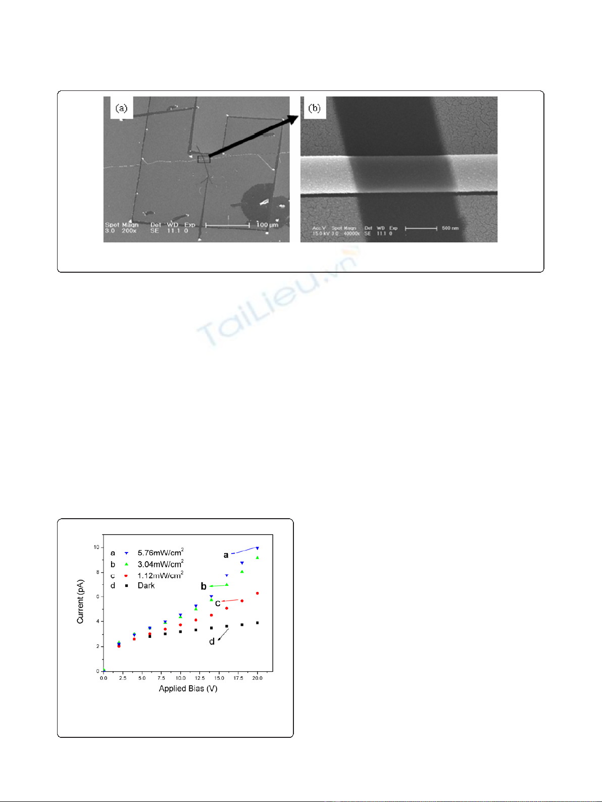
The I-V characteristics of the device were measured
under light illumination from a Xe lamp with different
intensities at room temperature in the shielded box.
Figure 7 shows that the photocurrent of the composite
nanofiber obviously increases with increasing the light
intensity from 0 to 5.76 mW/cm
2
.TheI-V curves in
Figure 7 show the non-ohmic character, which is consis-
tent with the I-V curves of PPV and its composite film
devices [17]. Under light illumination of 5.76 mW/cm
2
and voltage of 20 V, the photocurrent is over three
times larger than the dark current, which indicates that
the composite nanofibers have high and sensitive opto-
electronic response. The reason for the improvement
(or enhancement) of the opto-electronic response of the
composite nanofiber should be attributed to the follow-
ing factor: there is the coordination effect to a certain
extent between the Ag atom and the πsystem of PPV
mentioned in “FTIR spectra.”
It is noticed that the two I-V curves under light
illumination of 3.04 and 5.76 mW/cm
2
are close to each
other before 10 V, e.g., the phenomenon of photore-
sponse saturation happens, which could be related to
the charge accumulation and de-trapping by light effects
at the contacts.
Conclusions
The Ag nanoparticles/PPV composite nanofibers with
an average diameter of 500 nm were prepared by elec-
trospinning. The TEM image shows that the Ag nano-
particles with an average diameter of 25 nm were
dispersed uniformly in the PPV matrix. It was deduced
from the FTIR spectra that there was a complexation
between the Ag atom and the πsystem of PPV, which
should be significantly favorable for the charge separa-
tion of photoexcitons and the charge transport at the
interface between the Ag particles and the PPV. The J-V
measurement of the device under light illumination with
different intensities shows that the Ag nanoparticles/
PPV composite nanofibers have high and sensitive opto-
electronic response and will have good potential applica-
tion in the micro/nano organic opto-electronic field.
Acknowledgements
We thank Prof. Wenping Hu and Dr. Yajie Zhang (Beijing National Laboratory
for Molecular Sciences and Key Laboratory of Organic Solids, Institute of
Chemistry Chinese Academy of Sciences) for the fabrication of the Au top
Figure 6 top electrode device of the single composite fiber.aSEM image of Au top electrode device (the part closed by polygon indicated
by the red arrows). bSEM image of the device’s channel (insulate gap) and crossed nanofiber.
Figure 7 Photocurrent of the composite nanofiber.J-V curves of
the single Ag/PPV composite nanofiber under light illumination
with different intensities.
Chen et al.Nanoscale Research Letters 2011, 6:121
http://www.nanoscalereslett.com/content/6/1/121
Page 4 of 5

electrode device and the measurement of I-Vcurves. The research described
herein was supported by the National Natural Science Foundation (grant no.
2077 4017), Natural Science Foundation of Heilongjiang Province of China
(grant no. B200606), and Analysis and Testing Foundation of Northeast
Normal University.
Author details
1
Faculty of Chemistry, Northeast Normal University, Changchun, 130024,
People’s Republic of China
2
Bilingual Teaching Training Center, Changchun
Normal University, Changchun 130032, People’s Republic of China
3
Faculty
of Chemistry and Materials Science, Heilongjiang University, Harbin 150080,
People’s Republic of China
Authors’contributions
JC and PY carried out the preparation of the fibers and the devices. CW
drafted the manuscript. SZ and LZ participated in sequence alignment. ZH
contrubited to the redaction of the manuscript and in the design of the
study. WL and CW and CS participated in the certain measurements. All
authors read and approved the final manuscript.
Competing interests
The authors declare that they have no competing interests.
Received: 28 April 2010 Accepted: 7 February 2011
Published: 7 February 2011
References
1. Gu FX, Zhang L, Yin XF, Tong LM: Polymer single-nanowire optical
sensors. Nano Lett 2008, 8:2757.
2. Yu KH, Chen JH: Enhancing Solar Cell Efficiencies through 1-D
Nanostructures. Nanoscale Res Lett 2009, 4:1.
3. Kjelstrup-Hansen J, Norton JE, da Silva Filho DA, Brédas J-L, Rubahn H-G:
Charge transport in oligo phenylene and phenylene-thiophene
nanofibers. Org Electron 2009, 10:1228.
4. Chang M-Y, Wu C-S, Chen Y-F, Hsieh B-Z, Huang W-Y, Ho K-S, Hsieh T-H,
Han Y-K: Polymer solar cells incorporating one-dimensional polyaniline
nanotubes. Org Electron 2008, 9:1136.
5. Li D, Xia YN: Fabrication of titania nanofibers by electrospinning. Nano
Lett 2003, 3:555.
6. Li ZY, Huang HM, Wang C: Electrostatic Forces Induce Poly(vinyl alcohol)-
Protected Copper Nanopartocles to Form Copper/Poly(vinyl alcohol)
Nanocables via Electrospinning. Macromol Rapid Commun 2006, 27:152.
7. Xin Y, Huang ZH, Peng L, Wang DJ: Photoelectric performance of poly(p-
phenylene vinylene)/Fe
3
O
4
nanofiber array. J Appl Phys 2009, 105:086106.
8. Burroughes JH, Bradley DDC, Brown AR, Marks RN, Mackay K, Friend RH,
Burns PL, Holmes AB: Light-emitting diodes based on conjugated
polymer. Nature 1990, 347:539.
9. Prasad PN, Williams DJ: Introduction to Nonlinear Optical Effect in
Molecules and Polymers. New York: Wiley; 1991, 284.
10. Marks RN, Halls JJM, Bradley DDC, Friend RH, Holmes AB: The photovoltaic
response in poly(p-phenylene vinylene) thin-film device. J Phys Condens
Matter 1994, 6:1379.
11. Jiang ZJ, Huang ZH, Yang PP, Chen JF, Xin Y, Xu JW: High PL-efficiency
ZnO nanocrystallites/PPV composite nanofibers. Compos Sci Technol 2008,
68:3240.
12. Wang C, Yan EY, Huang ZH, Zhao Q, Xin Y: Fabrication of Highly
Photoluminescent TiO2/PPV Hybrid Nanoparticle-Polymer Fibers by
Electrospinning. Macromol Rapid Commun 2007, 28:205.
13. Lee JH, Park JH, Kim JS, Lee DY, Cho K: High efficiency polymer solar cells
with wet deposited plasmonic gold nanodots. Org Electron 2009, 10:416.
14. Nah YC, Kim SS, Park JH, Park HJ, Jo J, Kim DY: Enhanced electrochromic
absorption in Ag nanoparticle embedded conjugated polymer
composite films. Electrochem Commun 2007, 9:1542.
15. Halliday DA, Burn PL, Friend RH, Bradley DDC, Holmes AB: Determination
of the average molecular weigth of poly(P-phenylenevinylene). Synthetic
Met 1993, 55:902.
16. Jiang L, Gao JH, Wang EJ, Li HX, Wang ZH, Hu WP, Jiang L: Organic Single-
Crystalline Ribbons of a Rigid “H"-type Anthracene Derivative and High-
Performance, Short-Channel Field-Effect Transistors of Individual Micro/
Nanometer-Sized Ribbons Fabricated by an “Organic Ribbon Mask”
Technique. Adv Mater 2008, 20:2735.
17. Álvaro M, Corma A, Ferrer B, Galletero MS, García H, Peris E: Increasing the
Stability of Electroluminescent Phenylenevinylene Polymers by
Encapsulation in Nanoporous Inorganic Materials. Chem Mater 2004,
16:2142.
doi:10.1186/1556-276X-6-121
Cite this article as: Chen et al.: Ag nanoparticles/PPV composite
nanofibers with high and sensitive opto-electronic response. Nanoscale
Research Letters 2011 6:121.
Submit your manuscript to a
journal and benefi t from:
7 Convenient online submission
7 Rigorous peer review
7 Immediate publication on acceptance
7 Open access: articles freely available online
7 High visibility within the fi eld
7 Retaining the copyright to your article
Submit your next manuscript at 7 springeropen.com
Chen et al.Nanoscale Research Letters 2011, 6:121
http://www.nanoscalereslett.com/content/6/1/121
Page 5 of 5