
RESEARC H Open Access
Co-evolution of cancer microenvironment reveals
distinctive patterns of gastric cancer invasion:
laboratory evidence and clinical significance
Chun-Wei Peng, Xiu-Li Liu, Xiong Liu, Yan Li
*
Abstract
Background: Cancer invasion results from constant interactions between cancer cells and their microenvironment.
Major components of the cancer microenvironment are stromal cells, infiltrating inflammatory cells, collagens,
matrix metalloproteinases (MMP) and newly formed blood vessels. This study was to determine the roles of MMP-9,
MMP-2, type IV collagen, infiltrating macrophages and tumor microvessels in gastric cancer (GC) invasion and their
clinico-pathological significance.
Methods: Paraffin-embedded tissue sections from 37 GC patients were studied by Streptavidin-Peroxidase (SP)
immunohistochemical technique to determine the levels of MMP-2, MMP-9, type IV collagen, macrophages
infiltration and microvessel density (MVD). Different invasion patterns were delineated and their correlation with
major clinico-pathological information was explored.
Results: MMP2 expression was higher in malignant gland compared to normal gland, especially nearby the
basement membrane (BM). High densities of macrophages at the interface of cancer nests and stroma were found
where BM integrity was destroyed. MMP2 expression was significantly increased in cases with recurrence and
distant metastasis (P=0.047 and 0.048, respectively). Infiltrating macrophages were correlated with serosa invasion
(P= 0.011) and TNM stage (P= 0.001). MVD was higher in type IV collagen negative group compared to type IV
collagen positive group (P= 0.026). MVD was related to infiltrating macrophages density (P= 0.040). Patients with
negative MMP9 expression had better overall survival (OS) compared to those with positive MMP9 expression
(Median OS 44.0 vs 13.5 mo, P= 0.036). Median OS was significantly longer in type IV collagen positive group than
negative group (Median OS 25.5 vs 10.0 mo, P= 0.044). The cumulative OS rate was higher in low macrophages
density group than in high macrophages density group (median OS 40.5 vs 13.0 mo, P= 0.056). Median OS was
significantly longer in low MVD group than high MVD group (median OS 39.0 vs 8.5 mo, P= 0.001). The difference
of disease-free survival (DFS) between low MVD group and high MVD group was not statistically significant (P=
0.260). Four typical patterns of cancer invasion were identified based on histological study of the cancer tissue,
including Washing pattern, Ameba-like pattern, Spindle pattern and Linear pattern.
Conclusions: Proteolytic enzymes MMP9, MMP2 and macrophages in stroma contribute to GC progression by
facilitating the angiogenesis. Cancer invasion patterns may help predict GC metastasis.
Background
Tumor progression represents the greatest threat to
patients with gastric cancer (GC). The 5-year survival is
significantly decreased from over 80% in early GC to
below 28% in advanced GC [1]. Over the past 25 years,
the majority of cancer studies have focused on func-
tional consequences of activating and/or inactivating
mutations in critical genes and signal pathways that reg-
ulate cell proliferation and/or cell death as cancer is
often defined as a disease of cell proliferation [2]. How-
ever, such studies have largely ignored the fact that
interactions between cancer cells and stroma are critical
for growth and invasion of epithelial tumors [3]. It has
been recognized that invasion is regulated not only by
* Correspondence: liyansd2@163.com
Department of Oncology, Zhongnan Hospital of Wuhan University, Hubei
Key Laboratory of Tumor Biological Behaviors & Hubei Cancer Clinical Study
Center, Wuhan 430071, China
Peng et al.Journal of Translational Medicine 2010, 8:101
http://www.translational-medicine.com/content/8/1/101
© 2010 Peng et al; licensee BioMed Central Ltd. This is an Open Access article distributed under the terms of the Creative Commons
Attribution License (http://creativecommons.org/licenses/by/2.0), which permits unrestricted use, distribution, and reproduction in
any medium, provided the original work is properly cited.

intrinsic genetic changes in cancer cells as ‘initiators’of
carcinogenesis, but also regulated by stroma cell as ‘pro-
moter’[4,5]. A seminal event in cancer progression is
the ability of cancer cells to mobilize the necessary
machinery to break surrounding extracellular matrix
(ECM) barriers while orchestrating a host stroma
response that ultimately supports tissue-invasive and
metastatic processes [6]. Proteolytic ECM remodeling is
considered both prerequisite and consequence of inva-
sive cell migration [7]. The cancer cell and stroma both
modulate the process of invasion by remodeling the
ECM with tumor-associated proteases such as matrix
metalloproteinase (MMPs), which subsequently break-
down proteins of the ECM such as collagens and release
the cryptic information [8,9]. Many studies have focused
on the role of extracellular proteases. It was supposed
that cancer cells break through the ECM barriers and
invade surrounding tissues in two fashions: a protease-
independent and Rho kinase (ROCK)-dependent amoe-
boid migration mode and a protease-dependent and
ROCK-independent mesenchymal migration mode [10].
Further more, the process of pericellular proteolysis
leads to ECM degradation and realignment during cell
movement and integrate it into established steps of cell
migration [11].
It has long been recognized that the behavior of
tumor systems is complex, which means that under-
standing the individual component like pericellular pro-
teolysis in more detail does not necessarily explain the
collective behavior of many individuals, and thus usually
evokes Aristotle’s quote in that ‘The whole is more than
the sum of its parts’[12]. Therefore, instead of investi-
gating a single component of cancer matrix, this study
focused on the whole tumor microenvironment related
to GC invasion, by evaluating tissue destructive proteo-
lytic enzymes MMP9 and MMP2, tissue barriers against
invasion like type IV collagen, tumor infiltrating macro-
phages, and tumor angiogenesis, all of which are essen-
tial components of tumor stroma and involved in the
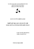
process of invasion (Figure 1.). Furthermore, the interac-
tions between cancer cells and tumor stroma termed as
‘invasion pattern’corresponding to the dynamic stroma
remodeling were also delineated so as to formulate new
concepts on cancer invasion at the histological level.
Methods
Patients and tissue samples
Tumor specimens were obtained from 37 GC patients at
the Department of Oncology, Zhongnan Hospital of
Wuhan University (Wuhan, China) from January 2004
to January 2008. Written informed consent was obtained
from the patients and the study protocol was approved
by the ethics committee of Zhongnan Hospital of
Wuhan University. Major clinico-pathological features
of these patients were listed in Table 1. The patients
underwent curative gastrectomy with D2 lymph nodes
dissection for stages I to III cases and palliative surgery
for some stage IV cases. Tumor staging was based on
TNM classification system of American Joint Committee
on Cancer (AJCC) staging criteria (version 6). All
patients beyond stage II received platinum and 5-flur-
ouracil (5-FU) based adjuvant chemotherapy beginning
21 days after surgery. The last follow-up was on Decem-
ber 1, 2009.
Immunohistochemistry
Immunolocalization of MMP9, MMP2, type IV Col-
lagen, macrophages and CD105 were performed using
streptavidin-biotin peroxidase complex method (SP).
Briefly, tissue slides were first deparaffinized in xylene,
ethanol and water, then the slides were pretreated in
0.01 M citrate buffer (pH 6.0) for MMP9, MMP2,
macrophages or 1 mM EDTA (pH 9.0) for CD105, and
heated in a microwave oven (98°C) for 10 min. For
staining, endogenous peroxidase activity was blocked by
immersing in 3% H
2
O
2
in methanol for 10 min to pre-
vent any nonspecific binding. After blocked with 2%
BSA, the slides were incubated with the primary antibo-
dies for MMP9 (sc13595, Santa Cruz, USA, dilution 1/
300), MMP2 (sc-6840, Santa Cruz, USA, dilution 1/300),
type IV collagen (ab6586, Abcam, England, dilution 1/
300), macrophages (MA1-38069, ABR, USA, dilution 1/
300), and CD105 (sc-23838, Santa Cruz, USA, dilution
1/300) for 90 min at 37°C, then incubated with the cor-
responding secondary antibody for 15 min at 37°C, and
finally incubated with peroxidase-labeled streptavidin
(Maixin Biotechnology, China) for 15 min. The reaction
products were visualized with diaminobenzidine
(DAKO, Denmark). All slides were counterstained with
haematoxylin. As a negative control, primary antibody
was replaced with Tris-buffered saline on sections that
were proven to be positive for MMP9, MMP2, type IV
collagen, macrophages and CD105 in preliminary
experiments.
Evaluation of Immunohistochemical Variables
Positive cells were stained brownish granules. The infil-
trating macrophages were counted in five high power
fields selected at the tumor invasion front, and the
mean cells counts were documented. Because CD105 is
a specific marker of newly formed and activated small
blood vessels, the MVD was calculated as the average
count from the three hotspot fields of view and used for
analysis of angiogenesis. The percentage of immunor-
eactive positive cells and intensity for MMP9, MMP2,
type IV collagen in GC were assessed. All slides were
independently observed by two investigators. The stain-
ing score of each slide was calculated by staining
Peng et al.Journal of Translational Medicine 2010, 8:101
http://www.translational-medicine.com/content/8/1/101
Page 2 of 11
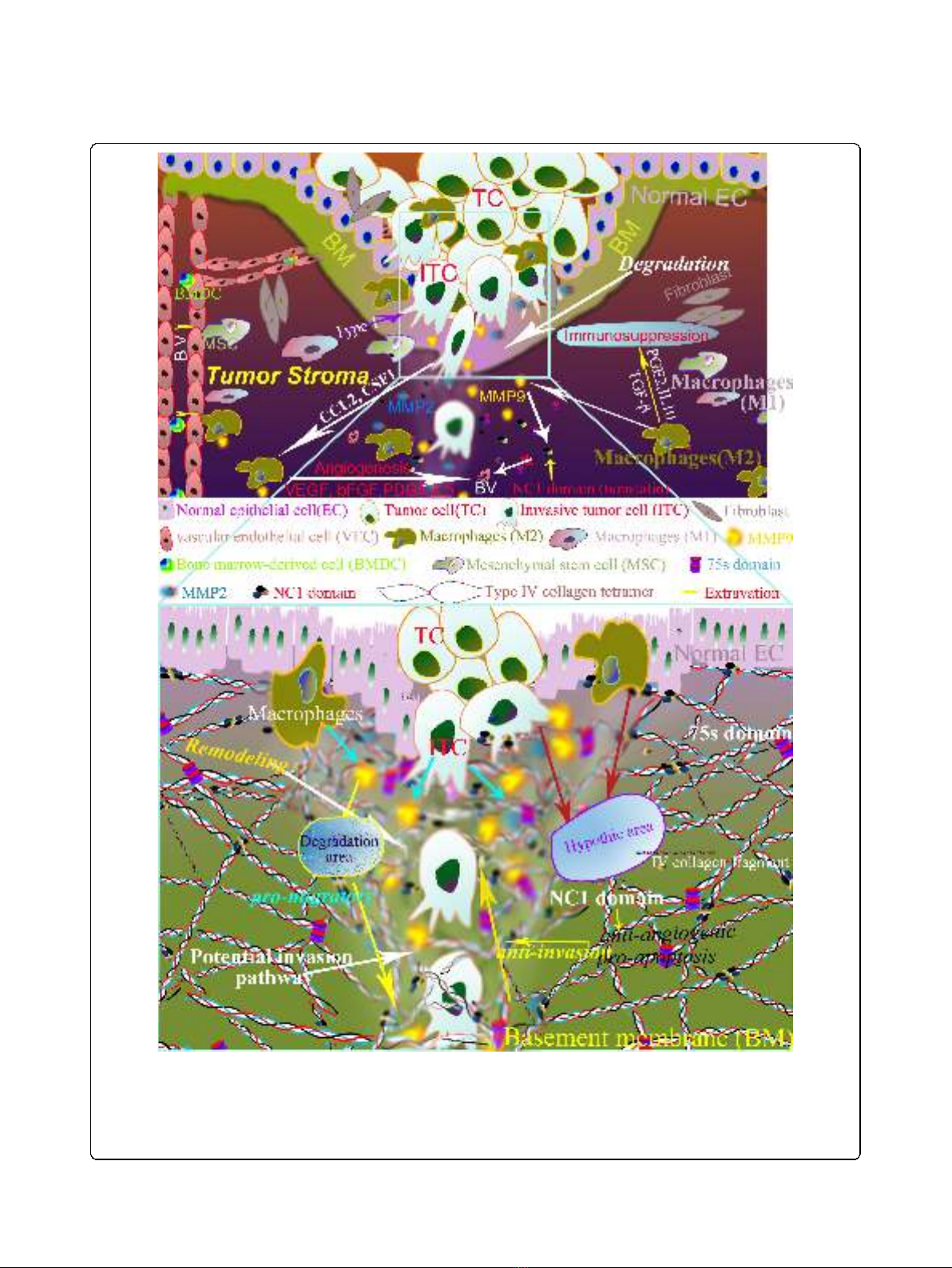
Figure 1 Co-evolution of tumor cells and their microenvironment in cancer invasions. Both of tumor cells and their microenvironment are
involved in cancer invasions. Invasion is the first observable step of cancer progression process that tumor cells cross the ECM barrier by
proteolytic enzyme such as MMPs after acquiring invasive phenotypes (upper graph). In addition, tumor infiltrating macrophages and type IV
collagen also play an important role in cancer invasion. In this process, cancer invasion networks capture “temporal evolution”and “spatial
evolution”between tumor cells and microenvironment before mechanical macrotrack can be observed as stroma remodelling at the histological
level (lower graph).
Peng et al.Journal of Translational Medicine 2010, 8:101
http://www.translational-medicine.com/content/8/1/101
Page 3 of 11

intensity and percentage of positive cancer cells. The stain-
ing intensity was scored as 1 (very weak), 2 (weak), 3
(moderate), 4 (intense) and 5 (very intense). Positive rate
score of cancer cells was: 0-10% was recorded as 0; 10-
30% was recorded as 1; 30-50% was recorded as 2; 50-75%
was recorded as 3; > 75% were recorded as 4. The expres-
sion of MMP9, MMP2 and type IV collagen, and macro-
phages infiltration in each slide were scored as the sum of
intensity and positive rate scores. Negative was defined as
the score ≤3 for MMP9, MMP2 and type IV collagen.
Statistical Analysis
Statistical analyses were performed with SPSS software
version 13.0 (SPSS Inc. Chicago, IL). Cumulative survi-
val was calculated by the Kaplan-Meier method and
analyzed by the Log-rank test. A secondary analysis was
performed to assess the relationship among immunohis-
tochemical variables and clinicopathological characteris-
tics. For the comparison of individual variables, Fisher’s
exact test, t test and Mann-Whitney Test were con-
ducted as appropriate. Two-tailed P< 0.05 was judged
to be significant.
Results
Immunohistochemical characteristics
Immunohistochemical analysis showed the linearity of
type IV collagen was disrupted indicating BM
destruction (Figure 2A). The characteristic distribution
pattern of MMP9 was diffused expression in tumor tis-
sue, although small areas of scattered expression were
also observed (Figure 2B). Furthermore, MMP2 expres-
sion was higher in malignant gland compared to normal
gland, especially nearby the BM (Figure 2C). High density
of macrophages was observed at the juncture of cancer
cells and stroma where BM integrity of gastric gland had
been broken (Figure 2D). CD105 was expressed in the
endothelium of blood vessels, but not in GC cells. The
number of CD105-positive vessels was increased at the
tumor front (Figure 2E). And CD105 is highly expressed
on proliferating endothelial cells of both the peri- and
intratumoral blood vessels (Figure 2F).
Correlation of Immunohistochemical Variables with
clinicopathologic features
Serosa invasion, lymph node status, TNM stages, recur-
rence status and distant metastasis were the variables
investigated in this study, all of which were not related
to the level of MMP9 and IV collagen, but IV collagen
expression was significantly decreased in older patients
(P= 0.042). MMP2 expressions were significantly
increased in cases with recurrence and distant metasta-
sis (P= 0.047 and 0.048, respectively). Moreover, the
expression of MMP2 expression was highest in distant
recurrence and lowest in local recurrence (P= 0.024).
Table 1 Clinicopathological characteristics in relation to MMP9, MMP2, Type IV collagen and Macrophages
immunoreactivity
Variables N MMP9 Positive
(%)
P* MMP2 positive
(%)
P* Type IV collagen Positive
(%)
P* Macrophages counts
(M ± SD)
P**
Age (yr)
≤58 18 13 (72.2) NS 9 (50.0) NS 12 (66.7) 0.042 19.9 ± 10.6 NS
> 58 19 17 (89.5) 11 (57.9) 18 (94.7) 19.4 ± 7.3
Recurrence
No 13 10 (76.9) NS 4 (30.8) 0.047
#
10 (76.9) NS 17.3 ± 7.9 NS
Yes 24 20 (83.3) 16 (66.7) 20 (83.3) 21.0 ± 9.4
Serosa invasion
No 8 7 (87.5) NS 4 (50) NS 7 (87.5) NS 12.7 ± 9.2 0.011
Yes 29 23 (79.3) 16 (55.2) 23 (79.3) 21.6 ± 8.0
Lymph node metastasis
No 10 8 (80.0) NS 3 (30.0) NS 10 (100) NS 16.3 ± 8.3 NS
Yes 27 22 (81.5) 17 (63.0) 20 (74.1) 20.9 ± 9.0
Distant Metastasis
M0 29 23 (79.3) NS 13 (44.8) 0.048 25 (86.2) NS 18.9 ± 8.3 0.09
M1 8 7 (87.5) 7 (87.5) 5 (62.5) 22.6 ± 11.0
TNM Stage
Early 11 9 (81.8) NS 3 (27.3) NS 10 (90.9) NS 12.8 ± 7.1 0.001
Advanced 26 21 (80.8) 17 (65.4) 20 (76.9) 22.6 ± 8.1
* Fisher’s exact test (two-tailed), bold face representing significant data (P< 0.05), NS: No statistically significant.
** t-test (two-tailed), bold face representing significant data (P< 0.05), NS: No statistically significant.
# The differences of MMP2 expression among different recurrence area (distant recurrence, local recurrence and ovarian recurrence) are statistically significant,
too (P= 0.024).
Peng et al.Journal of Translational Medicine 2010, 8:101
http://www.translational-medicine.com/content/8/1/101
Page 4 of 11
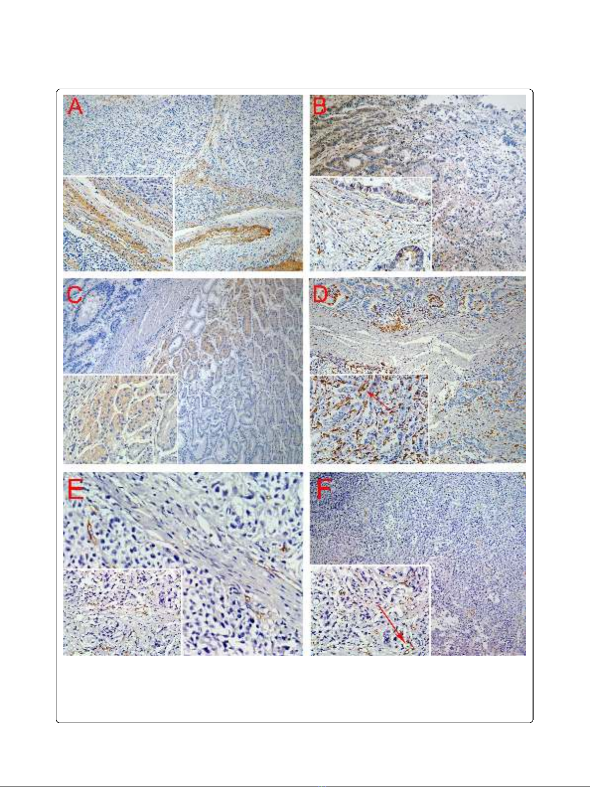
Figure 2 Positive staining of type IV collagen, MMP9, MMP2, macrophages, and microvessels. A. BM was revealed by type IV Collagen
staining. B. MMP9 was secreted by GC cells and mesenchymal. C. MMP2 expression is higher in malignant gland versus normal gland, especially
nearby the BM. D. Macrophages are mainly located in the margin of the tumor nest, and phagocytosis of cancer cells by macrophage was
observed (red arrow). E. New microvessels were increased at the tumor front. And CD105 is highly expressed on proliferating endothelial cells of
both the peri- and intratumoral blood vessels (red arrow). Magnifications: A, B, C, D, E, F: 100×; Inserts in lower left corner show the sub-cellular
localization of immunostaining at higher magnification (400×). All tissues were adenocarcinoma of GC.
Peng et al.Journal of Translational Medicine 2010, 8:101
http://www.translational-medicine.com/content/8/1/101
Page 5 of 11


![Bệnh Leptospirosis: Khóa luận tốt nghiệp [Nghiên cứu mới nhất]](https://cdn.tailieu.vn/images/document/thumbnail/2025/20250827/fansubet/135x160/63991756280412.jpg)