


Báo cáo khoa học: "A rare case of isolated wound implantation of colorectal adenocarcinoma complicating an incisional hernia: case report and review of the literature"
lượt xem 2
download
 Download
Vui lòng tải xuống để xem tài liệu đầy đủ
Download
Vui lòng tải xuống để xem tài liệu đầy đủ
Tuyển tập báo cáo các nghiên cứu khoa học quốc tế ngành y học dành cho các bạn tham khảo đề tài: A rare case of isolated wound implantation of colorectal adenocarcinoma complicating an incisional hernia: case report and review of the literature
Bình luận(0) Đăng nhập để gửi bình luận!
Nội dung Text: Báo cáo khoa học: "A rare case of isolated wound implantation of colorectal adenocarcinoma complicating an incisional hernia: case report and review of the literature"
- World Journal of Surgical Oncology BioMed Central Open Access Review A rare case of isolated wound implantation of colorectal adenocarcinoma complicating an incisional hernia: case report and review of the literature Aninda Chandra*, Lester Lee, Fahad Hossain and Harnaik Johal Address: Department of General Surgery, Queen Mary's Hospital Sidcup, Sidcup, UK Email: Aninda Chandra* - aninda_chandra@hotmail.com; Lester Lee - lester.lee@doctors.org.uk; Fahad Hossain - f.hossain@doctors.org.uk; Harnaik Johal - harnaik.johal@doctors.org.uk * Corresponding author Published: 17 January 2008 Received: 4 August 2007 Accepted: 17 January 2008 World Journal of Surgical Oncology 2008, 6:5 doi:10.1186/1477-7819-6-5 This article is available from: http://www.wjso.com/content/6/1/5 © 2008 Chandra et al; licensee BioMed Central Ltd. This is an Open Access article distributed under the terms of the Creative Commons Attribution License (http://creativecommons.org/licenses/by/2.0), which permits unrestricted use, distribution, and reproduction in any medium, provided the original work is properly cited. Abstract Background: The reported case illustrates an instance of colonic adenocarcinoma presenting as an isolated tumour 3 1/2 years after open surgery. The presentation was in some respects unique as it was complicated by an incisional hernia and occurred in the anterior abdominal wall. A literature review was performed. Case presentation: An 83 year old lady initially underwent an extended right open hemicolectomy for a mid-transverse colonic adenocarcinoma (T4N2M0). No adjacent structures were involved. After adjuvant chemotherapy, she was kept under regular surveillance. A CT scan and colonoscopy at one year were normal. At 18 months investigations including an ultrasound scan of the liver and a radioisotope bone scan were all negative. Over three and half years later the patient presented with an incisional hernia. Repeat CT scan and tumour markers were reported as negative. At operation, a mass was found within the anterior abdominal wall complicating the incisional hernia. This mass was widely resected and a laparotomy performed. Histology confirmed an adenocarcinoma of colonic origin extending to one of the lateral margins. A post-operative PET scan confirmed the absence of intra-abdominal pathology. Conclusion: The literature regarding recurrence of colonic tumours after open surgery reports low incidences of this occurring within abdominal incisions. The literature indicates prognosis is poor, but the numbers are small and distinction is often not made between isolated recurrence and those with other sites of tumour recurrence. In order to avoid missing isolated wound implantation, careful consideration should be given to those who present with new pathology related to previous cancer surgery incisions, both clinically and radiologically. Page 1 of 6 (page number not for citation purposes)
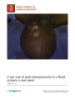
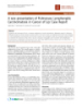
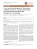
- World Journal of Surgical Oncology 2008, 6:5 http://www.wjso.com/content/6/1/5 5FU & Folinic acid. This was well tolerated with only Background The prognosis associated with colorectal cancer has signif- grade I nausea and mild hair loss and was completed at six icantly improved due to advances in early diagnosis and months post-operation. therapeutic techniques. The post-operative follow-up of such patients remain an integral part of management due The patient was seen regularly in clinic on a three monthly to the potential for recurrent disease. The prevalence of basis. At one year, the surveillance CT scan (chest, abdo- loco-regional recurrence or metastatic disease, especially men and pelvis) was unremarkable as was colonoscopy. to the liver and lung, is well recognised and hence forms At 18 months, the patient complained of lower back pain the main focus of follow-up imaging investigation. in April 2005. In view of her history a chest X-ray, tumour markers and ultrasound scan of the liver were ordered. The question of wound recurrences after laparotomy has These were all negative. A radioisotope bone scan was per- been infrequently addressed in the literature [1,2], in con- formed. The scan showed only lumbro-sacral arthritis and trast to port-site recurrences. This was due to a high inci- her pain resolved with simple analgesia. dence of early port-site/wound recurrences being reported after laparoscopic resection of colorectal malignancy At three and a half years post-surgery, she reported some [2,3]. Prospective randomised trials [4,5] showed how- mild abdominal discomfort and distension. She attrib- ever no difference between open and laparoscopic groups uted this to her incisional hernias, at the site of the mid- with less than a 1% wound recurrence rate, with at least a line scar. These had progressively worsened in size as had four year follow-up. Isolated wound recurrences of color- her symptoms. On examination, she was found to have ectal adenocarcinoma presenting after open surgery is two incisional hernias which lay 20 mm above and 20 rare: the literature reports an incidence of 0% to 0.4% of mm below her umbilicus and were 30 mm and 40 mm all resections when followed prospectively [6-8]. Isolated respectively in diameter. A contrast enhanced staging CT port-site recurrence after laparoscopic resection in large of the chest, abdomen and pelvis was performed. A mid- trials is also rare [4,5,8-10]; with one group [10] reporting line ventral hernia was noted on transverse slices of the CT an incidence of 0.2%. image but no focal lesion was reported. The anastomotic site appeared normal with no recurrent growth or lym- CT imaging is an effective modality in diagnosing recur- phadenopathy otherwise seen. Tumour markers were not rences; however it may be limited in cases where isolated elevated (CEA = 3, CA 19-9 = 3, CA125 = 5). An incisional wound recurrences following open surgery co-exist with hernia repair was subsequently arranged and a specialised other benign pathologies. The case report relates to a mesh was ordered. The provisional plan was to place the patient presenting with an anterior abdominal wall hernia mesh behind the anterior abdominal wall (anterior to the 3 1/2 years after open surgery, who was found to have an peritoneum). As there were two large defects which were closely related, a 20 cm × 15 cm Bard Composix-Mesh® (C. incidental anterior abdominal wound tumour at opera- tion, despite a pre-operative CT scan reported as normal. R. Bard, Inc., 730 Central Aves Murray Hill, New Jersey, 07974, USA) was ordered Case presentation An 83 year old lady initially underwent via a midline ver- At operation in 2007, a further midline incision was per- tical incision, an extended right hemicolectomy in 2003. formed. Following division of skin and subcutaneous tis- She had presented with weight loss with no previous med- sue the anterior abdominal wall was visualised. The two ical or surgical history. Functionally she was independent incisional hernia sacs were each identified and freed from and self-caring. Pre-operative radiology (including a stag- their attachments to the anterior abdominal wall allowing ing CT scan) showed a mid-transverse colonic lesion. pre-peritoneal access. At this point it became apparent, Colonoscopy revealed no other intra-colonic lesions and that the tissue in between the two incisional hernias was tumour markers were normal. not dense scar tissue. On palpation a hard mass measur- ing 20 mm × 20 mm in diameter was found situated At operation, there was no invasion into other structures within the anterior abdominal wall. This was not attached or the anterior abdominal wall. Histology demonstrated a to peritoneum. Thus it appeared as if it may be an isolated T4 N2 Mx adenocarcinoma in the transverse colon. The recurrence (Figure 1). The mass was excised with a wide serosa had been breached but the tumour had been com- margin and sent for histology. A formal laparotomy was pletely excised. The apical node was clear but 4 out of 11 performed and no intra-abdominal recurrence or perito- nodes were involved. The case was discussed pre- and neal seedlings were noted. post-operatively in the Gastro-intestinal (GI) multi-disci- plinary meeting (MDM) and staged as T4 N2 M0 (Dukes As defect following the wide excision was closed using the Bard Composix-Mesh®. This was attached with 3/0 Pro- C1). Adjuvant chemotherapy was offered to the patient, who subsequently underwent a weekly course of bolus lene to parietal peritoneum using continuous sutures as a Page 2 of 6 (page number not for citation purposes)
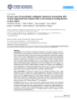
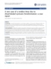
- World Journal of Surgical Oncology 2008, 6:5 http://www.wjso.com/content/6/1/5 the potential complications, she requested to be referred to an oncologist for consideration of palliative chemo- radiotherapy. Discussion After open surgery, tumour recurring within a surgical wound is uncommon but probably underestimated [7]. Two large prospective trials which looked at recurrence of colonic tumours after open surgery reported low inci- dences of abdominal scar recurrence; Hughes et al [6] reported a figure of 11 out of 1603 patients (0.7%) while Reilly et al [7] documented 9 cases from 1711 patients (0.5%). Isolated wound recurrence is an even rarer phe- nomena with laparotomy or radiology often demonstrat- Figure 1 incisional wound complicated by two incisional diameter) abdominalhernia view of tumour recurrence in anterior Sagital schematic20 mm above umbilicus (30 mmhernias: A – ing tumour recurrence at other sites [6,7,11]. Isolated Sagital schematic view of tumour recurrence in anterior occurrence occurred in the study by Reilly et al [7] in only abdominal wound complicated by two incisional hernias: A – 3 patients with abdominal or perineal wound recurrences incisional hernia 20 mm above umbilicus (30 mm diameter). B (0.2%). Hughes et al [6] stated that isolated recurrences – incisional hernia 20 mm below umbilicus (40 mm diameter). were found in only 6 abdominal scar cases (0.4%). As the study was from 1950 to 1980, this predates CT scan usage, therefore the actual incidence of isolated recurrence modified sub-lay technique. The rectus sheath was would probably have been lower if current imaging approximated but not apposed with 1/0 nylon to allow a modalities had been applied. tension free repair. A vacuum drain was placed superficial to the anterior rectus sheath. Closure was with interrupted In comparison to open surgery, wound recurrences at port subcutaneous 3/0 Vicryl sutures and clips to skin. The sites after laparoscopic surgery [12,13] were initially post-operative course was uncomplicated. thought to be more common [7]. Subsequently more objective prospective randomised trials [13,14] have The mass which measured 40 mm × 40 mm × 30 mm. His- showed no significant difference in recurrence compared tologically, it consisted of fibro-connective tissue infil- to open surgery. Two large studies [4,5] showed less than trated by a moderately differentiated adenocarcinoma. 1% wound recurrence in both laparoscopic resections and The tumour cells were seen to involve one of the lateral open colectomies, with a median follow-up of at least 4 surgical margins. There was no superior or inferior exten- years. Hartley et al., [8] found that all wound recurrences sion of the tumour. Subsequent immuno-histochemistry in their prospective study, comparing laparoscopy and was positive for CK20 and CDX2 and negative for CK7 open resection, were associated with advanced intra-peri- (Figure 2). This was characteristic of tumour cells arising toneal disease. Isolated port-site recurrence after laparo- from a colorectal origin and in keeping with the original scopic resection in large trials is rare [4,5,8-10]; Silecchia pathology. et al., [10] reported an incidence of 0.2% when cases were followed prospectively. The case was discussed again in the GI MDM. On review of the scans, a 3.6 × 1.6 cm nodule was seen in the midline Isolated tumour occurring at a point distal arises from a on the anterior abdominal wall just inferior to the hernia combination of different factors. An important factor is (Figure 3). The absence of intra-abdominal recurrence was considered to be residual viable tumour cells left in the reconfirmed, postoperatively with a repeat PET scan. The abdomen. These can be cells exfoliated from the tumour patient was subsequently seen in outpatients' clinic and [15] or by contamination of surgical equipment used the possible management strategies were outlined in the intra-operatively [16]. These cells can then disseminate to presence of the colorectal specialist nurse and the patient's the site of recurrence or spread may occur by direct iatro- surgical consultant. genic implantation. The presence of tumour cells at a site does not necessitate implantation and other local factors The presentation and case above was novel to the depart- need to be involved [17]. ment. As such an extensive literature search was per- formed using EMBASE and MEDLINE to find similar cases The trauma of surgery results in an inflammatory response and related articles. The prognosis obtained from the lit- which has been shown to enhance the successful implan- erature following surgery to attempt clearance was not sig- tation of exfoliated tumour cells in animal models [18]. Inflammatory cytokines such as TNF-α, IL-1 and IL-6 are nificantly better then adjuvant therapy. In view of this and Page 3 of 6 (page number not for citation purposes)
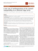
- World Journal of Surgical Oncology 2008, 6:5 http://www.wjso.com/content/6/1/5 Figure 2 nuclei B) Photomicrograph showing malignant glands typical of adenocarcinoma lined by atypical cells with hyperchromatic A and A and B) Photomicrograph showing malignant glands typical of adenocarcinoma lined by atypical cells with hyperchromatic nuclei. There is an increase in mitotic activity within the cells and the presence of necrotic material. Stained with haematoxylin & eosin. C) Immunohistochemistry with CK20 showing tumour cell cytoplasm stained. D) Immunohistochemistry with CDX2 staining showing prominent nuclei of tumour cells. CK20 and CDX2 are consistent with cells of colorectal origin. Note: Orig- inal magnifications a – d 20×. involved in angiogenesis, which is fundamental step in There were a number of clinical issues arising from this tumour development. These inflammatory cytokines case. Although disease recurrence had been the indication together with VEGF can be found in surgical wounds. for performing the preoperative investigations, the rela- They can also increase the expression of adhesion mole- tively rare occurrence of an isolated tumour within the cules and the adhesion of tumour cells becomes more suc- surgical wound (in the absence of intra-abdominal dis- cessful after the infliction of surgical trauma [17]. The ease or chest metastasis) was not appreciated by the con- environment of a healing incision can therefore not only sultant radiologist when reporting on the CT scan. The assist in the development of tumour cells, but also to their complexity of the incisional hernia with its components adhesion to cell surfaces. Wound implantation therefore lying above and below the tumour also contributed to the may be more likely in the early post operative period dur- difficulty in picking up the lesion (Figure 1). This was ing healing. The relatively late presentation of tumour compounded by normal tumour markers which included recurrence 3 1/2 years after initial surgery [1,4] as a normal CEA result. The identification of the tumour was described in the case report was an additional confound- complicated by the presence of the incisional hernia. In ing factor in the tumour not being detected pre-opera- the majority of reported cases in the literature (>90%), tively. recurrence was manifested within 2 years of surgery [1,4] Page 4 of 6 (page number not for citation purposes)
- World Journal of Surgical Oncology 2008, 6:5 http://www.wjso.com/content/6/1/5 Figure of CT scan3 abdomen showing soft tissue mass in the anterior abdominal wall (white arrow) CT scan of abdomen showing soft tissue mass in the anterior abdominal wall (white arrow). The ventral incisional hernia is seen on this slice and was arising cranially but lies superiorly to the mass. where as in the case reported it presented after 3 1/2 years. in diameter. However if this were to be used routinely as In light of the intra-operative findings, the case and the CT an imaging modality to exclude recurrence, it would be scan were presented at a joint radiological/surgical/onco- expensive. logical meeting. The lesion was retrospectively identified on the pre-operative CT images (Figure 3). This finding if Given the involvement of the surgical margins, the it had been noted pre-operatively would have altered options available were either radical re-excision or radio- management especially with regards to pre-operative therapy. Hughes et al [6] described a 5 year survival of 0% chemo-radiotherapy and the surgical approach. and Reilly et al [7] of 27% in their surgical incisional recurrences. The former study based from 1950 to 1980 In the case report, there was no clinical evidence of may have not benefited from the advances in adjuvant tumour within the wound pre-operatively. A combined chemotherapy in the last few decades. Reilly et al [7] could PET/CT scan was found by Goshen et al [11] to be not detect a significant difference in survival (or of time to extremely sensitive in detecting abdominal wound recur- recurrence) between the group with isolated recurrence rences in patients with advanced disease as small as 1 cm versus those with other sites of involvement, although the Page 5 of 6 (page number not for citation purposes)
- World Journal of Surgical Oncology 2008, 6:5 http://www.wjso.com/content/6/1/5 numbers were noted to be small. Based on the literature Written patient consent was sought and gained prior to the publication of this article the prognosis was deemed as poor even with resection. Excision and current adjuvant chemo-radiotherapy may References improve outcome but there is little definitively published. 1. Reymond MA, Bonjer HJ, Kockerling F: Port-Site and Wound Recur- rences in Cancer Surgery: Incidence, Pathogenesis. Springer 2000. Conclusion 2. Schaeff B, Paolucci V, Thomopoulos J: Port site recurrences after laparoscopic surgery. A review. Dig Surg 1998, 15:124-134. The case reported illustrates an instance of colonic adeno- 3. Jacquet P, Averbach AM, Jacquet N: Abdominal wall metastasis carcinoma recurring as an isolated tumour after open sur- and peritoneal carcinomatosis after laparoscopic-assisted gery. Its presentation was unique as it was complicated by colectomy for colon cancer. Eur J Surg Oncol 1995, 21:568-570. 4. Lacy AM, Garcia-Valdecasas JC, Delgado S, Castells A, Taura P, Pique an incisional hernia and presented in the anterior abdom- JM, Visa J: Laparoscopy-assisted colectomy versus open colec- inal wall. Tumour markers were negative and there was no tomy for treatment of non-metastatic colon cancer: a ran- domised trial. Lancet 2002, 359:2224-2229. intra-abdominal pathology. Wound implantation in an 5. The Clinical Outcomes of Surgical Therapy Study Group: A compar- incisional scar after open surgery is rare, particularly when ison of laparoscopically assisted and open colectomy for it is isolated and presentation is more than two years after colon cancer. N Engl J Med 2004, 350:2050-2059. 6. Hughes ES, McDermott FT, Polglase AL, Johnson WR: Tumor the original surgery. recurrence in the abdominal wall scar tissue after large- bowel cancer surgery. Dis Colon Rectum 1983, 26:571-572. 7. Reilly WT, Nelson H, Schroeder G, Wieand HS, Bolton J, O'Connell The literature indicates prognosis is poor, but the num- MJ: Wound recurrence following conventional treatment of bers are small and distinction is often not made between colorectal cancer. A rare but perhaps underestimated prob- isolated incisional wound implantation and those with lem. Dis Colon Rectum 1996, 39:200-207. 8. Hartley JE, Mehigan BJ, MacDonald AW, Lee PW, Monson JR: Pat- other sites of tumour recurrence or co-existent intra- terns of recurrence and survival after laparoscopic and con- abdominal malignancy. Further studies on this would ventional resections for colorectal. Ann Surg 2000, 232:181-186. shape current practice. 9. Mehta PP, Griffin J, Ganta S, Rangraj M, Steichen F: Laparoscopic- assisted colon resections: long-term results and survival. JSLS 2005, 9:184-188. There were a number of factors which arose in this case 10. Silecchia G, Perrotta N, Giraudo G, Salval M, Parini U, Feliciotti F, Lezoche E, Morino M, Melotti G, Carlini M, Rosato P, Basso N, For including the CT scan report, which may have been the Italian Registry of Laparoscopic Colorectal Surgery: Abdominal altered by a higher index of suspicion. In order to avoid wall recurrences after colorectal resection for cancer: missing isolated wound implantation, careful considera- results of the Italian registry of laparoscopic colorectal sur- gery. Dis Colon Rectum 2002, 45:1172-1177. tion should be given to those who present with new 11. Goshen E, Davidson T, Aderka D, Zwas ST: PET/CT detects pathology related to previous cancer surgery incisions, abdominal wall and port site metastases of colorectal carci- noma. Br J Radiol 2006, 79:572-577. both clinically and radiologically. 12. Berends FJ, Kazemier G, Bonjer HJ, Lange JF: Subcutaneous metas- tases after laparoscopic colectomy. Lancet 1994, 344:58. Abbreviations 13. Lacy AM, Delgado S, Garcia-Valdecasas JC, Castells A, Pique JM, Grande L, Fuster J, Targarona EM, Pera M, Visa J: Port site metas- CEA: Carcinoembryonic Antigen; CT: Computerized tases and recurrence after laparoscopic colectomy. A rand- Tomography; GI: Gastro-intestinal; MDM: Multi-Discipli- omized trial. Surg Endosc 1998, 12:1039-1042. nary Meeting; PET: Positron Emission Tomography. 14. Basha G, Penninckx F, Mebis J, Filez L, Geboes K, Yap P: Local and systemic effects of intraoperative whole-colon washout with 5 per cent povidone-iodine. Br J Surg 1999, 86:219-226. Competing interests 15. Umpleby HC, Fermor B, Symes MO, Williamson RC: Viability of exfoliated colorectal carcinoma cells. Br J Surg 1984, The author(s) declare that they have no competing inter- 71:659-663. ests. 16. Alagaratnam TT, Ong GB: Wound implantation – A surgical hazard. Br J Surg 1977, 64:872-875. 17. Oosterling SJ, van der Bij GJ, van EM, van dS Jr: Surgical trauma Authors' contributions and peritoneal recurrence of colorectal carcinoma. Eur J Surg Each author performed an independent literature search. Oncol 2005, 31:29-37. AC, and LL operated upon the patient initially, critically 18. Raa ST, Oosterling SJ, van der Kaaij NP, van den Tol MP, Beelen RH, Meijer S, van Eijck CH, van der Sijp JR, van Egmond M, Jeekel J: Sur- appraised the literature and conceived the case report; HJ gery promotes implantation of disseminated tumor cells, reviewed the literature and revised the final manuscript; but does not increase growth of tumor cell clusters. J Surg Oncol 2005, 92:124-129. FH reviewed the literature and helped in drafting the man- uscript. All authors read and approved the final manu- script. Acknowledgements Special thanks to the Department of Surgery at Queen Mary's Hospital, Sid- cup and in particular to Mr Hamid Khawaja for his support and as lead con- sultant responsible for the patient. Thanks to Dr Nana Ibrahim, Histopathology consultant for reviewing the histology and providing the immunohistochemistry annotations and pictures and to Dr Nick Maisey, Oncology consultant for correspondence regarding the case. Page 6 of 6 (page number not for citation purposes)
CÓ THỂ BẠN MUỐN DOWNLOAD

-

Báo cáo khoa học: "A rare case of “switch on and off” multi-system Langerhans cell histiocytosis in an adult patient"
 5 p |
5 p |  61
|
61
|  6
6
-
Báo cáo khoa hoc:" A patient with testicular pseudolymphoma – a rare condition mimicking malignancy: a case report"
3 p | 55
| 5
-
báo cáo khoa học: " A rare case of giant leiomyosarcoma in a filarial scrotum: a case report"
5 p | 55
| 4
-

Báo cáo y học: " A rare case of low-grade myofibroblastic sarcoma of the femur in a 38-year-old woman: a case report"
5 p | 65
| 4
-

Báo cáo khoa học: "A rare tumoral combination, synchronous lung adenocarcinoma and mantle cell lymphoma of the pleura"
5 p | 91
| 4
-

báo cáo khoa học: " A rare cause of chronic mesenteric ischemia from fibromuscular dysplasia: a case report"
9 p | 69
| 4
-

báo cáo khoa học: "A rare combination of an endocrine tumour of the common bile duct and a follicular lymphoma of the ampulla of Vater: a case report and review of the literature"
4 p | 82
| 3
-
Báo cáo y học: " A rare case of neuroleptic malignant syndrome presenting with serious hyperthermia treated with a non-invasive cooling device: a case report"
5 p | 60
| 3
-
Báo cáo y học: "A rare case of a swollen knee due to disseminated synovial chondromatosis: a case report"
5 p | 66
| 3
-
báo cáo khoa học: "A rare midbrain infarction presenting with plusminus lid syndrome with ataxia: a case report"
2 p | 44
| 3
-

báo cáo khoa học: " A rare occurrence of a steroid cell tumor of the pelvic mesentery: a case report"
4 p | 65
| 3
-
Báo cáo khoa học: "A rare disease"
2 p | 45
| 3
-
báo cáo khoa học: "A rare presentation of Pulmonary Lymphangitic Carcinomatosis in cancer of lip: case report"
3 p | 69
| 3
-
báo cáo khoa học: "A rare case of xanthogranuloma of the stomach masquerading as an advanced stage tumor"
3 p | 48
| 3
-
Báo cáo y học: "A rare case of term viable secondary abdominal pregnancy following rupture of a rudimentary horn: a case report"
3 p | 60
| 3
-
Báo cáo khoa học: "A rare coexistence of adrenal cavernous hemangioma with extramedullar hemopoietic tissue: a case report and brief review of the literature"
4 p | 72
| 2
-
Báo cáo khoa học: "A rare case of locally advanced fibrosarcoma of diaphysal humerus managed successfully with limb-sparing procedures after neoadjuvant chemotherapy"
4 p | 58
| 2


Chịu trách nhiệm nội dung:
Nguyễn Công Hà - Giám đốc Công ty TNHH TÀI LIỆU TRỰC TUYẾN VI NA
LIÊN HỆ
Địa chỉ: P402, 54A Nơ Trang Long, Phường 14, Q.Bình Thạnh, TP.HCM
Hotline: 093 303 0098
Email: support@tailieu.vn
Giấy phép Mạng Xã Hội số: 670/GP-BTTTT cấp ngày 30/11/2015 Copyright © 2022-2032 TaiLieu.VN. All rights reserved.

